Post-Ischemic Tau Protein: Comparison
Please note this is a comparison between Version 1 by Ryszard Pluta and Version 2 by Jessie Wu.

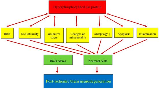
Recent data suggest that post-ischemic brain neurodegeneration in humans and animals is associated with the modified tau protein in a manner typical of Alzheimer’s disease neuropathology. Pathological changes in the tau protein, at the gene and protein level due to cerebral ischemia, can lead to the development of Alzheimer’s disease-type neuropathology and dementia. Some studies have shown increased tau protein staining and gene expression in neurons following ischemia-reperfusion brain injury. Recent studies have found the tau protein to be associated with oxidative stress, apoptosis, autophagy, excitotoxicity, neuroinflammation, blood-brain barrier permeability, mitochondrial dysfunction, and impaired neuronal function. In this review, we discuss the interrelationship of these phenomena with post-ischemic changes in the tau protein in the brain. The tau protein may be at the intersection of many pathological mechanisms due to severe neuropathological changes in the brain following ischemia. The data indicate that an episode of cerebral ischemia activates the damage and death of neurons in the hippocampus in a tau protein-dependent manner, thus determining a novel and important mechanism for the survival and/or death of neuronal cells following ischemia. In this review, we update our understanding of proteomic and genomic changes in the tau protein in post-ischemic brain injury and present the relationship between the modified tau protein and post-ischemic neuropathology and present a positive correlation between the modified tau protein and a post-ischemic neuropathology that has characteristics of Alzheimer’s disease-type neurodegeneration. 

  • brain ischemia
  • protein

1. Post-Ischemic Tau Protein versus Blood-Brain Barrier

Hyperphosphorylation of the tau protein after ischemic brain injury [1][2][3][4][5][6][7][8][9][10][11][48,49,60,62,63,64,65,70,71,79,80] triggers the development of neurofibrillary tangles [5][8][9][63,70,71], which are one of the major components of pathology in the brains of Alzheimer’s disease patients. An ischemic brain injury causes a pathological permeability of the blood-rain barrier [12][13][14][15][16][81,82,83,84,85], which also affects the hyperphosphorylation of the tau protein [1][2][3][5][6][7][17][18][8][9][10][11][48,49,60,63,64,65,67,68,70,71,79,80], and the modified tau protein may cause an additional exacerbation of blood-brain barrier dysfunction (Figure 1), which induces harmful feedback [19][86]. An accumulation of amyloid in the brain, associated with the ischemic permeability of the blood-brain barrier [20][21][87,88], may, in a roundabout manner, allow the onset of tau protein dysfunction, supporting the automatic link between amyloid accumulation and tau protein modification at some stage of blood-brain barrier breakdown [19][86]. Moreover, both oxidative stress [22][89] and neuroinflammation [23][24][16,17] cause damage to the blood-brain barrier that may cause hyperphosphorylation of the tau protein and the development of neurofibrillary tangles post-ischemia [5][8][9][25][63,70,71,90]. Moreover, after ischemia, the plasma-derived tau protein [26][27][52,53] crosses the ischemic blood-brain barrier in two directions and can enhance its own pathology in the brain [28][91]. In summary, ischemic blood-brain barrier failure may exacerbate in the brain tau protein neuropathology in post-ischemic brain injury and also suggests that ischemic brain pathology may be part of the cause responsible for the increase in the serum tau protein concentration [26][27][28][29][52,53,91,92].
Figure 1. Interrelationships between hyperphosphorylated tau protein and post-ischemic brain neurodegeneration. ↓—decrease. BBB—blood-brain barrier.

2. Post-Ischemic Tau Protein versus Excitotoxicity

Excitotoxicity has been identified as one of the most important pathological mechanisms associated with calcium changes in post-ischemic brain injury [30][31][32][23,93,94]. The existing data suggest that tau protein phosphorylation can be inhibited by reducing calcium influx into neurons [33][95]. It has been revealed that impaired glutamate homeostasis or the elevated activity of calcium-dependent kinases may induce tau protein phosphorylation [34][35][96,97], and consequently, glutamate-induced cytotoxicity may exacerbate the dysfunctional appearance of the tau protein (Figure 1) [36][74]. Conversely, many studies have shown that the tau protein also plays a significant role in enhancing excitotoxicity [37][38][39][40][41][42][58,98,99,100,101,102]. In P301L tau protein mice, KCl evoked an increase in glutamate release and decreased glutamate clearance in the hippocampus [42][102]. The exact mechanisms underlying tau protein-induced excitotoxicity require further elucidation. One study shows that the tau protein increases excitotoxicity without increasing calcium influx through the kainic acid receptor [43][103]. On the other hand, other studies suggest that reducing tau protein phosphorylation at Y18 may reduce N-methyl-d-aspartic acid receptor-mediated excitotoxicity in neurons [44][45][104,105]. Overall, the phenomenon of excitotoxicity with the phosphorylation of the tau protein leads to a vicious circle with respect to neuronal death in post-ischemic neurodegeneration (Figure 1).

3. Post-Ischemic Tau Protein versus Oxidative Stress

Oxidative stress is involved in neuropathological processes in the brain after ischemia in animals and humans. In experimental models of ischemic neurodegeneration, it has been established that the hyperphosphorylation of the tau protein may be a product of oxidative stress (Figure 1) [36][46][47][74,106,107]. Thus, tau protein hyperphosphorylation might be reduced using antioxidants [36][48][49][50][74,108,109,110]. There is no definite opinion about the causal interaction between oxidative stress and tau protein hyperphosphorylation. Some studies have shown that products of thiobarbituric acid, polyunsaturated lipids, and 4-hydroxynonenal, resulting from cell lipid peroxidation, are significantly increased, which can cause tau protein hyperphosphorylation [36][46][49][74,106,109]. Recently, it has been suggested that the hyperphosphorylation of the tau protein is due to the direct influence of reactive oxygen species, which is generated by 1,2-diacetylbenzene as a result of the phosphorylation of activated glycogen synthase kinase 3β [36][47][74,107]. Moreover, high levels of the hyperphosphorylated tau protein have been documented to initiate the production of reactive oxygen species (Figure 1). Ultimately, oxidative stress and the hyperphosphorylated tau protein may be two critical elements of the vicious cycle in the development of post-ischemic brain neurodegeneration (Figure 1).

4. Post-Ischemic Tau Protein versus Mitochondria

The activity of neurons is closely related to energy deficiency. Thus, the task of the mitochondria is to continually supply energy to neuronal and neuroglial cells. Consequently, impaired mitochondrial activity is an important neuropathological process in the brain following ischemia with subsequent recirculation. Dysfunctional mitochondrial activity is closely related to neuronal autophagy, necrosis, and apoptosis [51][111]. Mitochondrial stability conditioned by fusion and fission is a major issue in the development of mitochondrial dysfunction. Earlier data showed that protein 1 is related to dynamin, a mitochondrial fission protein, and may work together with the phosphorylated tau protein to induce mitochondrial dysfunction (Figure 1) [52][53][112,113]. A reduction in dynamin-related protein 1 protects against the hyperphosphorylated tau protein-induced dysfunction of mitochondria [54][114]. In a murine model of tauopathy, tau protein deposits undermine the distribution of mitochondria in neuronal cells [55][115]. The unusual behavior of mitochondria can be improved by reducing the level of soluble tau protein in their environment [56][55][51,115]. Tau protein accumulation can both damage normal activity and mitochondrial allocation by increasing mitofusins, which can cause ATP depletion, the development of oxidative stress, and synaptic abnormalities [57][58][59][116,117,118]. The pathway studies used axonal protein phosphatase 1, glycogen synthase kinase 3, and the retention of the C-Jun amino-terminal kinase-interacting protein 1 kinesin motor protein complex by phosphorylated tau protein, which may be involved in neuropathological interactions [60][61][119,120]. It should also be noted that tau protein phosphorylation can also be enhanced by reactive oxygen species, mimicking mitochondrial oxidative stress in neurons [62][121]. In summary, the dysfunction of the tau protein may disrupt the function and dynamics of mitochondria, and such altered mitochondria may be an indicator of tau protein phosphorylation and aggregation (Figure 1).

5. Post-Ischemic Tau Protein versus Autophagy

It is well known that autophagy plays a key role in the maintenance of normal levels of tau protein in neuronal cells [63][64][65][122,123,124]. Autophagy has been shown to be an important neuropathophysiological process in brain neurodegeneration after an ischemic stroke [66][125]. Previous research has shown that a decrease in the tau protein is correlated with an increase in an autophagy marker such as microtubule-associated protein 1A/1B-light chain 3B-II in a 3xTg mouse model of Alzheimer’s disease after reversible hypoperfusion, indicating that autophagy may be a way to reduce the dysfunctional tau protein levels in the brain [67][126]. In contrast, another study reported a significant reduction in microtubule-associated protein 1A/1B-light chain 3B protein growth and a reduction in infarct size in the P301L-Tau mouse model after ischemia [68][127]. It might be probable that autophagy insufficiency is triggered by a mutant tau protein with increased levels of its aggregates [68][127]. In addition, it has been documented that autophagy can induce tau protein expression in neuronal cells that overexpress the human P301L-Tau mutant [69][128]. In human tauopathies, p62 is an autophagy regulatory protein and its immunostaining co-localizes with tau protein inclusions [70][129]. In transgenic mice, the activity of autophagy may increase the clearance of the tau protein [71][130] and thus, reduce the aggregation of the seeded tau protein [72][131]. The phosphorylation of the tau protein is believed to be due to seeded aggregation [73][132]. The P62 and nuclear dot 52 protein are among the autophagy cargo receptors playing an important role in protecting against the aggregation of the seeded tau protein in neurons [69][74][128,133]. It is, therefore, highly likely that autophagy, not proteasomes, reduces the aggregation of the seeded tau protein (Figure 1) [69][128].

6. Post-Ischemic Tau Protein versus Apoptosis

Apoptosis is naturally programmed cell death, acting as the most important and dangerous neuronal killer following brain ischemia [75][134]. Tau protein hyperphosphorylation and apoptosis are believed to be two self-contained, self-sufficient, and overlapping neuropathological processes during neuronal death (Figure 1), although most researchers have found no significant relationship between these phenomena [76][77][135,136]. However, some studies have shown an ischemic accumulation of cyclin-dependent kinase-5 [5][63], which regulates tau protein phosphorylation, and may initiate neuronal apoptosis through degradation of the endoplasmic reticulum [78][137]. It has also been documented that hyperphosphorylation of the tau protein can be prevented by knocking down cyclin-dependent kinase-5, which may protect neuronal cells by alleviating endoplasmic reticulum stress from apoptosis [78][137]. Recent studies indicate that after cerebral ischemia, hyperphosphorylated tau protein accumulates in cortical neurons and is associated with their apoptosis (Figure 1) [1][2][3][4][5][6][48,49,60,62,63,64]. The above data clearly indicate that neuronal apoptosis after cerebral ischemia is associated with the hyperphosphorylation of the tau protein (Figure 1).

7. Post-Ischemic Tau Protein versus Neuroinflammation

Neuroinflammation is considered a pathway that influences neuronal death in the acute and chronic phase following cerebral ischemia with reperfusion [57][116]. Some previous studies have suggested that the dysfunctional tau protein is directly related to the neuroinflammatory cascade (Figure 1). It should also be noted that neuroinflammatory mediators can significantly affect the function and structure of the tau protein post-ischemia [79][80][81][73,138,139]. In addition, it has been suggested that the dysfunctional tau protein may be a trigger of the neuroinflammatory cascade (Figure 1) [79][80][81][73,138,139]. The exact role of neuroinflammatory processes in the post-ischemic neuropathology of the tau protein or the dysfunctional tau protein in neuroinflammation still needs to be clarified. Some researchers consider neuroinflammation as a worsening factor [78][137], but another study has found that neuroinflammation can lower the level of oligomeric tau protein by improving phagocytosis via microglia [82][140]. The first direct evidence for the involvement of neuroinflammation in tau protein pathology was presented in an in vitro study and showed that neuroinflammatory mediators, i.e., interleukin-1β, can promote tau protein hyperphosphorylation (Figure 1) by the stimulation of p38 mitogen-activated protein kinases [83][141]. This was also confirmed in the 3xTg model of Alzheimer’s disease in vivo with the development of plaques and tangles [84][142]. Recent studies have also shown that various stressors such as lipopolysaccharide, infection, and tumor necrosis factor-α can initiate an exacerbation of tau protein hyperphosphorylation [85][86][87][143,144,145]. As a consequence, lowering tau protein levels or inhibiting neuroinflammatory mediators may act as a treatment for tauopathies [88][146]. A study by Kovac’s group revealed a new toxic form of the misfolded tau protein, i.e., the formation of a truncated tau protein [89][72]. The truncated tau protein may increase the permeability of the blood-brain barrier (Figure 1) [89][72]. In addition, studies have also provided evidence that the truncated tau protein had a cytotoxic effect on astrocyte-microglia culture as manifested by increased levels of extracellular adenylate kinase. The blood-brain barrier damage induced by the truncated tau protein was mediated by the pro-inflammatory cytokine tumor necrosis factor α and the chemokine monocyte chemotactic protein 1 [89][72]. It should also be noted that the pro-inflammatory cytokine interferon-γ has been found to have an opposite effect on tau protein phosphorylation and dephosphorylation, and, ultimately, induced neurogenesis [90][147]. Microglial cells and macrophages play a very important role in neuroinflammation. Extracellular tau protein oligomers can be moderately phagocytosed by both microglia and macrophages under normal conditions [82][140]. Microglial internalization has been shown to be effective for both aggregated and soluble tau protein in vitro and in vivo [91][148]. Overall, the inhibition of neuroinflammation in the parenchyma of the brain may paradoxically be involved in the development of the neuropathology of the tau protein. In assessing the above information, more research is needed to elucidate these molecular phenomena.