Macrophage lncRNA in Lung Cancer: Comparison
Please note this is a comparison between Version 1 by Annika Karger and Version 2 by Vivi Li.

Ever since RNA sequencing of whole genomes and transcriptomes became available, numerous RNA transcripts without having the classic function of encoding proteins have been discovered. Long non-coding RNAs (lncRNAs) with a length greater than 200 nucleotides were considered as “junk” in the beginning, but it has increasingly become clear that lncRNAs have crucial roles in regulating a variety of cellular mechanisms and are often deregulated in several diseases, such as cancer. Lung cancer is the leading cause of cancer-related deaths and has a survival rate of less than 10%. Immune cells infiltrating the tumor microenvironment (TME) have been shown to have a great effect on tumor development with macrophages being the major cell type within the TME. Macrophages can inherit an inflammatory M1 or an anti-inflammatory M2 phenotype. Tumor-associated macrophages, which are predominantly polarized to M2, favor tumor growth, angiogenesis, and metastasis.

  • lncRNA
  • macrophage
  • TAM
  • lung cancer
  • tumor microenvironment

1. Lung Cancer

Lung cancer is one of the most frequent and most deadly types of solid cancers worldwide, and a highly complex, very heterogeneous disease. Histological classification divides lung cancers into non-small cell lung carcinoma (NSCLC, approximately 85% of the cases) and small cell lung carcinoma (SCLC, approximately 15% of the cases). NSCLC can be classified further into adenocarcinoma, squamous cell carcinoma, and large cell carcinoma. Although lung cancer is treatable with early-stage radical interventions, it remains challenging because >70% of patients relapse and expire [1][2][3][4][1,2,3,4]. In addition, the broad use of cytotoxic chemotherapies in lung cancer has reached a plateau [5]. In the past decade, exploring cancer treatment options has led to alternative therapies, such as immunotherapies and targeted therapies [6]. Cancer immunotherapies mainly target particular immune system components, such as immune-suppressive networks in the tumor microenvironment (TME), to enhance antitumor activity. Cancer cells shape their microenvironment and generate immune-suppressive networks, which finally overwhelm immunity and tumor progression [7]. Tumor cells and stromal cells, such as tumor-associated macrophages (TAMs), create an immunosuppressive environment by secreting immune-suppressive factors, such as programmed cell death ligand (PDL) 1 [8]. Additionally, the TME disturbs production of tumor-specific T cells like cytotoxic T cells (CTLs), and generates immunosuppressive leukocytes, regulatory T cells (Tregs), and myeloid-derived suppressor cells [9][10][9,10]. Various immune checkpoint inhibitors, such as CTL-associated protein 4 (CTL-4) and programmed cell death 1 (PD1), have been developed in immune-based therapies in different types of cancer [11][12][11,12]. Although immune checkpoint inhibitors provide durable responses to various cancers, they are effective in only a subset of patients [13]. Furthermore, recent advances in cancer research have led to molecular targeted therapies targeting identified gene mutations and molecular alterations to treat cancer [14]. Genomic profiling with advanced techniques like next-generation sequencing has identified molecular alterations and driver mutations in lung cancer [15]. The majority of genetic aberrations are in the epidermal growth factor receptor (EGFR), Kirsten rat sarcoma viral (KRAS), tyrosine-protein kinase MET (MET), anaplastic lymphoma (ALK), PI3KCA, ERBB2, and BRAF. EGFR and KRAS mutations are the most frequently found mutations in lung cancer. These specific mutations support tumor growth and proliferation by activating several signaling pathways [16]. These findings recently led to the development of targeted agents like T tyrosine kinase inhibitors (TKI) to target EGFR-activating mutation and to use targeted-based therapies in lung cancer. However, only about 20% of patients benefit from these targeted therapies in lung cancer patients with drug-sensitive mutations. Furthermore, drug resistance caused by genetic alterations is a major impediment to long-term therapeutic outcomes [17][18][17,18]. As a result, more in-depth research is required to develop new cancer-targeted medicines.
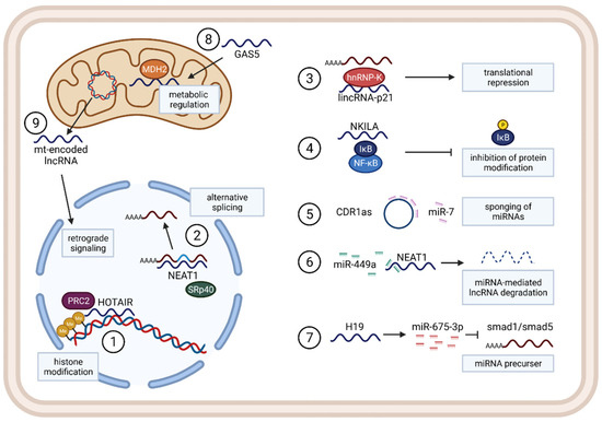
Non-coding RNAs, such as microRNAs (miRNAs), long non-coding RNAs (lncRNAs), and circular RNAs (circRNAs), are commonly dysregulated in cancer through the transcriptional, post-transcriptional, and epigenetic changes and have been shown to have significant roles in cancer initiation and progression [19][20][19,20]. Deep sequencing and microarray profiling investigations have further shown that deregulation of lncRNA expression is also a critical factor in initiation and progression of lung cancer [21][22][21,22]. Some recent studies also found that lncRNAs can influence TAM function in various ways [23][24][25][23,24,25]. However, understanding lncRNA dysregulation and function in cancer is still in its early stages. In this present review, we will first briefly discuss activation and polarization of TAMs and their role in lung cancer progression, and then specially focus on lncRNAs’ functions and mechanisms, influence of lncRNAs on TAMs, their roles in TME inflammation, and regulation pathways in lung cancer. Finally, we will discuss the therapeutic potential of lncRNAs in diseases, particularly lung cancer.

2. Long Non-Coding RNAs

The ability to sequence whole genomes and transcriptomes identified all kinds of transcripts and showed that approximately 90% of the human genome is actively transcribed. However, only about 2% of those transcripts are translated into proteins, with the rest remaining as non-coding RNAs [26][27][82,83]. The most prominent non-coding RNAs are microRNAs (miRNAs) and long non-coding RNAs (lncRNAs). MiRNAs are short, single-stranded RNA sequences (21–24 nucleotides) and mainly function via binding mRNAs, leading to their degradation and therefore translational inhibition. On the other hand, the large group of lncRNAs that comprise all ncRNAs > 200 nucleotides long are much more heterogeneous because they form complex secondary and tertiary structures and interact with proteins, DNA, or other RNAs.

2.1. Functions and Mechanisms

As a large and diverse group of regulatory non-coding RNAs, lncRNAs seem to be poorly evolutionarily conserved between species and often show tissue and cell-type-specific expression patterns. They are known to regulate through various mechanisms, for example, functioning as a guide for target proteins such as transcription factors to specific regions in the genome or by building a scaffold, binding different proteins, and bringing them closer together. Additionally, lncRNAs can be localized in different compartments of the cell and regulate on the transcriptional level, by alternative splicing, by regulating translation, or by directly interacting with proteins to influence their modification and activation (Figure 2).
Figure 2. Representative examples for general functions and mechanisms in lncRNA regulations in different cellular compartments. 1: lncRNA HOTAIR regulating histone modification by binding the PRC2 complex, recruiting it to specific genomic regions. 2: NEAT1 scaffolding SRp40 together with mRNAs, leading to regulation of alternative splicing. 3: cytoplasmatic lincRNA-p21 functions as a translational repressor, binding hnRNP-K together with a mRNA. 4: lncRNA NKILA binding the NF-κB complex inhibits phosphorylation of IkB, leading to loss of NF-κB activation. 5: CircRNA CDR1as contains miR-7 target sequences, functioning as a miRNA sponge. 6: lncRNA NEAT1 is degraded after miR-449a binding. 7: lncRNA H19 serves as a precursor RNA for several miRNAs, such as miR-675-3p that inhibits smad1 and smad5. 8: nuclear-encoded lncRNA GAS5 is able to enter the mitochondria, binding MDH2 protein and influencing TCA flux and cellular metabolism. 9: mitochondrial genome-encoded lncRNAs can traffic between the mitochondria and nucleus and provide retrograde signaling functionally.
Nuclear lncRNAs are known to regulate gene expression: e.g., through chromatin modification, such as the HOX antisense intergenic RNA (HOTAIR). HOTAIR is highly expressed in NSCLC tissue and recruits the PRC2 complex, leading to histone methylation and transcriptional repression [28][84]. Nuclear paraspeckle assembly transcript 1 (NEAT1), on the other hand, is localized in nuclear speckles, and associates with SRp40, affecting alternative splicing of transcripts (Figure 2) [29][85].
Cytoplasmatic RNAs can affect gene expression on the post-transcriptional level, such as lncRNA 1/2-sbs-RNA binding to BACE1-AS-mRNA, increasing its stability and enhancing expression or such as lincRNA-p21, binding to a target mRNA sequence, and enhancing interaction with the post-transcriptional repressor RCK and FMRP, consequently inhibiting translation [30][86]. Other lncRNAs can interact with proteins and influence protein modification, such as lncRNA NKILA, binding the NF-κB complex and repressing phosphorylation of IκB, thus inhibiting NF-κB activation [31][87]. Additionally, lncRNAs and miRNAs can influence each other due to their possible sequence complementarity, and lncRNAs can contain binding sequences and function as miRNA sponges, serving as endogenous competitors for miRNA–mRNA binding, thereby interfering with the miRNA-targeted degradation of a specific mRNA [32][88]. As endogenous competitors, LncRNAs can have the form of a circRNA, a newly identified large class of RNA that is predominantly localized in the cytoplasm of cells. CDR1as, for instance, is a circRNA containing 74 binding sequences for miR-7 [33][89]. On the other hand, miRNAs can regulate the stability of lncRNAs by binding to them and causing degradation, such as in the case of miR-449a in lung cancer, which binds to NEAT1 to inhibit the lncRNA-function [32][88]. It has also been found that lncRNA transcripts can serve as precursors for miRNAs, such as lncRNA H19, which gives rise to miR-675-5p and miR-675-3p that inhibit smad1 and smad5, among others (Figure 2) [34][90].
More recently, several mitochondrial lncRNAs were identified as regulators of cellular metabolism. Some examples are nuclear-encoded lncRNA growth arrest specific 5 (GAS5) that enters the mitochondria, binds to MDH2, and regulates TCA flux under stress conditions [35][91], or even mitochondrial genome-encoded lncRNAs, which are mostly described as sending retrograde signals to the nucleus (Figure 2) [36][92].

2.2. LncRNAs in Cancer

Given the abilities of lncRNAs for controlling all kinds of processes within the cell, it is not surprising that disruption can lead to aberrant gene expression and is associated with multiple diseases, especially cancer. In previous years, more lncRNAs have been found to regulate the occurrence and progression of many aspects of tumors by targeting genomic mutations, DNA damage, metabolic disorders, and EMT or cancer cell stemness.
Accumulation of DNA damage plays an important role in cancer development. By regulating proteins that are involved in DNA damage repair or stress response, lncRNAs can influence the mutational burden of cells. Some examples are lncRNA MEG3 that activates p53 to trigger its tumor-suppressive function [37][93], and lncRNAs CUPID1 and CUPID2, which are associated with the progression of breast cancer, modulating the DNA damage response [38][94]. Another hallmark of cancer, cellular metabolic disorders, can be regulated by lncRNAs. It has been shown that under energy stress, lncRNA NBR2 activates AMPK via direct binding, and absence of this lncRNA leads to changed metabolism and subsequent enhanced tumor cell proliferation [39][95]. MALAT1 has been extensively investigated for its function in tumorigenesis in NSCLC by promoting EMT and enhancing tumor progression and metastasis through the miR-124/STAT3 axis [40][41][42][96,97,98]. Additionally, HOTAIR is known to promote metastasis, such as in breast cancer, liver cancer, and pancreatic cancer, by activating the SMAD cascade signaling pathway, which induces EMT [43][44][45][46][99,100,101,102]. Cancer stemness is another important factor for tumor metastasis, since cancer cells with high stemness are able to survive and colonize other tissues. Several studies have shown that lncRNAs are involved in signaling pathways associated with stemness. In liver cancer cells, two lncRNAs, lncBRM and lncSox4, have been shown to participate in self-renewal through the YAP1 and the STAT3 pathways [47][48][103,104]. Altogether, lncRNAs have been shown to play a pivotal role in tumorigenesis, tumor progression, and metastasis.

2.3. LncRNAs in Immunity, Inflammation, and the TME

Although lncRNAs have been extensively described in a cancer and disease context, their role in immunity and inflammatory response is still not completely understood. The immune system consists of various cell types that mediate response to infections while maintaining tissue homeostasis [49][50][105,106]. Several studies have revealed that lncRNAs can influence proliferation, differentiation, and activation of immune cells such as monocytes, macrophages, dendritic cells, neutrophils, T cells, and B cells. For example, linc-Ccr2-5′AS is a Th2-associated lncRNA that regulates expression of Th2 genes in immune cells and has the ability to influence recruitment of Th2 cells to the lung [51][107]. Additionally, RNA sequencing analysis revealed a large number of lncRNAs specifically expressed in CD8+ and CD4+ T cells [52][53][108,109]. LncRNA Lethe was shown to be highly expressed in mouse embryonic fibroblasts, influencing NF-κB-dependent inflammatory response [54][110].
Within the tumor microenvironment, lncRNAs can act as modulators and communicators between immune cells and tumor cells. CASC2c was identified to regulate macrophage infiltration and polarization in glioblastoma by negatively regulating the expression of coagulation factor X [55][111]. Furthermore, lnc-EGFR was shown to promote differentiation of T reg cells, enhancing tumor progression of hepatocellular carcinoma [56][112].

2.4. LncRNAs in Macrophages in Lung Cancer

Although various lncRNAs seem to function in different cancer types and in all cell types of the TME, in the next chapters, we aim to focus on several examples of lncRNAs that, to our knowledge, are known to influence macrophage activation and polarization states and are associated specifically with lung cancer (Figure 3).
Figure 3. Influence of lncRNAs on modulation of TAMs of lung cancer. 1: GAS5 binds miR-21 in macrophages, upregulating PTEN and ultimately leading to an increase of proinflammatory cytokine and chemokine markers such as TNF-a, IL-6, iNOS, and IL-12. 2: Xist expression is upregulated in anti-inflammatory macrophages by transcription factor TCF4 and increases production of IL-10, CD163, and Arg-1. 3: GNAS-AS1 acts as a miRNA sponge binding miR-433-3p and miR-4319, leading to the upregulation of GATA3 and NECAB3, promoting CD206, IL-10, and Arg-1. 4: lincRNA-p21 interacts with MDM2 and inhibits the p53 complex, repressing transcription of the p53-associated gene PUMA, upregulating IL-10, IL-4, and Arg-1 and downregulating TNFa, IL-6, and iNOS. 5: Linc00662 is found in lung cancer patient plasma exosomes, lung cancer cells, and anti-inflammatory macrophages, suggesting that linc00662 is secreted into exosomes by cancer cells, activating the Wnt/ß-catenin signaling pathway in macrophages, training them to an anti-inflammatory M2 phenotype, and increasing CD163, IL-10, and Arg-1.

2.4.1. GAS5

Growth arrest specific 5 (GAS5) is a long intergenic non-coding RNA (lincRNA) that has been shown to regulate the cell cycle in various systems [57][113], and its high expression inhibits tumor progression of several cancer types [58][59][60][114,115,116]. Therefore, it is widely accepted that GAS5 acts as a tumor suppressor. In NSCLC, GAS5 seems to be downregulated, whereas overexpression leads to decreased proliferation, enhanced apoptosis, and even reduced resistance to chemo- and radiotherapy [61][62][63][117,118,119]. Mechanistically, GAS5 acts as a sponge for several miRNA molecules such as miR-21 and miR-23a, resulting in upregulation of PTEN and increased sensitivity to cisplatin treatment [63][119]. Aside from its role in cancer cells, GAS5 also has been shown to have a regulatory function on immune response and on macrophages in the TME. High expression of GAS5 was found in human IFNɣ-stimulated M1 macrophages, again accompanied by elevated expression of PTEN and upregulation of inflammatory genes such as IL-12 and TNF-α [64][120]. Downregulation of GAS5 promotes macrophages toward an M2 phenotype, suppresses inflammation, and inhibits inflammatory cytokine release [64][65][120,121], supporting its role in macrophage polarization. Additionally, GAS5 overexpression in TAMs leads to decreased tumor cell migration, whereas GAS5 knockdown reverses this effect. PTEN knockdown in GAS5 overexpressed macrophages abolishes the effect on inflammatory cytokine expression and increased Arg-1 expression [64][66][120,122], whereas overexpression of PTEN inhibits M2 polarization [67][123], again highlighting the influence of this lncRNA on macrophages through the PTEN axis. This evidence suggests that GAS5 is an important regulator promoting the proinflammatory and anti-tumorigenic macrophage activation within the TME and that targeting GAS5 could serve as an option for lung cancer treatment.
Latest insights have shown that GAS5 can also regulate the metabolic state of cells by influencing energy production through the mitochondrial respiratory chain [35][91]. Since macrophage metabolism has also been shown to affect their activation state [68][76], further research is needed to evaluate the effect of GAS5-associated metabolic regulation on macrophage phenotype within the TME.

2.4.2. Xist

The lncRNA X-inactive specific transcript (Xist) has been initially described in dosage compensation in mammalian cells by transcriptional silencing of the second X-chromosome in female cells [69][124]. More recently, functions beyond X-chromosome inactivation have been identified, including deregulation in several diseases and cancer, particularly lung cancer. Xist expression has been shown to be upregulated in NSCLC tissue and to increase cisplatin resistance in A549/DDP and H460/DDP lung adenocarcinoma cell lines [70][71][125,126] by sponging miR-144-3p and therefore leading to upregulation of the MDR1 and MRP1 chemoresistance genes. On the other hand, shRNA-mediated knockdown led to miR-144-3p–mediated downregulation of MDR1, MRP1, and reduced tumor-cell migration and invasion of lung cancer cells [71][126]. Emerging evidence also has shown Xist to be a regulator in macrophages, but its role is not yet completely clear. In some cases, high expression seems to be associated with inflammatory M1 polarization of macrophages such as in an osteoarthritis model and in IFNɣ/LPS-treated THP1 macrophages, whereas knockdown leads to a switch in polarization to a more M2 phenotype regulated via the mir-101-3p/KLF6/C/EBPa axis [72][73][127,128]. In the present study, downregulation of Xist in M1 macrophages led to higher proliferation and migration of breast and ovarian cancer cells. In lung cancer on the other hand, opposing studies have been shown which indicate that lncRNA Xist positively regulates M2 polarization of THP1 macrophages [74][129]. Furthermore, expression of Xist has not only been analyzed in the system of artificial cytokine-mediated polarization, but has also been shown to be increased in A549-conditioned macrophages, demonstrating the role of Xist in promotion of a pro-tumorigenic phenotype in TAMs associated with lung cancer. In this study, the transcription factor TCF-4 was found to be responsible for lncRNA-expression activation. Moreover, TCF-4 overexpression was able to restore downregulation of Xist-knockdown-associated downregulation of anti-inflammatory genes in macrophages such as IL-10, Arg-1, and CD163.
These findings support the characterization of Xist as an oncogenic factor in lung cancer not only from a tumor-cell site, but also in relation to macrophages in the TME. However, more studies are necessary to fully understand the underlying mechanisms and to clarify all aspects of Xist-regulation within macrophage activation.

2.4.3. GNAS-AS1

GNAS Antisense RNA 1 (GNAS-AS1) is a non-coding RNA described only very recently that seems to be upregulated especially in cancerous tissue, such as NSCLC, promoting EMT and invasiveness of cancer cells [75][76][130,131], but its expression is also associated with M2 macrophage polarization [77][132]. Two studies have demonstrated that GNAS-AS1 overexpression in macrophages led to an anti-inflammatory phenotype by promoting CD206, IL-10, and Arg-1 expression [75][77][130,132]. Additionally, overexpression in macrophages led to higher proliferation, migration, and invasion of cancer cells. In tumor cells, GNAS-AS1 has been proposed to function via regulating the WNT/β-catenin pathway, whereas in macrophages, GNAS-AS1 seems to function as a miRNA-sponge. Both miR-433-3p and miR-4319 have been shown to bind to the lncRNA sequence, inhibiting degradation of GATA3 and NECAB3, which was already shown to have oncogenic functions [78][133].
Overall, these studies suggest an interesting role of GNAS-AS1 in tumor-promoting macrophages associated with NSCLC.
Video Production Service