Hematopoietic stem cell transplant (HSCT) is a multi-step process with a high risk for complications during marrow ablation, during engraftment, or afterwards. Successful transplantation depends on the selection of the hematopoietic stem cell source, host preparation (conditioning regimen), and modulation of immune cell engraftment to minimize graft-versus-host disease (GVHD).
- post-HSCT
- PERDS
- DAH
- IPS
- bronchiolitis obliterans syndrome
1. Introduction
Hematopoietic stem cell transplant (HSCT) involves replacing a patient’s bone marrow with hematopoietic stem or progenitor cells from peripheral blood, bone marrow, or umbilical cord from another person or the same individual to restore immune–hematopoietic function after the underlying disease is eliminated [1]. Pulmonary complications post-HSCT affect between 45% and 60% of recipients [2][3][2,3] with a mortality rate exceeding 60% in mechanically ventilated patients after autologous HSCT [4]. This review is intended to form a framework for diagnosing and treating non-infectious and infectious pulmonary complications post-HSCT for the general clinician.
2. Hematopoietic Stem Cell Transplant Overview
Hematopoietic stem cell transplant (HSCT) is a multi-step process with a high risk for complications during marrow ablation, during engraftment, or afterwards [5][6][7][5,6,7]. Successful transplantation depends on the selection of the hematopoietic stem cell source, host preparation (conditioning regimen), and modulation of immune cell engraftment to minimize graft-versus-host disease (GVHD) [7].
Advantages of allogeneic transplant include the ability to correct congenital or acquired defects and the immunologic, anti-tumor effects that can occur in response to persistent disease referred to as the graft versus tumor (GVT) effect [8][9]. Allogeneic transplant requires that the products be HLA-matched, as poorly HLA-matched products can lead to immune dysregulation and increased risk of GVHD. Furthermore, immune recovery is slower, and opportunistic infections are more common.
After selecting the source of stem cells, the host is prepared via high dose myeloablation therapy, reduced intensity myeloablation, or non-myeloablative conditioning [9][10]. Once a patient undergoes HSCT, engraftment occurs within a conditioning and host-dependent timeframe.
While there are several definitions of successful engraftment, patients typically develop a neutrophil count greater than 500/mm 3, a platelet count greater than 20,000/microliter without any transfusions for one week, and a hematocrit greater than 25% for at least 20 days without any transfusions [10][11]. For autologous HSCT, white blood cell recovery typically occurs within two weeks while red blood cell and platelet recovery varies from patient to patient [6]. For allogeneic HSCT, peripheral blood granulocyte counts usually show signs of recovery within three weeks while platelet recovery is often delayed, taking on average 5–7 weeks [6]. Neutrophil engraftment is also dependent on the immunosuppressive regimen used, and recovery can average 10–30 days [11][12].
3. Timeline of Complications Following HSCT
The diagnosis and management of post-HSCT pulmonary manifestations requires a multidisciplinary approach. Diagnosis can be challenging as post-HSCT syndromes often have nonspecific clinical presentations. Utilizing common timelines of illness presentation post-HSCT ( Figure 1 ) in combination with exposures to infectious agents, prior anti-microbial use, and unique radiographic findings aid in the diagnosis. Use of non-invasive tests including upper respiratory cultures, respiratory pathogen PCR, and serologies are useful adjuncts and can take the place of invasive testing to identify infectious pathogens [12][13]. Timely coordination and open communication between primary service, hematology–oncology, pulmonary, radiology, pathology, and infectious disease specialists is vital to the management of these patients.
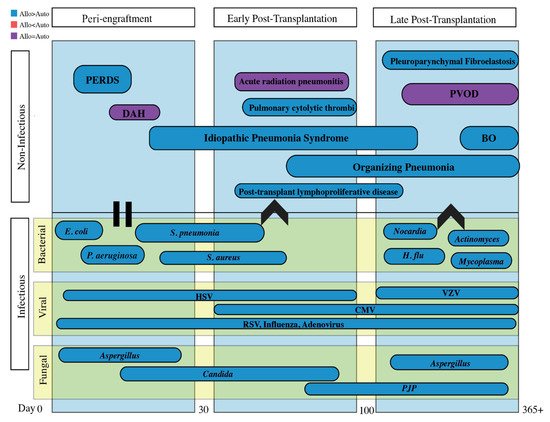
Infectious and non-infectious pulmonary complications are classified by etiology and temporality in reference to HSCT, as it reflects the immunologic state of the patient. The often-cited time course for non-infectious entities consists of the pre/peri-engraftment phase (first 30 days), early post-transplantation phase (30–100 days), and the late post-transplantation phase (after 100 days). Infectious complications will be discussed separately.
4. Infectious Complications
The peri-engraftment phase is hallmarked by mucositis due to conditioning therapy and prolonged neutropenia due to non-functional transplanted marrow [13][72]. The risk of infection is highest when absolute neutrophil count (ANC) is less than 500 and increases with increasing duration of neutropenia before engraftment [14][73]. During the early post-transplantation phase, infections are related to the defects in cellular immunity caused by immunosuppressive and conditioning regimens [13][72]. In the late post-transplantation phase, infection rates decline as immunosuppressant medications are tapered unless needed for cGVHD. Because of differences in immune recovery, autologous HSCT recipients are at higher risk of infectious complications during the peri-engraftment and early post-transplantation phase while allogeneic HSCT recipients are at higher risk of infection throughout the late post-transplantation phase due to cGVHD and prolonged immunosuppressive therapy [15][13][8,72].
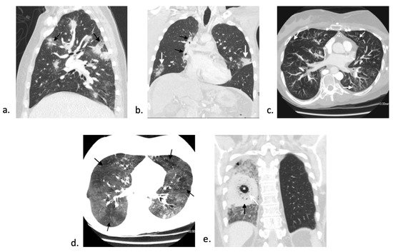
Zygomycosis is the second most common mold infection in HSCT with an increasing incidence over the past few decades [16][85]. Rare infections with fusarium and scedosporium can occur as well [17][88]. Invasive fungal infections with mucormycosis generally occurs > 3 months post-transplant and can make up to 8% of fungal infections in this patient population [18][89]. Although there are various imaging appearances of mucormycosis, one nearly pathognomonic imaging manifestation is the “bird’s nest” sign ( Figure 25 e) [19][90]. While mucormycosis infections are rare, they are frequently fatal and require early, aggressive management. Treatment involves combination of medical (with amphotericin B for zygomycosis) and surgical therapy [17][88].
Viral pneumonia from reactivation is common during the peri-engraftment phase and should be closely monitored while patients are immunosuppressed. Viral infection from herpes simplex virus (HSV)-1 and -2 occur in peri-engraftment and early post-transplantation phase, those from cytomegalovirus (CMV) and human herpes virus-6 (HHV-6) are often present in early post-transplantation phase, and infection from varicella zoster virus (VZV) is more common in the late post-transplantation phase [20][94]. The incidence of reactivation of viruses such as HSV, CMV, and VZV has drastically decreased since prophylaxis became routine.
RSV is one of the more common CARV that is associated with high mortality rate. In a single center retrospective study of 280 allo-HSCT recipients with RSV infection, 29% had progression to lower respiratory tract infection [21][113]. Older age, smoking history, conditioning with high-dose total body irradiation, and lymphopenia were associated with an increased risk of progression from upper respiratory to lower respiratory tract infection [21][22][113,114]. RSV is associated with a wide mortality rate, ranging from 5% to 43%, in HSCT patients [23][21][24][25][26][112,113,115,116,117]. Older age, male gender, bone marrow or cord blood as transplant source, corticosteroid use, lower respiratory tract infection, and oxygen requirement are all associated with increased overall mortality [21][25][26][27][113,116,117,118].