Polyhydroxyalkanoates (PHAs) constitute a family of biopolyesters that are synthesized and accumulate within the cellular structure of prokaryotic cells by bacteria, and they act as carbon and energy reserve materials under conditions of limited nutrient, such as nitrogen, oxygen, phosphorous or magnesium. As naturally derived materials, PHAs have been used for multiple cell and tissue engineering applications; however, their widespread biomedical applications are limited due to their lack of toughness, elasticity, hydrophilicity and bioactivity. To overcome this challenge combination of PHA with different polymers and inorganic materials are used to form hybrid composites with improved structural and functional properties.
Polyhydroxyalkanoates (PHAs) constitute a family of biopolyesters that are synthesized and accumulate within the cellular structure of prokaryotic cells by bacteria, and they act as carbon and energy reserve materials under conditions of limited nutrient, such as nitrogen, oxygen, phosphorous or magnesium. As naturally derived materials, PHAs have been used for multiple cell and tissue engineering applications; however, their widespread biomedical applications are limited due to their lack of toughness, elasticity, hydrophilicity and bioactivity. To overcome this challenge combination of PHA with different polymers and inorganic materials are used to form hybrid composites with improved structural and functional properties. This work presents an up-to-date overview of ongoing research strategies that make use of PHAs as versatile and prospective biomaterials.
1. Introduction
Polyhydroxyalkanoates (PHAs) constitute a family of biopolyesters that are synthesized and accumulate within the cellular structure of prokaryotic cells by bacteria, and they act as carbon and energy reserve materials under conditions of limited nutrient, such as nitrogen, oxygen, phosphorous or magnesium. The carbon source is the main influencing factor for the PHA production at industrial scale, because it affects the cell growth, productivity, molecular mass, quality, and composition of a polymer
[1][2][1,2]. There are published works, including review papers, addressing utilization of various types of carbon sources, such as whey, waste plant oils, waste animal fats, starch, wheat and rice bran, molasses, wastewater as a cheap carbon sources for PHA production
[3][4][5][6][3,4,5,6].
PHAs are biodegradable, biocompatible, piezoelectric, and thermoplastic and show good barrier properties and controllable thermal and mechanical properties depending on the polymer composition
[7][8][7,8].
Biocompatibility is the ability of a material to perform a desired function without causing any local or systemic adverse responses in the recipient of the material. The biological rejection of an implant leads to an inflammatory response mediated by immune cells, and it may require the removal of the implant. Although PHA has good biocompatibility
[9][10][9,10], many authors have shown that hybrid materials based on PHA have greater biocompatibility with different cells
[11][12][13][14][15][16][17][11,12,13,14,15,16,17].
The thermoplasticity, barrier properties and degradability characteristics of PHAs indicate that they can be recycled, which makes them very attractive for use as bioplastics for packaging purposes; however, the high cost of PHAs limits their use as a “green plastic”. In spite of this, interest in PHAs as bioplastics for fighting plastic pollution challenge continues to grow worldwide
[18]. PHA production increased from 5.3 million tons to 17 million tons within the year 2013 to 2020
[19]. PHA market size is estimated to be USD 62 million in 2020 and is projected to reach USD 121 million by 2025, at a CAGR of 14.2% between 2020 and 2025
[20]. The market is mainly driven by the rising demand for PHA industries such as food and packaging services, agriculture, biomedical, and some others. Factors such as consumer awareness about the toxicity of the petroleum based and sustainable ecofriendly bioplastics will drive the PHA market. There are key markets for PHA, which are Europe, followed by North America and Asia, in terms of value and volume. With an objective to reduce the total cost of PHAs production, new approaches of utilization of different cheap and eco-friendly carbon sources are employed
[21][22][23][21,22,23].
The nontoxicity, biodegradability and biocompatibility characteristics of PHAs suggest their potential uses in the biomedical field, especially in tissue engineering and as implants. Gradual biodegradation of PHA-based scaffolds creates a structure for the formation of new tissue and promotes cell growth; moreover, a second surgery is not required to remove the implant
[9]. PHA applications include cardiovascular tissue engineering, bone tissue engineering, nerve tissue engineering, and drug delivery systems.
Only a few members of the PHA family are commercially available and produced on a large scale, including poly(3-hydroxybutyrate) (PHB), poly(3-hydroxybutyrate-co-3-hydroxyvalerate) (PHBV) and poly(3-hydroxybutyrate-co-3-hydroxyhexanoate) (PHBHHx). PHB is the most investigated member of the PHA family. PHB is piezoelectric, crystalline, water insoluble and relatively resistant to hydrolytic degradation; however, it reveals poor mechanical properties and is a highly brittle and stiff material
[10]. The copolymer PHBV has better mechanical properties than PHB and is tougher, less stiff, and more flexible. PHBV exhibits both a lower crystallinity and melting temperature and increased elongation to break. PHBV copolymer does not cause inflammatory reactions when implanted in mice and rats
[24]. PHBVs have also been shown to support in vitro osteogenesis, which makes them suitable for bone regeneration
[25].
PHBHHx is another member of the PHA family with improved mechanical properties compared with both PHB and PHBV. PHBHHx promotes enhanced osteogenic differentiation of mesenchymal stem cells (MSCs)
[26] and possesses good biocompatibility with fibroblasts, chondrocytes, nerve cells and osteoblasts compared with polylactic acid (PLA), PHB and PHBV
[27].
PHB and PHBV are nontoxic because their degradation products are water, carbon dioxide and D-3-hydroxybutyric acid, which are natural constituents of human blood, and PHA-based biomaterials cause less-severe inflammatory reactions compared to other biopolymers, such as PLAs
[28]. D-3-Hydroxybutyric acid increases calcium influx in cultured cells and suppresses their death
[29]. Oligo(3-hydroxybutyrate-co-3-hydroxyhexanoate), oligo(3-hydroxybutyrate) and 3-hydroxybutyrate, the main degradation products of PHBHHx, are nontoxic and cause low inflammatory effects
[30]. However, the application of PHAs is limited due to their weak mechanical and thermal properties, slow degradation rate, lack of bioactivity, and poor hydrophilic properties. To overcome these disadvantages and improve PHA properties and make it more suitable for biomedical applications, many hybrid PHA-based composites have been investigated
[28][31][32][33][34][35][28,31,32,33,34,35]. Several reviews have been published describing production, properties, biocompatibility, and potential applications of pure PHAs
[36][37][38][39][40][36,37,38,39,40]. Unfortunately, there is a lack of systematic and thorough overview addressing the performance of PHA hybrid materials for tissue engineering and biomedical applications.
2. The Most Important Properties of the Hybrids Based on PHAs
2.1. Wettability of the Composites
Hydrophilicity is an important property of scaffolds and defines cell adhesion, proliferation and differentiation in vitro and tissue ingrowth in vivo
[41][42][43][44][45][41,42,43,44,45]. It has been reported that mammalian cells prefer to adhere and proliferate on the surface with moderate hydrophilicity with a water contact angle in the range of 50–70°
[46]. In addition, some serum proteins, such as fibronectin and vitronectin, which are well known to play an important role in cell adhesion, are more susceptible to moderate surface wetting
[47]. Hydrophilic surfaces absorb proteins more easily than hydrophobic surfaces, thus making them more suitable for cell spreading and proliferation
[48]. The hydrophilicity influences not only the amount and type of serum protein adsorption but also the conformation of these proteins on the surface of the scaffolds, which in turn affects the degree of cell adhesion
[49]. Therefore, the improved hydrophilicity could facilitate adsorption of more serum proteins to the surface, which improves cell adhesion. The hydrophobic nature of PHAs limits their applications in the biomedical field. The wettability of the PHA surface can be enhanced by the addition of different fillers as well as surface treatment, thus providing better cell adhesion, spreading and proliferation.
Silk fibroin (SF) is a natural biopolymer used in the human body as a suture material. SF has been employed as a versatile material for tissue-engineered scaffolding due to its biocompatibility and the presence of easily accessible chemical groups for functional modifications. SF have been used to impart hydrophilicity to PHAs. For instance, the water contact angle (WCA) of the PHBHHx/SF electrospun films decreased as the SF content in the blends increased. Human umbilical cord-derived mesenchymal stem cells (hUC-MSCs) showed better adhesion on electrospun PHBHHx/SF (1:1, 1:3) and SF films than on electrospun PHBHHx and PHBHHx/SF (3:1) films
[31]. The cell layer was more homogenously widespread and adhered completely onto the electrospun film surface. The addition of SF to PHB decreased the WCA of the PHB/SF nanofibrous scaffold. The PHB/SF composite scaffold (50/50 PHB/SF) showed excellent attachment behavior to L929 and HaCaT cells
[50]. Fibroblasts demonstrate better adhesion on PHBV/SF nanofibrous scaffolds than pure PHBV scaffolds since the hydrophilicity of the materials is helpful for the absorption of fibronectin, which is essential for fibroblast adhesion in vitro
[51].
Chitosan (CTS) is a natural polymer that is biocompatible and nontoxic with highly availability and low cost and possesses antibacterial activity, and it presents a high mass-loss rate and hydrophilicity
[32]. CTS has hydrophilic functional groups on its backbone
[43][44][43,44] that may increase the hydrophilicity of materials through blending. It has been shown that the addition of CTS to PHB increased the hydrophilicity of PHB and decreased its WCA to ~67° for PHB/20 wt% CTS composites
[32]. Contact angle measurements carried out on aligned and random electrospun PHB/CTS revealed that the fibrous scaffolds containing CTS were more hydrophilic than the pure fibers and that the aligned fibers had a lower WCA than random scaffolds. The WCAs of PHB, PHB/15 wt% CTS and PHB/20 wt% CTS are 124°, 62°, 43° for random fibers and 110°, 54°, 43° for aligned electrospun fibers, respectively
[52]. Other works have reported that the addition of CTS improves the hydrophilicity of PHAs
[53][54][55][53,54,55].
Synthetic hydroxyapatite (HA) is the most widely used bioceramic material and has a similar composition and morphology to the inorganic component of natural bone, which can provide a favorable environment for cell adhesion, osteoconduction and osteoinduction
[56]. It has been shown that the addition of mHA to PHB scaffolds decreases the WCA of the composite compared to pure PHB scaffolds
[33]. HA deposition on the surface of both nanofibrous and cast flat PHB films turned it hydrophilic
[57]. The investigation
[58] showed that there are no differences in WCA between pure PHB and PHB/nHA composites. There are some conflicting reports of the effects of HA, including the positive and negative effects, on cell adhesion or proliferation
[59][60][61][62][63][59,60,61,62,63]; thus, additional trials are required in this field.
The hybrid fibrous PHB/polycaprolactone (PCL) membrane possesses a hydrophilic surface
[64]. Modification of PHB/PCL fiber mats with silica decreases the WCA
[34]. The addition of graphene oxide (GO) nanosheets significantly enhanced the wettability of the surface of the PHBV biopolymer films
[65]. The nanofiber PHB scaffold turned from hydrophobic into hydrophilic in surface characteristic with WCA decreasing from 124° to 44° upon addition of soybean protein nanoparticles (SPN)
[66].
The COOH functional groups of carbon nanotubes (CNTs) increase the amount of oxygen on the surface, increasing the quantity of C–O. Thus, the wettability of the PHB scaffolds increases by adding CNTs
[16]. In another work, addition of 1 wt% of CNT into electrospun PHB nanofibers decreased WCA by 40°
[67]. The PHB/carboxyl multiwalled carbon nanotubes (CMWCNT) composite and PHB–calcium alginate/CMWCNT nanofiber membrane have improved hydrophilicity compared to pure PHB
[68][69][68,69].
The influence of the nanobioglass (nBG) and microbioglass (mBG) particles on the hydrophilic property of the PHB scaffold was investigated
[35]. Hybrid films had significantly increased wettability compared to neat polymer. However, the decrease in WCA was more prominent for the nBG composites than for the mBG composites.
The oxidation of the PHBV membrane in an ozone environment generates polar functional groups such as peroxides, hydroxyl, and carbonyl groups on the surface of the PHBV films. Further grafting of methyl methacrylic acid and covalent immobilization of type I collagen on the surface of PHBV led to WCA reduction and better hydrophilicity
[17]. The addition of collagen to nanofibrous PHBV/GO scaffolds with a WCA of 110° made the scaffold hydrophilic with a WCA of 52°
[11]. Collagen-coated electrospun PHBV nanofiber films demonstrated better hydrophilic behavior than uncoated films
[70].
Plasma surface modification is one of the most promising techniques for enhancing hydrophilicity because it does not alter the bulk properties of the treated material
[71][72][71,72]. Plasma particles interact with the material and introduce polar functional groups such as hydroxyl, carboxyl and carbonyl groups onto the surface of the substrate depending on the plasma gas. However, hydrophilicity decreases over time due to the “hydrophobic recovery” effect, which is the effect of rearrangement of polar groups towards the bulk of the material to reduce the surface energy
[73]. This effect has to be inhibited by covalent immobilization of various bioactive molecules, such as silk, gelatine or collagen
[74][75][74,75]. The oxygen and nitrogen plasma treatment of PHBV nanofiber mats with and without further immobilization of SF was investigated
[76]. Unmodified plasma-treated PHBV mats showed hydrophobic recovery after 14 days. SF-modified nitrogen plasma-treated PHBV mats had stabilized WCA at 70°, while oxygen plasma-treated PHBV presented hydrophobic recovery even after SF immobilization, which is probably due to the repulsion of negatively charged SF with negatively charged oxygen-containing groups on the mat surface. Plasma treatment of SF-coated PHBHHx film improves its hydrophilicity, leading to a larger amount of extracellular matrix (ECM) secretion and better cell migration of human smooth muscle cells
[74]. PHB/polyaniline (PANi) electrospun scaffolds surface modification with air plasma for 60 s reduced the water contact angle of the composite from 106° to 29.3°
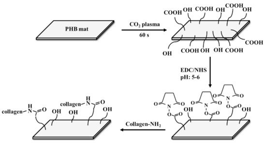
[77]. Collagen cross-linked plasma-modified PHB nanofibrous mats showed a better hydrophilicity of the modified nanofibers compared to the nonmodified mats with an 85° difference in WCA
[13]. A scheme of the covalent coupling of the protein with PHB mat fibers is shown in .
Figure 1. Schematic of the covalent coupling reaction for the attachment of protein to PHB fibers. Re-designed based on
[13].
The hydrophobic character of PHAs can be altered to be hydrophilic with the addition of different materials, as shown in detail above. Hydrophilic surfaces provide attachment of cells to the surface of materials as well as cell spreading and proliferation. The addition of different fillers should not impair other important properties of materials, such as mechanical properties, biocompatibility, and biodegradability, which are necessary for their use in biomedicine. The physico-mechanical properties of the PHA-based hybrids will be discussed in the next section.
2.2. Physico-Mechanical Properties
Scaffolds in tissue engineering applications must have sufficient mechanical strength during in vitro culturing to maintain the required space for cell infiltration and formation of ECM. Scaffolds should also provide sufficient temporary mechanical support, matching the mechanical properties of the host tissue as closely as possible, to bear in vivo loading and stress conditions. Thus, scaffolds should be designed with appropriate mechanical properties and degradation rates so that they match the mechanical properties of the injured tissue until the newly grown tissue is remodeled by the host tissue and is able to support in vivo stresses
[78][79][78,79].
Although the different fillers favorably affect the hydrophobic character of PHA, some of them act simultaneously with an improvement in hydrophilicity and can worsen the mechanical properties of the hybrids. For example, SF-modified PHBHHx films have the maximum tensile strength, and the elongation at break is slightly lower than that of PHBHHx films
[12]. The combination of SF with PHB led to an increase in elongation at break for the PHB/SF composite and a decrease in tensile and yield strength in comparison to pure PHB
[50]. The increase in CTS in the PHB/CTS composite decreases the tensile strength of the scaffolds
[32]. All the PHB/CTS blend scaffolds exhibited lower Young’s moduli than pure PHB scaffolds. These changes in tensile strength and tensile Young’s modulus are related to the lower mechanical properties of CTS in comparison to PHB. The toughness and maximum strain of the scaffolds were enhanced with increasing CTS in the blended scaffolds. This increase is due to the tough nature of CTS in comparison to PHB. In another work, it was reported that the CTS addition in PHB causes a reduction in tensile strength and that the Young’s modulus and tensile strength for aligned PHB/CTS electrospun fibers are greater than those for random fibers
[52]. Collagen immobilization decreases the tensile strength of PHBV/collagen electrospun nanofibers
[14]. To overcome this disadvantage, the addition of one more filler into the composite may be a prospective option. For example, in the case of the PHBV/GO/collagen composite, collagen did not play any significant role in the mechanical properties of the material
[11]. The addition of biphasic calcium phosphate (BCP) particles into the PHB/CTS membrane is beneficial to mechanical properties
[80]. Compared to the PHB/CTS and pure PHB membranes, mechanical properties, such as the initial Young’s modulus and ultimate tensile strength, were enhanced after incorporation of BCP. The mechanical properties of the PHB/CTS scaffolds were improved significantly after the addition of multi-walled carbon nanotubes (MWCNTs). MWCNT addition resulted in a significant increase in the scaffold’s elastic Young’s modulus, tensile strength and yield strength
[54]. It was found that incorporation of curcumin up to 20 wt% into PHB/MWCNT electrospun scaffolds had a significant effect on increasing ultimate strength values as compared to the neat PHB nanofibers
[81].
The addition of mechanically strong materials into the PHA matrices leads to an improvement in the mechanical properties of the composites. The tensile properties of PHBV were significantly increased by the addition of HA nanoparticles (NPs)
[24]. The addition of HA to the PHB scaffold allows an increase in the compressive Young’s modulus and compressive strength of the PHB, while the PHBHHx/mHA composite has a decreased compressive elastic Young’s modulus with the same compressive strength compared to the pure PHBHHx scaffold
[82]. PHB/10 wt% nHA composite scaffolds have an improved compressive Young’s modulus and compressive strength compared to neat PHB scaffolds
[83]. The significant increase in mechanical properties of the composite scaffolds compared to the pure PHB scaffold was due to the homogeneous dispersion of nHA in the matrix. HA NP incorporation within PHB/nHA (blend) fibers significantly improved the mechanical properties of the PHB mats
[58]. In contrast, the mechanical properties of the PHB/nHA (spray) framework deteriorated in comparison with those of the neat PHB mat. The tensile strength and strain as well as the elastic Young’s modulus decreased dramatically in the PHB/nHA (spray). PHBV fibers containing 10 wt% nHA or 10 wt% nHA/bredigite (BR) showed higher mechanical strength and Young’s modulus than PHBV fibers incorporated with 10 wt% BR
[84]. PHBV nanofibers containing the highest amount of NPs (15 wt%) showed reduced Young’s modulus and strength, which was probably because of the agglomeration of the NPs. The tensile Young’s modulus and tensile strength of the hybrid PHB/nHA scaffold were higher than those of the PHB scaffold
[85]. With the integration of gelatine with the electrospun PHB/nHA, both the tensile Young’s modulus and the tensile strength slightly decreased compared to those of the PHB/nHA mat. The addition of HA–NPs to the polymer matrix up to 15 wt% resulted in a significant increase in the compressive Young’s modulus and compressive strength of the scaffolds
[86]. When the nHA content of the scaffolds reached 20 wt%, a significant decrease was observed due to HA agglomeration. The mechanical properties of laminated nHA/PHB scaffolds are significantly improved in comparison to traditional nHA/PHB and PHB scaffolds
[87].
Blending PHB with poly(l-lactide-co-ε-caprolactone) (PLCL) significantly reduced the brittleness of the electrospun fibers and significantly increased the extension to break
[88]. The addition of GO to PHBV significantly enhanced the tensile strength, Young’s modulus and percent elongation of the nanofibrous scaffold in comparison with pure PHBV
[11]. Compression modulus of PHBV film increased by 25% with the addition of GO nanosheets
[65]. Incorporation of 0.7 wt% graphene nanoplatelets in the PHB matrix with uniform dispersion resulted in the enhancement of tensile stress from 7.5 to 12.2 MPa
[89]. Addition of mechanically stable GO into the polymeric matrix bestowed the flexibility of the PHBV based Fe
3O
4/GO-g-PHBV composite enhancing tensile strength and elongation at break
[90]. The ductility of the PHB nanofiber scaffold significantly improved with addition of 1 wt% SPN increasing the elongation at break by 190% compared to pure PHB scaffold
[66].
PHBV/poly lactic-co-glycolic acid (PLGA) and PHBV/PCL membranes have better mechanical properties than pure PHBV membranes. The highest values of tensile strength, elongation at break and Young’s modulus are in 50/50 hybrid membranes
[91]. It has been shown that PCL is able to increase the tensile strength and elongation of PHB, although if the mass ratio of PHB/PCL was higher than 40:60, the effect was not considerable
[64]. CTS-g-PCL/PHBHHx fibers possess increased tensile strength, elongation at break and Young’s modulus values compared to pure PHBHHx fibers
[92].
PHB electrospun nanofibers have improved tensile strength after MWCNT incorporation and hot-stretching treatment
[93]. Maximum values of the tensile strength, breaking elongation rate, initial Young’s modulus and fracture energy of the CMWCNT-g-PHB/PHB composite nanofiber scaffolds are achieved at a CMWCNT content of 6 wt%
[68]. The tensile strength and breaking elongation rate of composite nanofiber scaffolds were more than twice those of pure PHB nanofiber scaffolds. CNTs can significantly increase the tensile strength and Young’s modulus of scaffolds. The highest strength and Young’s modulus values are obtained for PHB/0.5wt% CNT nanocomposite scaffolds
[16]. A significant tensile strength increase in PHBV/MWCNT nanocomposites was observed upon the addition of MWCNTs with the maximum tensile strength at 1 wt% MWCNT content
[94]. The tensile strength of the composite PHB/CNT scaffold was significantly increased in the presence of 1% CNTs compared to pure PHB scaffold
[67]. Small amount of the humic acid loaded CNT (HACNT) greatly improved the ductility of the HACNT/PLA/PHB composite, with the maximum tensile strength increased by 236% and the elongation at break improved by 790%
[95].
The Young’s modulus and elastic modulus values increased after the addition of nBG to the PHB film but decreased after the addition of mBG
[35]. The reduction in Young’s modulus is due to poor mixing of mBG particles with the polymer matrix, leading to large agglomerations. The maximum tensile strength and Young’s modulus were obtained for the PHB/nBG scaffolds containing 7.5 wt%. nBGs. A further increase in nBG content worsened the tensile strength of the nanocomposites due to the agglomeration of nBGs in the polymer matrix at 10 wt%. and 15 wt%. Mesoporous bioglass (MBG) increases the compressive strength of the PHBHHx film with increasing MBG content
[96]. The compressive strength of PHBV/mBG composite scaffolds was significantly higher than that of pure PHBV scaffolds
[97]. It has also been reported that MBG did not obviously influence the compressive strength of PHBHHx scaffolds
[98].
The orientation of the electrospun fibers directly affects the mechanical strength of the scaffolds
[99]. Overall, aligned electrospun nanofibers reveal better mechanical properties than random nanofibers.
Deposition of NPs into a polymer matrix also enhances the mechanical properties of the composites
[24][82][83][84][85][24,82,83,84,85]. The homogeneous dispersion of NPs provides a high interfacial surface, which may enhance the load transfer between the polymer matrix and the NPs, which results in improvement of the mechanical properties of the composite scaffolds. When the concentration of the NPs is low, the matrix can transfer the concentrated stress to the NPs effectively, thus improving the strength of the material. However, as the concentration of the NPs increased, the NPs agglomerated in the polymer matrix, which might weaken the stress transference
[84][86][84,86]. They act as weak points in the structure and can easily break when stress is applied to the composite. Broken agglomerates then act as stress concentrators leading to the formation of microcracks, consequently leading to a significant decrease in the Young’s modulus and strength of the composites
[100][101][100,101].