Placenta in SARS-CoV-2 Infection: Comparison
Please note this is a comparison between Version 1 by Paweł Kowalczyk and Version 2 by Bruce Ren.

A pandemic of acute respiratory infections, due to a new type of coronavirus, can cause Severe Acute Respiratory Syndrome 2 (SARS-CoV-2) and has created the need for a better understanding of the clinical, epidemiological, and pathological features of COVID-19, especially in high-risk groups, such as pregnant women. Viral infections in pregnant women may have a much more severe course, and result in an increase in the rate of complications, including spontaneous abortion, stillbirth, and premature birth—which may cause long-term consequences in the offspring. In this review, we focus on the mother-fetal-placenta interface and its role in the potential transmission of SARS-CoV-2, including expression of viral receptors and proteases, placental pathology, and the presence of the virus in neonatal tissues and fluids. 

  • COVID-19
  • lactoferrin
  • pregnant women
  • oxidative stress
  • mother’s placenta

Note: The following contents are extract from your paper. The entry will be online only after author check and submit it.

1. Introduction SARS-CoV-2 Infection

Coronaviruses (CoV) are single-stranded RNA viruses belonging to the order Nidovirales, family Coronaviridae and subfamily Coronaviridae [1]. In November 2019, a new type of coronavirus was identified in the Chinese city of Wuhan. It was named SARS-CoV-2, due to respiratory infections (COVID-19) it caused [2]. Several genetically different types of SARS-CoV-2 have been distinguished so far [3]. Clinical manifestations of respiratory infections vary from asymptomatic, mild upper and lower respiratory tract infection to life-threatening pneumonia with acute respiratory distress syndrome (ARDS) [4][5][4,5].

Receptor Recognition Is the First Step of Viral Infection That Determines a Cell/Tissue Tropism

SARS-CoV-2 S-protein recognizes angiotensin-converting enzyme 2 (ACE2) [6][7][6,7], and by attaching to it may bind to other proteins: dipeptidyl peptidase 4 (DPP4), glucose regulated protein 78 (GRP78), carcinoembryonic antigen-related cell adhesion molecule 1 (CEACAM1), aminopeptidase N (APN), and recognize various sialosides and different glycosaminoglycans (GAGs) as cellular targets via lectin-type interactions. The cell entry of SARS-CoV-2 is possible after protein S activation by cellular proteases, including transmembrane serine protease 2 (TMPRSS2), cathepsin L, and furin [7][8][9][7,8,9]. TMPRSS2 and ACE2 co-expression is observed in several tissues, such as nasal epithelial cells, lungs, and bronchial branches [10][11][10,11].
SARS-CoV-2 infection begins in the nasal epithelial cells that initiate genetically innate and adaptive immune responses [10]. After cell invasion, a virus is recognized by the host’s innate immune system through pattern recognition receptors (PRRs), including C-type lectin-like receptors; toll-like receptor (TLR), NOD-like receptor (NLR), and RIG-I-like receptor (RLR) [12][13][14][12,13,14]. PRRs recognize molecules frequently found in pathogens’ pathogen-associated molecular patterns (PAMPs), or molecules released by damaged cells damage-associated molecular patterns (DAMPs), [15]. In addition, SARS-CoV-2 infection can cause host cell pyroptosis and the release of DAMPs. TLRs activation by DAMPs further enhances inflammation [15]. Consequently, the production of several anti-viral substances is activated, such as: “Lactoferrin” (LF), type I and III interferons, nitric oxide, b-defensins, and other chemokines and cytokines that recruit inflammatory cells, i.e., dendritic cells (DC), macrophages and influence adaptive immunity [16]. During SARS-CoV-2 infection, both Th1 and Th2 immunity pathways are activated almost at the same time during the infection course [17].
The course of COVID-19 varies significantly through the patients, strongly depending on immune responses. Elevated IL-6 levels are correlated with an increased risk of death in COVID-19 patients [18]; whereas, early activation of adaptive immunity is predicted for less disease development [19]. Moreover, patients with more severe disease (“cytokine-storm”) have increased plasma concentrations of interleukin (IL)-2, IL-7, IL-10, tumor necrosis factor α (TNFα), interferon-γ-inducible protein 10 (IP-10), granulocyte-colony stimulating factor (G-CSF), chemoattractant protein 1 (MCP-1) and macrophage inflammatory protein 1 alpha (MIP-1 alpha) [17]. The number of T-cells, such as helper T-cells, and memory helper T-cells, are decreased; whereas, naïve helper T-cells are increased in severe cases of COVID-19 compared to the group with mild symptoms [20]. The clinical outcomes, immune responses, and immunopathology are summarized in Table 1.
Table 1. Immune characteristics related to the clinical course of SARS-CoV-2 infection.
Clinical Course of Infection Asymptomatic/Mild COVID-19

(Appropriate Immune Response)
Severe COVID-19

(Defective Immune Response)
Immune response
  • Immune cell activation: Monocytes, neutrophils, B cells, CD4+ T, CD8+ T cells, and NK cells;
  • Physical activation of Cytokines/Chemokines secretion: TNFα, IL-2, IL-6, IL-7, IL-8, IL-17, G-CSF;
  • Virus inactivation by neutralizing antibodies;
  • Lowering of immune cells number: Monocytes, eosinophils, basophils, B cells, CD4+ T, CD8+ T cells, and NK cells;
  • Increased number of neutrophils;
  • Elevated levels of IL-6, IL2R, IL-10, and TNF-α;
  • Increased level of SARS-CoV-2 specific IgG;
Immunopathology None/Mild
  • Lymphopenia—infection susceptibility;
  • Systemic cytokine storm;
  • Lymphocyte dysfunction:
T cell depletion and exhaustion;

virus-specific T-cells central memory phenotype;
  • Antibody-dependent enhancement (ADE) of infection;
Clinical outcomes Infection resolution
  • ARDS
  • Respiratory failure
  • Multi-organ dysfunction
  • Sepsis

2. COVID-19 during Pregnancy—The Role of the Placenta

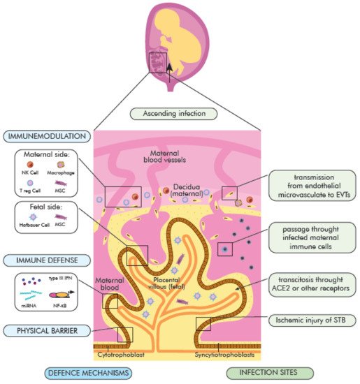
Pregnancy is a unique immunological state reflected by a combination of signals and responses from the maternal and the fetal–placental immune system. The maternal immune responses actively change throughout gestation from an anti-inflammatory state (first and third trimester) to pro-inflammatory state (second trimester) [21]. The immunological events show precise timing, named “immune clock” [22], and ensure the maintenance of maternal tolerance to fetus and protection against infectious agents. Physiological changes make pregnant women more susceptible to viral droplet-transmitted infections [23]; however, the virus infection appears to be milder compared to those caused by SARS-CoV and MERS-CoV [24][25][24,25] or H1N1 influenza [26]. The most common symptom of COVID-19 in pregnant women is fever (68%), then persistent dry cough (34%), malaise (13%), dyspnea (12%), and diarrhea (6%) [27]. An important role as a natural physical and immunological barrier that protects the fetus from various pathogens plays placenta [28][29][30][31][28,29,30,31]. In response to maternal infection, hypoxia, or nutrition status, it releases anti-microbial peptides and cytokines that activate and modulate maternal and placental immune response [32][33][34][32,33,34]. The effects of COVID-19 on the fetus are still largely unknown. The neonates’ outcomes following maternal COVID 19 may be diverse and sequel from immune-mediated events or direct cytopathic effect of the virus [35] (Figure 1).
Figure 1. Implications of SARS-CoV-2 virus infection at the maternal–fetal interface. The placental function disruption (histomorphological alterations) as a consequence of maternal clinical implications of COVID-19 (e.g., hypoxia, cytokine storm) and/or placenta infection, as well as probable fetal infection (vertical transmission) may result in pregnancy complications, compromise fetal health and long term adverse neonatal outcomes.
Pregnant women who were diagnosed with COVID-19 are 20 times more likely to die than healthy pregnant women. These are the conclusions of a global study published in the medical journal JAMA Pediatrics [36]. The study, led by doctors from the University of Washington’s School of Medicine and the University of Oxford, analyzed data from 2100 pregnant women from 43 maternity hospitals in 18 countries between April and August 2020. Additionally, the risk of a severe infection was greatest in women with obesity, high blood pressure, or diabetes. For each pregnant woman with COVID-19, the researchers selected two pregnant women who were cared for in the same hospital and at the same stage of pregnancy, but with no known viral infection for comparison. They then followed both groups—706 women with COVID-19 infection and 1424 women without infection—until delivery and after discharge from the hospital. Eleven women from the group with COVID-19 died, and only one died in the group without COVID-19. In contrast, pregnant women with asymptomatic or mild infections were not at increased risk of intensive care, preterm labor, or pre-eclampsia. In the study, about 40 percent of pregnant women had COVID-19. The study authors found that women with COVID-19 have between 60 to 97 percent higher risk of premature birth. In women with COVID-19 who have a fever and respiratory failure, they found a five-fold increase in neonatal complications, including lung immaturity, brain damage, and visual disturbances. Of the babies born to mothers with COVID-19, eleven percent tested positive for the coronavirus. However, infections passed on to babies do not appear to be related to breastfeeding. Rather, the examination links them with delivery by cesarean section. These results highlighted the importance of including pregnant women in priority groups for vaccination against SARS-CoV-2 [35][36][35,36].

2.1. Maternal Immune Changes during Pregnancy

The SARS-CoV-2 (COVID-19) pandemic is still in the early stages of research, and preliminary case reports of infections in pregnant women are available. Changes in the hormone levels during pregnancy can modulate immune responses against pathogens [37]. Innate immunity remains unchanged, while adaptive responses change during pregnancy and vary with gestational age.
Innate immunity provides interaction with fetal tissues to promote successful placentation and pregnancy course, as well as is the first line of host defense against infections [38]. The maternal immune phenotype is characterized by an increase in peripheral blood neutrophils (up to 60–95%) monocytes, DCs (producing interferon (IFN) l), and suppression of peripheral NK cells in number and function [39][40][41][39,40,41]. The neutrophils directly interact with other immune cells, such as macrophages, DCs, NK cells, B, and T cells, therefore up- or down-modulating both immunities—innate and adaptive [42].
In opposite, cytotoxic CD8+ adaptive immune responses are diminished, bypassed, or even abrogated; whereas, regulatory immunity is enhanced in pregnant women. Moreover, a Th2 (pro-inflammatory) to Th1 (anti-inflammatory) cytokine shift is observed. Promotion of Th1 humoral responses can result in an altered clearance of infected cells [43].

2.2. Inflammatory Response to SARS-CoV-2 during Pregnancy—Infection Outcomes

Immune characteristics among pregnant and non-pregnant women with COVID-19 seem to be similar [17][44][17,44]. During virus infections, an increased Th2–associated cytokines profile is observed [17][45][17,45]. It is feasible that elevated Th2 immunity during pregnancy seems to be associated with a milder virus infection course [44]. Similarly, Th1 pro-inflammatory pathway inhibition, probably decreases the “cytokine storm” and results in COVID-19 severity being similar in pregnancy and non-pregnancy [46]. Pregnant women compared with non-pregnant women showed milder or no symptoms [47]. Moreover, the TLRs alteration during virus infection enhances immune response, however, it is not known how pregnancy affects this aspect of the viral response [48]. Importantly, the immune system activation influences clinical outcomes of the virus in mothers, as well as modulates the scale of fetal complications [49]. However, the risk of adverse clinical outcomes in pregnant women with the virus is still unclear. At present, there are insufficient data on the possible impact of the virus in early pregnancy, and only a few reports are available showing conflicting information: Hydrops fetalis and fetal deaths in one case [50], and no significantly increased risk of pregnancy loss in the second case [51]. Most studies concern pregnant women in the third trimester, and the observations are divergent from a similar clinical course of the disease [52] to an increased mortality rate among pregnant women compared to the rest of the population [53][54][55][53,54,55]. The pregnancy raises the morbidity of the virus, especially in the presence of risk factors, such as advanced maternal age, obesity, being Black or Hispanic, elevated D-dimer, and IL-6, as well as medical comorbidities [53][54][55][53,54,55]. The prevalence of cesarean sections in pregnant women with SARS-CoV-2 varied between 69.4% and 84.7% in different studies, the most common mentioned maternal complication was preterm labor (33.3%) [56], and a maternal mortality rate (MMR) reached 1.6% in some studies, while the others reported none or single deaths [46][54][55][57][58][59][60][46,54,55,57,58,59,60]. The COVID-19 related MMR in the UK was 1% (5/427 pregnant women) and in France was 0.2% (1/617 pregnant women) [61][62][61,62]. A significant increase in MMR has been documented in pregnant women from Brazil (12.7% vs. 5% of the general population) [63]. That high mortality rate may be a result of the low quality of prenatal care [63].
The maternal infection severity, including hypoxia or “cytokine storm”, may exaggerate the maternal immune system and participate in placental and fetal complications like fetal growth restriction (FGR) (10%), miscarriage (2%), preterm labor (39%) [64] (Figure 1). In addition, maternal inflammation during pregnancy can affect fetal brain development, CNS dysfunction, and behavioral phenotypes that may be recognized later in the postnatal life [65]. The fever, one of the most common symptoms of the virus, could be associated with increased hyperactivity disorder/attention-deficit later in life [65]. An elevated level of IL-6 observed in virus infection may be responsible for autism, psychosis, and neurosensorial deficits development in the offspring [66]. Similarly, increased maternal IL-17a levels correlate with autism spectrum-like phenotype in offspring [66]; whereas, an increased level of TNFα in the maternal peripheral blood additionally may have a toxic effect on early embryo development [67].

3. COVID 19—Placenta

3.1. The Maternal–Fetal Physical and Immunological Barrier

The placenta is essential organ with various physiological, immune, and endocrine functions needed—to nourish and protect the fetus. It is composed of cells from two different individuals—mother and fetus [68]. The fetal part of the placenta forms from the chorionic sac—including the amnion, yolk sac, chorion, and allantois. The outer layer of the placenta is called the trophoblast and consists of two layers: The cytotrophoblast layer (inner) and the syncytiotrophoblast layer (outer). The maternal part comes from the endometrium and is called the decidua with maternal vessels [69]. Between these two regions is located the intervillous space filled with maternal blood [70]. The basic functional units of the placenta are fetus-derived chorionic villi (CV) with fetoplacental vessels. CV are formed and maintained by the fusion of: syncytiotrophoblast (STB), extravillous trophoblasts (EVTs), and cytotrophoblasts (CTBs) [68]. The placenta’s unique structure and function determine the protective properties against most pathogens [71]. Its role in infections is multi-directional and involves: (1) Physical blockade of viral entry; (2) active anti-viral function and in case of infection (3) immunomodulatory action (Figure 2). The most important elements of the placenta as a physical barrier are: (i) A dense network of branch microvilli and periodical regeneration of the most outer STB layer [72], the lack of intracellular gap junctions between STB cells [73]; (ii) dense actin cytoskeletal network, forming a brush border at the apical surface of the STB layer [74]; (iii) limited expression of TLR or internalization receptors as E-Cadherin at the STB layer [75]; (iv) little to no expression of caveolins at STB surface [76]; (v) the basement membrane beneath the villous cytotrophoblast [77]. The immunological role of the placenta in infections depends on may things, including its immunomodulatory property with trophoblast-immune crosstalk. It has been suggested as a crucial component of the innate immune response. Immune cells from the fetal and maternal compartments interact to provide an intricate balance between fetal tolerance (pregnancy maintenance) and anti-microbial defense in case of infection. Moreover, the breakage or breach of the decidual or syncytial barrier continuity initiates a strong innate immune reaction against pathogens. The maternal decidua is composed of stromal cells and leukocytes (40% of decidua) [78]. 50–70% of decidual leukocytes are NK cells, 20–30% are macrophages, 10–30% are T cells, including regulatory T cells (Treg), and approximately 2% are DC’s [79][80][81][79,80,81]. The proportion of immune cells vary throughout pregnancy, with an increase in the proportion of T cells at term [82]. During the first trimester of pregnancy, macrophages and NK cells accumulate around the trophoblastic cells [83][84][83,84]. The fetal part of the placenta—the chorionic villi contains, at the core part, fetal macrophages (Hofbauer cells), fetal endothelial cells, fibroblasts, and mesenchymal stem/stromal cells (MSCs) [85][86][87][85,86,87]. The trophoblast releases several immunomodulatory molecules, such as a secretory leukocyte protease inhibitor (SLPI), β-defensins, and expresses “maternal lactoferrin” [88]. During pregnancy, TLRs (TLR-3, TLR-7, TLR-8, and TLR-9) are expressed on the surface of trophoblast, decidua, Hofbauer cells, endothelial cells, and chorioamniotic membranes. Furthermore, a soluble form of TLR2 is also present in amniotic fluid [88]. The expression of TLRs by trophoblast varies through the gestation (first trimester: villous cytotrophoblasts (vCTBs) and EVTs; term: STB and EVTs) [89]. Its immunoregulatory function includes caspase activation, cytokine production, and inflammatory response induction, as well as the release of anti-microbial peptides and proteins into the amniotic fluid [90]. They also play an important role in bridging innate and adaptive systems [91]. Acquired viral infections may disturb the immune regulation at the border of the mother and the fetus, leading to fetal damage, even when the virus does not reach it directly [92]. The TLR-3 receptor in the first trimester of pregnancy may mediate a rapid anti-viral response [93][94][93,94], and induce the production of cytokines, type I and III IFN [95]. Similarly, TLR7 induces the synthesis of anti-viral cytokines and play a role in preventing intrauterine transmission of some viruses (e.g., HBV) [96]. Cytokines and interferons are important mediators in healthy pregnancies, due to their role in regulating cell function, proliferation, and gene expression. However, their deregulation may disrupt the developmental paths of the fetus and placenta [97]. Lactoferrin may also play a similar role to TLR and interferon receptors. Moreover, to ensure the maternal humoral protection of fetus and neonates, the maternal antibodies are actively transported to the fetus via the neonatal IgG receptor expressed on the STB surface [98].
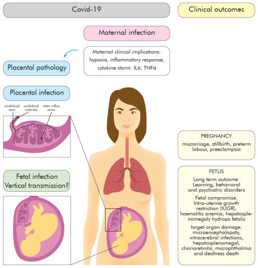
Figure 2. The defense mechanism of the placenta and potential infection sites of SARS-CoV-2. Placental properties that prevent SARS-CoV-2 infection include: Physical blockade, release/synthesis of anti-viral molecules (miRNA, IFN III, NF-κB), and stimulation of immune defense by decidual and fetal immune cells. The SARS-CoV-2 fetal infection may occur due to placental barrier breakage or via ascending route. Abbreviations: ACE2, Angiotensin converting enzyme 2; EVTs, extravillous trophoblasts; IFN, interferon; miRNA, microRNA; MSC, mesenchymal stromal cells; NF-κB, nuclear factor kappa-light-chain-enhancer of activated B cells; NK cell, natural killer cell; STB, syncytiotrophoblast; T reg cell, regulatory T cell.

3.2. The Role of the Placenta in COVID-19

The role of the placenta in SARS-CoV-2 infections remains poorly understood and requires further research, including vertical transmission mechanisms, fetal infection, and its consequences. The most important activated molecular signaling pathways against viruses (including SARS-CoV-2) are: Type III IFN signaling, autophagy-regulating microRNAs, and the NF-κB pathway (summarized, in detail, by Kreis et al. [99]). At the placenta level, the type III IFN provides a powerful anti-viral response. The IFN initiates a signaling cascade that activates transcription of IFN-regulated genes. Given that SARS-CoV-2 induces the release of type III IFN, this could be one of the possible mechanisms protecting the fetus against SARS-CoV-2 infection. Trophoblastic micro RNAs may constitute an important placental anti-viral defense mechanism on viral invasion restriction and trophoblast integrity maintenance [99]. Therefore, using a miRNA construct as one of the therapeutic targets or as a vaccine against SARS-CoV-2 was proposed [100]. On the other hand, inhibition of the nuclear factor kappa-light-chain-enhancer of activated B (NF-κB) pathway in COVID-19 mice led to a reduction of inflammation and lung pathology in infected animals [101][102][101,102], showing the importance of NF-κB pathway regulation for a controlled immune response [103]. The immunomodulatory action of placental immune cells may cause immune response mitigation, cytokine storm reduction, damages of cells, tissue limitation, and reduction of SARS-CoV-2 transmission (Figure 2).
Information on the effects of similar viruses on the embryo and fetus is very limited. Very recently, humanity has faced two severe diseases caused by viruses from the same viral family as SARS-CoV-2. In both cases, the route of spread of the infection and clinical symptoms were very similar to COVID-19, but those diseases were associated with higher mortality. In February 2003, SARS (Severe Acute Respiratory Syndrome), caused by the SARS-CoV virus also began in China, and the virus has spread to nearly 30 countries. More than 8000 people fell ill then, 770 of them died. Among the cases reported in the literature, 17 cases concerned pregnant women, of which 12 were the largest group, while the remaining reports referred to single cases. The second disease was MERS (Middle East Respiratory Syndrome), caused by the MERS-CoV virus. The disease first appeared in Saudi Arabia in 2012, then in other countries of the Arabian Peninsula, including the USA, and in 2015 in South Korea. So far, about 2500 people have fallen ill with MERS, more than 860 have died. MERS has been reported in 13 women at different stages of pregnancy. Both in the case of SARS and MERS infection, spontaneous abortions, premature births, and the birth of healthy children were observed in pregnant women. Because the observed groups of women were sparse, the percentage data were not provided in the literature. The effects of SARS-CoV-2 on the embryo and fetus are investigable in those countries with congenital disability registers. Such defect register also operates in Poland under the name of the Polish Register of Congenital Developmental Defects (PRWWR), which was established in 1997 and covered the entire country, being the register subjected to the Polish Ministry of Health. PRWWR is the largest register of defects in the EU and has been in the EUROCAT register consortium since 2001. PRWWR is conducted jointly by doctors of many specialties, especially neonatologists, clinical geneticists, and pediatricians. One of the important goals of defect registers is the constant monitoring of possible mutagenic and teratogenic threats in the population for the purpose of quick identification and elimination of detected harmful agents. Keeping the Registry for over 20 years, it makes it possible to identify well the frequency and structure of congenital malformations in the Polish population. In the case of SARS-CoV-2 infection in pregnant women, the question of whether it also poses a threat to the developing child could be answered. If a pregnant woman becomes ill with COVID-19, it is important to avoid prolonged high body temperature, especially during the first trimester of pregnancy. It should be noted that increased stress in the mother adversely affects the developing child and even the future children of that child. This is mediated through epigenetic mechanisms. The first 12 weeks of pregnancy are a special period—when all the organs of the baby start to develop. Interfering factors, including teratogenic factors, can cause congenital disabilities and sometimes also disorders that are detected later in life. Teratogens include physical, chemical disruptors, certain drugs, biological agents, including some viruses, especially the rubella virus. Viral diseases during pregnancy can directly affect the embryo and the fetus, but also through the harmful effects of the increased body temperature of the sick mother. It should also be remembered that under normal conditions, approx. 12–25 percent of diagnosed pregnancies end in spontaneous miscarriage (normal population risk). In cases of miscarriage, the common cause is a chromosomal aberration in the embryo/fetus, which is a severe genetic disease of the developing baby, and is not associated with any infection. Similarly, 2–3 percent of children are born with a developmental defect (population risk). Thus, not every spontaneous miscarriage or neonate with defects born by a woman with SARS-CoV-2 should be associated with the effects of the virus [104][105][106][107][108][104,105,106,107,108].

3.3. The Possible Mechanisms of SARS-CoV-2 Vertical Transmission

Based on the current knowledge on viral vertical transmission routes, some possible mechanism used by the SARS-CoV-2 virus to cross the placenta [109] are proposed:
(1)
direct infection of STB syncytiotrophoblasts and their rupture, virion transcytosis via immune receptors ACE2 and Fc (FcR),
(2)
passage through endothelial microcirculation into the intravascular extravascular trophoblasts (EVT) or other placental cells, as well as
(3)
passing through infected maternal immune cells and
(4)
ascending vaginal infection (placental barrier) (Figure 2).
Placental tissue appears to be a potential site for SARS-CoV-2 infection, since the expression of the receptor and priming proteases in various cell types of the maternal–fetal interface was detected (1).
The presence of ACE2 was demonstrated in the female genital tract and the placenta, including STBs, vCTBs, invasive and intravascular trophoblast, vascular smooth muscle cells in primary villi, decidual cells, and vascular endothelial cells in umbilical vessels [99][110][111][99,110,111]. The expression of ACE2 dominates, especially in the early gestation placenta [112]. However, co-expression of ACE2 and TMPRSS2, by the human placenta and chorioamniotic membranes throughout pregnancy is rare [113]. The presence of alternative receptors and proteases for SARS-CoV-2 entry into STB cells has been suggested [114]. Recently proposed alternative receptors are DPP4 (CD26) and CD147 [115][116][115,116]. Whereas, furin and trypsin, both expressed on placental tissues through gestation, have been suggested as SARS-CoV-2 entry proteases [113][117][118][113,117,118].
Moreover, several placental cell types can be used as replication and entry sites of pathogens (2, 3): EVTs, vCTBs, Hofbauer cells, giant trophoblast cells, or maternal immune cells of decidua [87]. It is possible that PBMCs can be infected by SARS-CoV-2 and transmit the virus through the placenta, however, the viral replication does not seem to occur within this compartment [119].

3.4. Placenta Pathology

Maternal–fetal interplay during COVID-19 includes histomorphological changes in the infected placenta although, some research revealed SARS-CoV-2 presence in the placenta without abnormalities in placental histopathology [120]. Currently, several reports suggesting placental infection with SARS-CoV-2 and the viral presence were confirmed by PCR (placental tissue/amniotic membrane), immunohistochemistry, and in-situ hybridization assays (formalin-fixed paraffin-embedded tissue sections) [121][122][123][124][125][126][121,122,123,124,125,126]. The available findings of placental pathology from COVID-19 patients came from the third trimester [15][17][31][127][128][15,17,31,127,128], and the most common findings are vascular malperfusion (FVM), fetal vascular thrombosis and maternal vascular malperfusion (MVM) (20–73%), massive infection with generalized inflammation (presence of M2 macrophages, cytotoxic and helper T cells, and activated B-lymphocytes) (13–20%), fibrin deposition and intervillous thrombosis [15][17][31][127][128][15,17,31,127,128]. These abnormalities result from direct infection of cells, systemic inflammation (“cytokine storm”), hypercoagulable state, and maternal hypoxia [129]. Consequently, adverse perinatal outcomes: MVW associated intrauterine growth restriction (IUGR), increased incidences of preterm births, higher rates of perinatal death, miscarriage, pre-eclampsia, cesarean section deliveries are observed [130]. The placenta abnormalities seem to be independent of maternal clinical manifestation, and even asymptomatic pregnant women with viral infection may develop obstetrical complications [131]. Placental transmission of proinflammatory cytokines is likely to stimulate hormone signaling dysregulation, enhancing poor neonatal outcomes, due to oxygen deprivation [105][132][105,132].

3.5. The Vertical Transmission Rate

The virus present in the placenta does not determine the incidence of vertical transmission.
In most studies [133], detection of SARS-CoV-2 is performed using RT-PCR analysis on neonatal airway swabs, less common on placental tissue (30.0%), umbilical cord blood (32.5%), and amniotic cavity (reported in 35.0% of publications). The maternal diagnostic material includes additionally vaginal, cervical, or rectal swabs to detect genital tract viral shedding during vaginal delivery (22.5% of cases) [134][135][136][137][138][139][140][141][134,135,136,137,138,139,140,141]. In few studies, IgG and IgM serology in the mother and neonate was performed [135][137][142][143][144][135,137,142,143,144]. The neonatal SARS-CoV-2 infection was reported by Mahyuddin et al. in 25% of papers [145]; whereas, the rate of positive SARS-CoV-2 test for neonates born to mothers with COVID-19 was estimated by Jafari et al. as 8% [146]. Kotlayard et al. determined that viral transmission from mother to fetus may reach 3.2% based on the nasopharyngeal swab (NPS) RT-PCR testing. The rate of SARS-CoV-2 RNA positive test may occur in approximately 7.7% of placental and 2.9% of cord blood samples. The IgM serology confirmed SARS-CoV-2 infection in 3.7% of neonates [147]. The vertical transmission rate was estimated as 2.23–5.3% (1.3–16) [146]. Although the strong evidence that vertical transmission of the virus may occur; intrapartum transmission (exposure to maternal blood, vaginal secretions, or feces) and early postnatal transmission cannot be excluded. To date, Vivanti et al. [133] showed the clearest evidence for transplacental transmission of the virus, due to the detection of viral genetic material and protein in the placenta, and viral RNA alone in the amniotic fluid and neonatal blood sampled at birth.
The vertical transmission of the virus in the third trimester is approximately 3.2% (22/936) by infant NPS testing, with severe acute respiratory syndrome coronavirus 2 RNA positivity in other test sites ranging from 0% (0/51) in amniotic fluid and (0/17) urine, 3.6% (1/28) in the cord blood, 7.7% (2/26) by placental sample analysis, 9.7% (3/31) by rectal or anal swab, and 3.7% (3/81) by serology [147].
The vertical transmission risk seems to be relatively low. However, the lack of a precise and universal definition of the term “vertical” transmission prevents comparison of described cases of neonatal viral infection. Standardized definitions, including diagnosis time of neonates, method, and analyzed biological material clarified the rate of “vertical” transmission and distinguishing it between intrapartum and postnatal transmission of the virus. This may have implications for future research describing clinical courses and long-lasting post-infection neonatal outcomes. Moreover, further research and observations of pregnant women and their children with the virus are needed to assess further long-lasting clinical implications which can appear in offspring. Furthermore, more assessment should be made regarding the rates of vertical transmission in the early trimester of pregnancy and the potential risk for consequent fetal morbidity and mortality [135][136][137][138][139][140][141][142][143][144][145][146][148][149][150][151][135,136,137,138,139,140,141,142,143,144,145,146,148,149,150,151]. To date, research is underway to check whether the SARS-CoV-2 virus can be transmitted from mother to fetus. Until now, only a few cases of COVID-19 infection through the placenta have been documented, however, these occurred in the second and third trimesters of pregnancy. There are no known reports of the first trimester of pregnancy and infection of fetal tissues with the virus to date. Damage to the placenta and organs of the fetus from early pregnancy miscarriage was analyzed, related to the multi-organ hyperinflammatory process identified in histology and immunohistochemistry as a result of maternal COVID-19 infection. Analyzes were performed by immunohistochemical qPCR, immunofluorescence, and electron microscopy. The SARS-CoV-2 nucleocapsid protein, viral RNA molecules in the placenta and fetal tissues were found, accompanied by RNA replication revealed by positive immunostaining against double-stranded RNA (dsRNA). In this study, the results indicate that congenital SARS-CoV-2 infection is possible in the first trimester of pregnancy and that fetal organs, such as the lungs and kidneys, are targeted by the coronavirus [152].
In addition, the processes leading to damage to the placenta include thrombosis or vasculopathy that have been found in the placenta of women with COVID-19 infection. This is further evidence of the mechanisms of macrophage action by initiating anti-viral responses associated with chronic granulomatous (ulcerative) enteritis, which has been identified as a common feature of the virus-exposed placenta, supporting these hypotheses [153][154][153,154].