Challenge of Antimicrobial Drug Resistance: Comparison
Please note this is a comparison between Version 1 by Tariq Khan and Version 2 by Peter Tang.

Antimicrobial resistance is mushrooming as a silent pandemic. It is considered among the most common priority areas identified by both national and international agencies. The global development of multidrug-resistant strains now threatens public health care improvement by introducing antibiotics against infectious agents. 

  • antimicrobial resistance
  • ESKAPE
  • bacteria
  • antibiotics

1. Introduction

In the mid-20th century, when the clinical practice of antimicrobial drugs was introduced, it revolutionized the public health sector [1]. The infectious microorganisms that had threatened human survival are now at the mercy of different chemical compounds. The introduction of antibiotics significantly reduced the risks linked with childbirth, injuries, and intrusive medical procedures [2]. On the other side, what has been observed in the last 70 years is ongoing microbial experimentation on a large scale and the haphazard use of antimicrobials in large amounts. This poses a genuine threat to human beings by pathogenic bacteria that acquire antimicrobial resistance. This alarms a coming time where common infections are as untreatable as in the pre-antimicrobial era [3]. It is assessed that by 2050, 10 million lives may be lost per year due to antimicrobial resistance. This exceeds the number currently lost due to cancer, 8.2 million lives [4]. To put this figure in perspective, every year, 700,000 people die globally due to acquired resistance against different antimicrobials, more than the total number of deaths caused by measles, cholera, and tetanus. The drivers of antimicrobial resistance are lesser knowledge about the best-applied practice of antibiotic stewardship and its education [5]; overuse of inappropriate antibiotics; unfair practices such as under or overdosing to treat minor bacterial, fungal, or viral infections; and most importantly the uncontrolled use of antibiotics in animal’s food to increase their meat production [6]. It is feared that if the current rise in antimicrobial resistance continues, the world economies will be hit by a loss of $100 trillion by the year 2050 [7]. As efforts are being made in research and development to find better antibacterial drugs, more research is performed in areas like CRISPER/Cas9, vaccines, and nanotechnology. The world health organization recognized these alternatives as essential and highly effective tools to mitigate antimicrobial resistance.

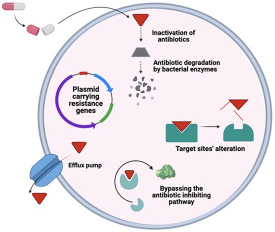
2. Drivers of Antimicrobial Resistance

During the 1960s, the first bacteria showing resistance to multiple drugs were Shigella, Salmonella, and Escherichia coli [8][9][10][8,9,10]. The increase in antimicrobial-resistant bacteria/pathogens poses a serious threat to the health sector and leads to extra-economic burdens. One of the significant contributors to this increasing antimicrobial use are the health care systems fighting against it, which allow inappropriate prescriptions and availability of antimicrobials without prescription to the patients, especially in developing countries. All this is then backed by the poor sanitation services, which aid the transmission, and low healthcare budgets have to rely on cheap antibiotics instead of the safer but more expensive ones [11].
We are not creating antimicrobial resistance; we are simply endorsing it by putting on selective evolutionary pressure, which will result in the evolution of numerous genetic mechanisms [12]. Mechanisms by which antibiotics imply selective pressure are poorly understood. We have represented the genetic mechanism of antimicrobial resistance in the ESKAPE pathogen in Figure 1. Routes associated with antimicrobial resistance are dynamic and less predictable. Problems related to antimicrobial resistance can be assessed by simply recognizing two components: the antimicrobials that inhibit an organism’s susceptibility and the resistant genetic determinants in the microorganism selected by antimicrobials [13][14][13,14]. Subsequently, the resistance emerges when these two components interact in an environment or hosts, leading to several clinical problems. Over the years, constant evolution has led to the emergence of that Enterobacteriaceae strains, which have both MDR (multidrug-resistant) and XDR (extensively drug-resistant) strains [15], to nearly all antibiotics available, without any promising treatment alternatives [16].
Figure 1.
Genetic mechanism of Antimicrobial resistance in ESKAPE pathogen.
Bacterial strains are tremendously effective vehicles to spread the antibiotic resistance traits, transferring them horizontally through mobile genetic elements (transposons and plasmids) or vertically to its daughter cells and other species [17]. These genes usually confer resistance against a single group or a family of antibiotics. A high level of resistance arises through sequential mutation in chromosomes, in the absence of plasmids and transposons, which typically mediate high-level resistance [18][19][20][18,19,20]. This scenario was the foremost reason for the initial emergence of penicillin and tetracycline resistance in Neisseria gonorrhoeae. Likewise, a group of Enterobacteriaceae acquired resistance to fluoroquinolones due to mutations in topoisomerase enzymes that alter gene expression and accelerate the membrane proteins that pump the drug out of the cell [18][20][21][18,20,21]. Resistant Staphylococcus aureus strains first appeared in response to vancomycin [22], followed by high-level resistant transposon from Enterococci [23][24][23,24]. An effective administration of contemporary antimicrobials, and the sustained development of the novel candidate, is crucial to protect human and animal health against bacterial pathogens [25].

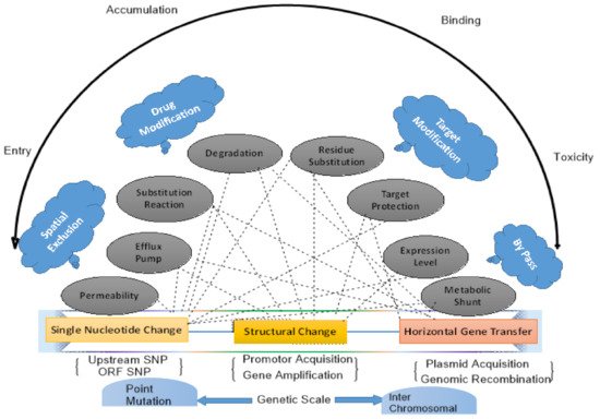
3. Global Dissemination of Antibiotic Resistance

Several studies have been conducted on different samples of resistome from various environments, including studies of human and animal gut microflora, soil, and wastewater microbial communities [26][27][26,27]. Meanwhile, it has become clearly understood that ARGs (antimicrobial-resistant genes) related to clinical sides are prevalent in the environment [28]. Studies utilize metagenomics approaches to directly recover DNA from all microorganisms in a biological sample to investigate the resistome properly. Massive data has been generated from the sequencing of metagenomes and placed in databases. Such data will help in resolving different public health concerns. However, these studies’ data is only limited to identifying genes or predicting novel sequence-based on the same homology to the known reported sequence. Annotation by using sequence-based studies and functional genomics revealed the already known ARGs, which are prevailing in diverse conditions and environments such as in microflora of animals [29] and humans [30][31][30,31] in soil [32][33][32,33] as well as in activated sludge [34]. Numerous examples show that ARGs in human pathogens originated from soil and wastewater bacteria. One of the most well-known examples is blaCTX−M genes, which are the significant root of extended-spectrum b-lactamases (ESBLs) diaspora in Enterobacteriaceae globally and the main starting point of clinical treatment complications [35]. These genes’ marks were identified from chromosomal DNA of different conservational Kluyvera species found in soil and sewage. This can be the origin from where they are disseminated to diverse bacterial species [36]. Likewise, plasmid-encoded qnrA genes, presumed to be originated from fresh marine water species i.e., Shewanella algae, which confers Quinolone resistance, with its various Vibrionaceae species might also be considered as reservoirs [37]. This spread in different Enterobacteriaceae species globally in some areas with a high prevalent rate [38]. Even more, beta-lactamase genes, i.e., OXA-48-type carbapenem-hydrolyzing, progressively reported in various Enterobacteriaceae species, were also found to be originated from environmental Shewanella species [39]. It is thus believed that many clinically relevant resistance genes are found to be originated from non-pathogenic bacteria underlining the colossal potential of horizontal gene transfer (HGT) for these pathogens in overcoming human use of antibiotics.

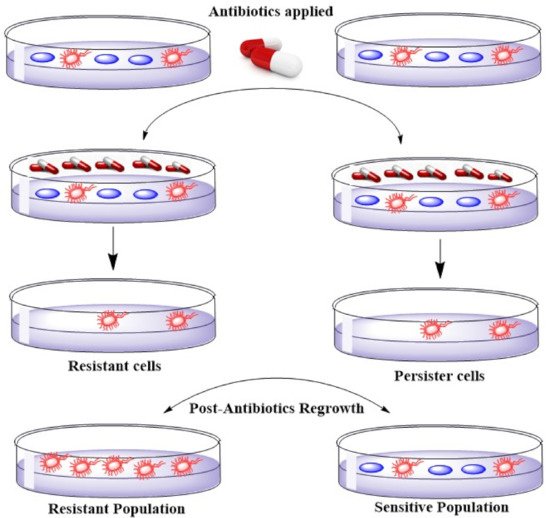
4. Emerging Resistance–Development of Resistant Strains

Resistance genes exist in association with genes specifying resistance to other antimicrobials on similar plasmids that lead to multiple drug resistance [40]. The occurrence of MDR plasmids assures the plasmid’s presence if any one of the resistances offers survival benefit to the host bacterium. This principle similarly implies every determining factor of resistance to biocides like quaternary ammonium compounds because plasmids bearing efflux genes exist that offer resistance to antibiotics in S. aureus [41]. Some studies show a decline in resistance frequencies when an antibiotic is removed [42]. A noteworthy coast-to-coast setback of macrolide resistance in Streptococcus pyogenes occasioned from a Finnish countrywide operation to reduce macrolide practice. In two years, the resistance dropped from about 20% to less than 10%. If a bacterium is resistant to a particular antimicrobial agent, then all the daughter cells would also be resistant (unless additional mutations occurred in the meantime). Persistence, however, describes bacterial cells that are not susceptible to the drug but do not possess resistance genes. The persistence is because some cells in a bacterial population may be in the stationary growth phase (dormant). Most antimicrobial agents do not affect cells that are not actively growing and dividing. These persister cells occur at around 1% in a culture in the stationary phase [43][44][43,44]. Figure 2 shows the difference between persistent and resistant bacterial cells. As depicted in Figure 2, persister cells tolerate the antibiotics by changing to a dormant state. These cells do not divide, and they develop tolerance to a high level of antibiotics. Unlike, resistant cells which develop resistance through accumulating mutations, tolerant persister cells are not antibiotic-resistant mutants. Antibiotic tolerance in persister cells is developed through going to a reversible physiological state in a small subpopulation of bacterial cells [45].
Figure 2.
Illustration of the comparison of Resistance and Persistence in the bacterial population.

5. ESKAPE, Healthcare Concomitant Bugs–Bad Bugs with No Drugs

ESKAPE is an acronym for the group of pathogens, including Gram-positive and Gram-negative species, comprising Enterococcus faecium, Staphylococcus aureus K. pneumoniae, Acinetobacter baumannii, P. aeruginosa, and Enterobacter species (Table 1). The Infectious Disease Society of America has started referring to this group of hospital-originated pathogens as ESKAPE [46][47][46,47]. These bacteria are usually the reasons behind most life-threatening nosocomial infections amongst immunocompromised and critically ill individuals [7]. Klevens [48] revealed that around 1.7 million people are affected by hospital-acquired infections (HAIs) in the US hospitals, which are responsible for nearly 99,000 deaths each year. A survey of HAI in the United States (US) in 2011 reported a total of about 722,000 reported cases, with 75,000 deaths associated with nosocomial infections [11]. It has also been shown that hospitals using antibiotics are where drug-resistant strains first appeared [46]. For instance, S. aureus, which is known to be resistant to penicillin, threatened London’s civilian hospitals soon after the penicillin drug was introduced in the 1940s [7].
Table 1. Narrative of pathogenic bacterial strains (ESKAPE) that instigated nosocomial infection [49].

Bacterial Strain

Gram Staining Type

Resistance

Type

Antibiotics

Treatment Option

Resistance Level

Acinetobacter

Negative

Multidrug

Ceftazidime, aminoglycoside, fluoroquinolones, carbapenems

Carbapenems, b-Lactamase inhibitors, Tigecycline,

Aminoglycosides

, Polymyxin therapy, Synergy, and combination therapy

High level

Inactivation of Drug

Use of hydrolysis or modification

b-lactamase for b-lactam resistance, acetyltransferases for aminoglycoside resistance

E. coli

Negative

Multidrug

Cephalosporins (ESBL-producers), fluoroquinolones, aminoglycosides

GyrB/ParE programme,

EV-035

High level

K. pneumoniae

[64]. However, there is a chance that the bacterial cells, to fight these reactive oxygen species, may produce more detoxification enzymes [65]. The metallic nanoparticles can interact with phosphorus and sulfur, present in biomaterials in bacterial cells like DNA bases. Hence, these can help destroy DNA resulting in killing the cell [66], (Table 3). Some of the possible action mechanisms of nanoparticle-induced death of bacteria are shown in Figure 4.
Figure 4. Suggested action mechanisms of metallic nanoparticles against gram-negative bacteria. Adopted from [67].
Table 3. Mechanism of bactericidal activity of Nanoparticles and synergic effect of antibiotic-conjugated metal oxide nanoparticles against ESKAPE Pathogen.

Nanoparticles (NP)

Mode of Action/Mechanism of Nanoparticles Against ESKAPE Pathogens

Antibiotic Used

Microorganism

Synergic Effects

(Antibiotics-Nanoparticles)

Ref.

[

53

]

AgNPs

Damage the bacterial cell membrane and disrupt the activity of membranous enzymes. Cell wall distraction by cell DNA was condensed to a tension state and could have lost its replicating abilities

[

Doxycycline

54]

[53,54]

K. pneumoniae

Observed

[68]

Alteration of Target

Gentamicin and Neomycin

Reduction of binding affinity to the drug by bypassing the drug target

DNA gyrase mutation for fluoroquinolone resistance

S. aureus

[

AgNPs + Gentamicin showed resistance in 50% strains while AgNPs + Neomycin showed synergy 45% of the strains.

55]

[

69

]

Negative

Multidrug

Drug influx Reduction

By decreasing permeability

Gram-negative outer membrane

Cephalosporins (ESBL-producers), fluoroquinolones, aminoglycosides, carbapenems


E. coli, S. aureus

[

Observed increase in activity was such that Erythromycin showed 18.9.6%, Kanamycin = 27.9.3%, Chloramphenicol = 18.1.3%, and Ampicillin = 74.8.9%

56]

[

POL7080 and ACHN-975 compounds

High level

69

]

P. aeruginosa

Negative

Multidrug

Piperacillin/tazobactam, ceftazidime, ciprofloxacin, aminoglycosides, carbapenems

POL7080 and ACHN-975 compounds

High level

Extrusion of Drug

β-Lactam, cefotaxime

Efflux pumps

E. coli, S. aureus

Accessory membrane fusion proteins

[

Synergistic increase in activity was such that 17.2%, 13.5% for 57]

Enterococcus spp.

Positive

Multidrug

Ampicillin, aminoglycosides, glycopeptides

Horizontal gene transfer

By resistance determinants from other microorganisms


[58]RX-04 lead series, 50S ribosomal subunit; inhibit translation by stabilizing a distorted mode of P-tRNA binding

High level

S. aureus

Positive,

Multidrug

β-lactam antibiotics (except new anti- methicillin-resistant S. aureus cephalosporins), macrolides, fluoroquinolones, aminoglycosides

RX-04 lead series, 50S ribosomal subunit; inhibit translation by stabilizing a distorted mode of P-tRNA binding

High level

6. General Mechanism of Antimicrobial Resistance

Many bacteria live as complex communities called biofilms in their natural habitat, including human hosts. These communities of bacteria offer enhanced resistance to environmental stress, including resistance to antibiotics [50]. The resistance that microorganisms obtain via biofilm formation can be approximately 1000 folds higher than the resistance obtained at the cellular level [50][51][50,51]. The development of resistance at a cellular level is endogenous gene mutations and horizontal gene transfer of resistance determinants through plasmids to other microbes (Figure 3). Apart from resistance, tolerance is also one way to evade antibiotics developed in persister cells, described previously [52]. Both types of resistance may be simultaneous, hence increasing the microbial community’s antimicrobial resistance [50] (Table 2).
Figure 3.
Illustration of the general mechanism of antimicrobial resistance in bacteria.
Table 2. Types of antimicrobial resistance at the cellular level.

Resistance

Proposed Mechanism

Examples

Ref.

7. Alternative Mechanisms for Combating Multidrug Resistance in ESKAPE Pathogens

7.1. CRISPR-Cas9

There are several applications of the cutting-edge technology known as Clustered Regularly Interspaced Short Palindromic Repeats and their associated Cas proteins (CRISPR/Cas system). As the CRISPR induces double-standard breaks, one could be the knocking out of a particular bacterial gene. This characteristic of CRISPR/Cas has led to its use to target specific genes for resistance located in plasmids. One of the advantages of using the CRISPR/Cas system is that it has the capability of multiplexing against different targets, which then enables it to target different resistance genes simultaneously. The question arises whether this approach can be effective in the removal of the resistant genes from MDR bacteria that are present in intestinal microbiota or not? The main limitation is to have a collection of appropriate temperate phages designed against multiple resistance genes, and that resistance genes carried by the bacteria should be known. This is feasible in the current situation. It has been observed that phages are well tolerated when they are orally administered [9]. The orally administered phase therapy for bacteria targeting present in the intestinal tract has been a success. However, to avoid bacteriophages’ deactivation by acid, the stomach must be passed before using the CRISPR/Cas approaches. However, there is a need to conduct further studies to confirm whether the phages will still be active then they reach the intestinal tract, and if not, how can we make sure of it? There is also a need to know the optimal dose that should be used.
Another advantage of this approach is that without compromising the patients’ normal microbiota, susceptibility to antibiotics is restored. Further development of the two approaches discussed above would be revolutionary in the fight against antimicrobial resistance. These techniques could be used for patients with MDR bacteria in various settings to prevent the spread of MDR bacterial strain [59]. On the other hand, the animals have also been shown to play an essential role in reservoirs of MDR bacteria. Therefore, these techniques can also be used for them.

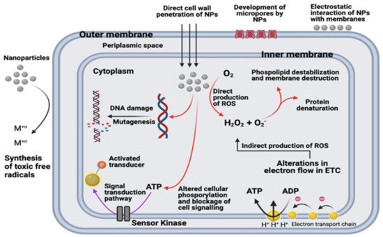
7.2. Nanotechnology and Nanoparticles to Combat Multidrug Resistance

Several hypotheses have been put forwarded for the mechanism of nanoparticles of metals and metal oxides. The hypothesis includes protein dysfunction, physically disrupting the cell structure, generation of reactive oxygen species and depletion of antioxidants, impairing of membrane and interfering with the nutrient assimilation and use of dephosphorylation of the peptide substrates on tyrosine residues which help to alter the signal transduction resulting in its inhibition and suppressing the bacterial growth [60]. The nanoparticles derived from zinc oxide and silver can penetrate the bacterial cell wall and result in changes of its cell membrane, which causes structural damage; hence, the integrity of the membrane is lost, leading to cell death [61][62][61,62].
Silver nanoparticles are also known to mount on the cell wall and form pits in it, while gold nanoparticles apply their antibacterial activities by disintegrating the bacterial cell membrane [63]. Apart from these mechanisms, there is another mechanism in which free radicals are produced to generate oxidative stress. These generated reactive oxygen species can destroy the bacteria by destroying its DNA, membrane, and mitochondria, hence ultimately killing the bacterial cell

E. coli

and

S. aureus

, respectively

[

70]

Ampicillin, chloramphenicol, and kanamycin

S. aureus, E. coli,

and

P. aeruginosa

Synergistic effects observed

[71]

Beta-lactam: cephem

S. aureus

Cephalothin and cefazolin showed a 30% increase in activity when used in combination with 20 μg/ mL AgNPs against Micrococcus luteus, and Bacillus subtilis

[72]

AuNPs

Disturb membrane potential by inhibiting ATPase activities; inhibit the subunit of the ribosome from binding tRNA. Cellular death induced by gold nanoparticles do not include reactive oxygen species-based mechanisms

Ampicillin, streptomycin, and kanamycin

E. coli and S. aureus

15%, 12%, and 34% increase in inhibition zone for E. coli with A/S/K+Au, respectively; 20%, 109%, and 18% increase in inhibition zone for M. luteus A/S/K+AuNPs, respectively; 12% and 34% increase in inhibition zone for S. aureus with A/ K+AuNPs, respectively

[73]

Beta lactams: cefaclor

S. aureus and E. coli

MICs of cefaclor reduced gold nanoparticles were 10 mg/mL and 100 mg/mL for S. aureus and E. coli, respectively

[74]

ZnONPs

Interactions between reactive oxygen species and membrane proteins result in cell damage. ZnO-NPs disrupt bacterial cell membrane integrity, reduce cell surface hydrophobicity, and down-regulate the transcription of oxidative stress-resistance genes in bacteria

Ceftriaxone

E. coli

Synergistic antibacterial effects against E. coli have been observed by ZnO nanorods with ceftriaxone

[75]

Ciprofloxacin

S. aureus and E. coli

Increase in inhibition zones in S. aureus = 27% and 22% in E. coli when ciprofloxacin and ZnONPs were applied in synergism

[76]

Beta lactams, aminoglycosides, and azolides

S. aureus

The highest increase was observed for penicillin G and amikacin, i.e., 10 mm increase in the zone of inhibition, whereas for clarithromycin, a 2 mm increase had been observed

[77]

TiO2NPs

Electrostatic interaction between TiO2 NPs and the bacterial cell surface results in suppression of cell division, degradation of the cell wall and cytoplasmic membrane due to the production of reactive oxygen species such as hydroxyl radicals and hydrogen peroxide

Penicillin G, amikacin, cephalexin, cefotaxime

MRSA

10 mm increase in zone size. TiO2 nanoparticles significantly improved antibiotic efficacy against S. aureus when combined with beta-lactams, cephalosporins, and aminoglycosides

[78]

Fe3O4NPs

Generation of reactive oxygen species from the disruption of the electronic transport chains owing to the resilient affinity of the iron-based nanoparticles for the cell membrane. Reactive oxygen species generated by Fe3O4 nanoparticles kill bacteria without harming non-bacterial cells

Streptomycin

S. aureus, E. coli, and P. aeruginosa

Zones of inhibition at concentrations (10, 20, 40, and 80): S. aureus (15 mm, 14 mm, 17 mm, 20 mm), E. coli (12 mm, 14 mm, 15 mm, 17 mm), P. aeruginosa (13 mm, 14 mm, 15 mm, 18 mm)

[79][80][81]

[79,80,81]

Kanamycin and rifampicin

E. coli and S. aureus

Kanamycin formed an inhibition zone against both, whereas rifampicin formed an inhibitory zone against S. aureus only

[81]

Amoxicillin

E. coli and S. aureus

A total of 9.9% and 8.9% increase in inhibitory effect observed in the presence of Cu NPs for E. coli and S. aureus, respectively

[80]

CuNPs

Generation of reactive oxygen species, lipid peroxidation, protein oxidation, and DNA degradation. Cu2+ ions released from nanoparticles penetrate bacterial cells and are subsequently oxidized intracellularly

Amikacin, ciprofloxacin, gentamicin, norfloxacin

E. coli, P. aeruginosa, Klebsiella spp. S. aureus

At 60 mg/mL, 18 mm for E. coli, 16 mm for Klebsiella

[82]

BiNPs

Production of reactive oxygen species

Ciprofloxacin, norfloxacin, tetracycline, and metronidazole

K. pneumoniae

A synergistic effect was observed between all antibiotics and BiNPs.

[83]

Cefotaxime, ampicillin, ceftriaxone, cefepime

E. coli, K. pneumoniae,

and

P.aeruginosa

Significant decrease in MIC decrease with cefotaxime and ZnO NPs against K. pneumoniae (85.7%), P. aeruginosa (70%), and E. coli (50%) has been observed. Meanwhile, a decrease in MIC due to ZnO NP with other antibiotics has been observed.

[84]

Norfloxacin, Ofloxacin, and Cephalexin

P. aeruginosa, E. coli

Significant increase in inhibition zone of antibiotics with ZnONPshave been observed against all isolates.

[67]