Myeloid-Derived Suppressor Cells in Cancer: Comparison
Please note this is a comparison between Version 2 by Vivi Li and Version 1 by David Gallego-Ortega.

The emergence of immunotherapy has been an astounding breakthrough in cancer treatments. In particular, immune checkpoint inhibitors, targeting PD-1 and CTLA-4, have shown remarkable therapeutic outcomes. However, response rates from immunotherapy have been reported to be varied, with some having pronounced success and others with minimal to no clinical benefit. An important aspect associated with this discrepancy in patient response is the immune-suppressive effects elicited by the tumour microenvironment (TME). Immune suppression plays a pivotal role in regulating cancer progression, metastasis, and reducing immunotherapy success. Most notably, myeloid-derived suppressor cells (MDSC), a heterogeneous population of immature myeloid cells, have potent mechanisms to inhibit T-cell and NK-cell activity to promote tumour growth, development of the pre-metastatic niche, and contribute to resistance to immunotherapy. Accumulating research indicates that MDSC can be a therapeutic target to alleviate their pro-tumourigenic functions and immunosuppressive activities to bolster the efficacy of checkpoint inhibitors.

  • Myeloid derived suppressor cells
  • tumour microenvironment
  • immunotherapy
  • immune system
  • immune checkpoint inhibitors

1. Introduction

The immune system is a complex and dynamic system that operates through an intricate network of cellular interactions and transient functional states. It is involved in various biological activities and is the sine qua non for natural defense of the human body against pathological processes. In cancer progression, the immune system plays a pivotal role where immune cells infiltrate tumours, co-evolving and cooperating with cancer cells to create an inflammatory and immunosuppressive microenvironment to facilitate tumour growth and dissemination.
In the early stages of carcinogenesis, immunologically vulnerable neoplasms are contained and abrogated by immune cells upon detection by immunosurveillance, a process where the immune system inhibits aberrant cell growth. The elimination of immunogenic neoplasms creates a selective pressure that drives the propagation of non-immunogenic clones with adapted mechanisms for immune evasion and survival. Paradoxically the immunosurveillance process against tumour cells promotes the immunoselection of poorly immunogenic variants. The continuous cycle of immune selection for resistant cancer variants leads to tumour escape through multiple mechanisms, including reduced expression of tumour-associated antigens and co-stimulatory molecules, including major histocompatibility complex (MHC) [1]. Tumour cells can also hijack mechanisms that confer survival advantages by increasing proliferation and/or reducing apoptosis [2]. This paradigm of cancer “immunoediting” describes the evolution and selection of cancer cells to develop clinically relevant tumours.
The development, survival, and spread of cancer cells involve a myriad of complex interactions between cancer and immune cells; in which immune cells are involved in both pro-tumourigenic and anti-tumourigenic roles [3,4,5,6][3][4][5][6]. The diverse immune milieu that exists within the tumour microenvironment (TME) secretes various signals that orchestrate the development and progression of cancer through the selection of pro-tumourigenic characteristics such as bypassing apoptotic pathways, immunoevasion, and maintaining inflammation and angiogenesis [3]. As the TME develops and evolves, immunosuppressive cells such as T-regulatory cells (Treg) and myeloid-derived suppressor cells (MDSCs) are co-opted to inhibit the proliferation and activity of killer T cells; thereby promoting tumour progression and metastasis. On the other hand, the immune system can be stimulated to elicit an immune response that targets the tumour for eradication. Thus, the main theory of immunotherapy resides on the plasticity of the immune system and its capacity to be re-educated into restoring a potent anti-tumourigenic response. Thus, immunosuppressive cells within the TME have become a major target for improving the efficacy of immunotherapy, and multiple therapeutic strategies have been developed in the last few years.

2. Targeting MDSCs in Cancer

The reduction in T-cell responsiveness by MDSCs is often associated with resistance against treatments, reducing the efficacy of immunotherapies, and ultimately in patient outcomes [55,191,195][7][8][9]. In breast cancer, circulating MDSCs were associated with cancer stage and metastatic burden, ultimately resulting in poor patient outcomes [72][10]. Clinical trials have also revealed the correlation between patient response to CTLA-4 and PD-1 checkpoint inhibitors, and the abundance of MDSC populations [150,196,197,198][11][12][13][14]. Studies on MDSCs have been more focused on assessing the dynamic roles of MDSC in immunosuppression and tumourigenesis, characterising their relationship with other cell species within the TME, and identifying new targetable pathways to deplete MDSC populations or inhibit their function [199][15]. MDSCs can be targeted by (1) depletion of circulating and tumour-infiltrated MDSCs; (2) prevention of MDSC recruitment and trafficking; (3) inhibition of MDSC immunosuppressive functions; and (4) differentiation of MDSCs into a non-suppressive immune state (Figure 1).
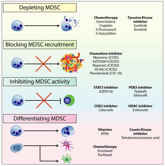
Figure 1. Treatments used to target different mechanisms associated with pro-tumourigenic MDSC. There are multiple therapeutic approaches against MDSC to restore anti-tumour functions in immune cells and improve immunotherapy, in particular checkpoint inhibitors. These approaches include: (1) depleting MDSC populations through low-dose chemotherapy and tyrosine kinase inhibitors; (2) preventing MDSC recruitment to the TME by targeting chemokine receptors responsible for the recruitment and migration of MDSCs; (3) attenuating the immunosuppressive mechanisms of MDSC by downregulating the expression of ARG1 and iNOS, and reducing ROS generation; (4) inducing the differentiation of MDSC into mature myeloid cells to reduce MDSC population and remove their immunosuppression.

2.1. Depleting MDSC Populations

Low dose chemotherapy has been shown to be effective in eliminating MDSC populations in tumour-bearing mice; treatments with chemotherapy such as 5-fluorouracil (5FU), paclitaxel, cisplatin, and gemcitabine were found to deplete MDSCs and enhance anti-tumour immune activity [200,201,202,203][16][17][18][19]. However, a contrasting effect in the use of chemotherapy against MDSC was observed where MDSCs were transiently induced following cyclophosphamide treatment in tumour-bearing mice and patients [204,205][20][21].
Signalling cascades involved in MDSC expansion has also been a target in reducing the amplification of MDSC populations. For example, VEGF promotes the expansion of MDSCs, recruitment of Tregs, angiogenesis, and tumour progression. To target this, the tyrosine kinase inhibitor Sunitinib has been used successfully to deplete MDSC in patients suffering from renal cell carcinoma via blockade of VEGF and c-KIT signalling [184,206,207][22][23][24]. In addition, Sunitinib was found to also inhibit STAT3, and renal cell carcinoma patients treated with Sunitinib showed a reversal in MDSC accumulation and consequently T-cell suppression [206][23]. Finally, through a unique peptibody, Qin et al. developed a novel therapeutic peptide-Fc fusion protein that targeted the S100A family proteins to selectively deplete MDSCs without targeting other proinflammatory immune cells [208][25].

2.2. Blockade of MDSC Migration

As mentioned previously, it is strategically more effective to use therapeutic blockade to target chemokine receptors on MDSCs owing to ligand redundancy. The chemokines receptor CCR5 plays a crucial role in the chemotaxis of MDSC into the TME via the ligands CCL3, CCL4, and CCL5 [51][26]. However, the expression is not ubiquitous to all MDSCs; in melanoma mouse models and patients, MDSCs that express CCR5 were found to have more potent immunosuppressive mechanisms compared to the ones that do not express CCR5 [77][27]. Blattner et al. demonstrated that the blockade of CCR5 inhibited the recruitment and immunosuppressive activity of MDSC and improved survival in melanoma [77][27]. Similarly, CCR5 antagonists inhibited the metastatic potential of basal breast cancer and reduced tumour growth [209,210][28][29].
Elevated levels of CCL2 and CCL5 are present in the TME to recruit MDSC through the chemokine receptor CXCR2 [118,211][30][31]. CXCR2+ MDSC promoted tumour expansion, metastasis, EMT, and T-cell exhaustion in breast cancer [212][32]. By targeting CXCR2, MDSC populations were reduced and reported to decrease metastasis, promote T-cell infiltration into the tumours, improve anti-PD1 therapy, and extend survival in pancreatic cancer [159][33]. Additionally, CXCR2 antagonists against MDSCs have been shown to enhance the therapeutic efficacy of PD-1 immunotherapy, T-cell transfer, and chemotherapy [150,213,214][11][34][35].
CSF-1R has also been a primary target to inhibit MDSC recruitment to the tumour site to constrain tumourgenesis. CSF-1R is a tyrosine kinase receptor that when bound with its ligand CSF-1 promotes the differentiation and expansion of myeloid cells into MDSC and TAMs in addition to promoting their migration to tumours [215][36]. CSF-1R has been found to be upregulated in several types of cancer, such as pancreatic and breast [216,217][37][38]. Treatments targeting the receptor or its ligand CSF-1R/CSF-1 has been found to improve T-cell responses and combining CSF-1R inhibition with checkpoint blockades or adoptive T-cell transfer therapy resulted in improved anti-tumour T-cell activity and tumour regression [215,218,219][36][39][40]. CSF-1R inhibition and CXCR2 antagonism has also been used in combination to reduce TAM and PMN-MDSC populations and improve anti PD-1 efficacy [220][41].
Currently, the following MDSC inhibitors are in clinical trials [221][42]: Reparixin (CXCR2) is in Phase 2 clinical trials for TNBC (NCT02370238); AZD5069 (CXCR2) is in Phase 1b/2 for advanced solid tumours and metastatic squamous cell carcinoma (NCT02499328); Plexidartinib (CSF-1R) is in Phase 2 for recurrent glioblastoma (NCT01349036), and Maraviroc (CCR5) is in Phase 1 for metastatic colorectal cancer (NCT01736813).

2.3. Attenuating MDSC Immunosuppressive Functions

Mitigating the potent immunosuppressive mechanisms of MDSCs have been a major therapeutic target to re-establishing T-cell activity and immunotherapy success. PGE2, as mentioned previously, is involved in inflammation, angiogenesis, tumour progression via recruitment of MDSC, and is also involved in the expression of one of the primary immunosuppressive mechanisms employed by MDSC: ARG1 [222,223,224][43][44][45]. Since cyclooxygenase-2 (COX-2) is upstream of the PGE2 synthesis pathway, therapies targeting COX-2, such as celecoxib, have been of great interest as a form of immunoregulatory treatment to suppress MDSC function whilst enhancing immunotherapy [225][46]. Disruption of the COX-2/PGE2 signalling has been successful in reducing MDSC recruitment and differentiation, repressing MDSC-associated suppressive factors such as ARG1 expression and ROS production, and shifting an inflammatory tumour profile to more anti-cancer immune rejection; consequently, COX-2 inhibition has resulted in improved CTL frequency and immune response, delayed tumour growth, and synergy between checkpoint inhibitors and dendritic cell-based immunotherapy [71,222,223,225,226][47][43][44][46][48].
Phosphodiesterase-5 (PDE-5) inhibitors are also able to abrogate MDSC immunosuppressive mechanisms by targeting MDSC expression and function of ARG1 and iNOS. Administration of PDE-5 inhibitors, such as sildenafil and tadalafil, have reportedly reduced inflammation in the TME, restabilised anti-tumour immune rejection through T-cell and NK cell activity, and prolonged survival in vivo [227,228,229][49][50][51]. Clinical trials with PDE-5 inhibitors have also shown positive results in head and neck squamous cell carcinoma and metastatic melanoma patients [230[52][53],231], abatement of MDSC and T-reg populations, enhanced intra-tumour T-cell activity, and improved patient outcome [231,232][53][54].
Anti-inflammatory triterpenoids have been used to activate the Nrf2 gene in MDSCs. Nrf2 is involved in modulating the expression of antioxidant enzymes, including NADPH, NQO1, and hemeoxygenase, and conferring cytoprotection against oxidative stress [233][55]. Selective activation of Nrf2 using synthetic triterpenoids, such as CCDO-IM and CCDO-Me, has reduced intracellular ROS production (abrogating MDSC-driven immunosuppression), reduced metastasis, and has shown promising anticancer results in Phase 1 clinical trials that are well-tolerated with patients [234,235,236][56][57][58]. Another target to reduce oxidative stress is NO. Nitroaspirin targets iNOS to reduce ROS build-up; treatments have resulted in improved T-cell proliferation, function, invasion into the tumour core, and suppressed tumourigenesis [180,237][59][60].
STAT3 inhibition is another promising target. The antisense oligonucleotide STAT3 inhibitor, AZD9150, has been used in conjunction with immune checkpoint inhibitors in Phase 1b clinical trials for the treatment of diffuse large B-cell lymphoma. Systemic administration of AZD9150 in patients showed a marked decrease in granulocytic MDSC within the peripheral blood mononuclear cells (PBMC) [238][61].

2.4. Inducing MDSC Differentiation

Promoting the differentiation of IMC is another successful strategy in reducing MDSC populations and abolishing their immunosuppressive functions. All-trans-retinoic acid (ATRA), an agonist of retinoid receptors, inhibits retinoic signalling to shift the differentiation of MDSC into mature myeloid cells, such as macrophages and dendritic cells. ATRA treatment has resulted in a reduction in T-cell suppression by directly inducing differentiation of MDSCs into mature antigen-presenting precursor cells [239][62]. This reduction in MDSCs and improvement in T-cell response have been observed in both mice and patients in various cancer types, such as renal cell carcinoma and small cell lung carcinoma [240,241][63][64]. The improvement by ATRA administration was reported to reduce circulating MDSC, enhance cancer vaccine treatments, improve dendritic cell function, and ameliorate antigen-specific T-cell response [240,241][63][64]. The mechanism of ATRA-induced differentiation of MDSC was reported to be mediated by glutathione synthase and neutralising ROS generation [242][65]. In addition, the casein kinase inhibitor tetrabromocinnamic acid was also shown to restore myeloid cell differentiation in tumour-bearing mice through improved Notch signalling [243][66].
Finally, epigenetic reprogramming is a novel avenue to target the pro-tumorigenic properties of MDSCs. The class I histone deacetylase inhibitor (HDAC), entinostat, has shown positive results in neutralising MDSC through epigenetic reprogramming in mouse models of pancreatic, breast, and lung cancers; and renal cell carcinoma [244,245][67][68]. The combination of entinostat with immune checkpoint inhibitors have resulted in prolonged survival, expansion of CD8+ T cells, and inhibition of immunosuppressive functions in both M-MDSC and PMN-MDSC via downregulation of ARG1, iNOS, and COX-2; overall, this resulted in a shift of the tumour dynamic into a more immune-susceptible TME [244,245][67][68]. Clinical trials involving entinostat are currently underway. However, clinical trials ENCORE 602 (NCT02708680) and ENCORE 603 (NCT02915523) for TNBC and ovarian cancer, respectively, have failed to increase progression-free survival. Another similar effect was observed with the use of the DNA demethylating epigenetic agent 5-azacytidine, which resulted in a reduction of MDSC and Arg1 expression [246][69].
Application of other chemotherapies was also reported to induce MDSC differentiation into non-immunosuppressive cell types. For example, docetaxel had a novel chemoimmunomodulatory effect by inhibiting STAT3 phosphorylation and polarising MDSC differentiation into M1-like macrophages [247][70]. Comparably, paclitaxel was also reported to reduce MDSC populations by promoting MDSC differentiation into dendritic cells that were independent of TLR-4 [248][71].

3. Combining MDSC-Targeted Treatments with Immunotherapy

To improve the success of immunotherapy, there has been a paradigm shift—both the innate and adaptive layers of the immune system are simultaneously targeted to alleviate the immunosuppressive TME and re-elicit the anti-tumour response [67][72]. As MDSCs are one of the primary immunosuppressive cells acting as an escape mechanism for cancer cells by subverting immunosurveillance and abrogating T-cell activity, treatment strategies have been shifting towards a combination of both targeting MDSCs and immunotherapy. Indeed, targeting MDSCs may be key in diminishing tumour expansion and resensitising tumours to immune governance, thus overcoming MDSC-driven immunosuppression (Figure 2). Targeting myeloid populations alone is often insufficient as an immune-based monotherapy; however, there is compelling research and clinical trials that have shown promising results for combination therapy.
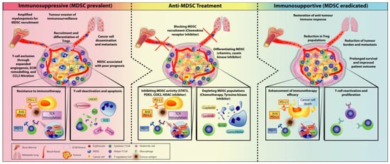
Figure 2. Treatment of MDSC to alleviate an immunosuppressive environment as an approach to enhancing immunotherapeutic treatments by shifting towards an immunosupportive TME. The immunosuppressive TME is propagated by various suppressive cells such as MDSCs and Tregs. Recruitment of MDSC within the TME can promote tumour expansion through various mechanisms (developing a pre-metastatic niche to help cancer cell metastasis, inducing resistance to immunotherapy by preventing the infiltration of T-cell into the tumour, suppressing and deactivating T-cell function, and inducing T-cell apoptosis) and recruitment of Tregs to further amplify immunosuppression. Thus, MDSC is often associated with poor prognosis in patients. Anti-MDSC treatments have become a major clinical target to re-establish immune control against cancer. By creating an immunosupportive environment, T-cell activity is restored, which leads to improved immunotherapy efficacy. Overall, this has resulted in prolonged survival and reduction of metastasis and tumour regression.

3.1. Checkpoint Inhibitors Combined with MDSC Depletion

Pre-clinical studies have demonstrated successful results when combining checkpoint inhibitor treatment with MDSC depletion. Kim et al. showed that co-treatment using the epigenetic modulatory drugs, entinostat and 5-azacytidine, with checkpoint inhibitors, anti-PD1 and anti-CTLA4, resulted in complete tumour regression and metastatic progression in the aggressive TNBC model 4T1, with over 80% survival rate 100 days post-implantation of the tumour [249][73]. Interestingly, when entinostat and 5-azacytidine were used together but not in combination with checkpoint inhibitors and vice versa, the primary tumours and metastasis remained, pointing to the synergistic effects of combination therapy in targeting MDSC and immune checkpoints. Similar results were observed in murine models of lung and renal cell carcinoma [244][67]. In an HER2 transgenic breast cancer and a metastatic pancreatic cancer mouse model, the entinostat-driven inhibition of MDSC activity with checkpoint inhibitor treatment resulted in an upregulation of granzyme B-producing CD8+ T-cells and improved the infiltration and function of adaptive immune cells. Tumour-free survival was significantly improved in these highly aggressive cancer types [245][68].

3.2. Immunotherapy Combined with Obstructing MDSC Trafficking Therapy

CXCR2+ MDSC were found to promote immune suppression when migrated to the TME; the efficacy of checkpoint inhibition in a mice model of rhabdomyosarcoma was severely limited by MDSC [150][11]. Disruption of CXCR2-mediated migration in MDSC was demonstrated to significantly improve anti-PD1 treatments. CXCR2+ MDSCs were also found to have potent immunosuppressive properties in human paediatric sarcoma, and thus CXCR2 may serve as a target to prevent MDSC recruitment to improve immunotherapeutic intervention. Furthermore, targeting CXCR2 improved T-cell infiltration and when combined with anti-PD1 treatment, mice bearing pancreatic cancer showed significantly extended survival [159][33]. SX-682, a small molecule CXCR1 and CXCR2 inhibitor, was reported to substantially reduce PMN-MDSC trafficking and infiltration to the tumour in mice [214][35]. Reduction in intratumour PMN-MDSC populations enhanced the accumulation of both endogenous T-cells and T-cells from adoptive transfer. Similarly to epigenetic agents, SX-682 had little anti-tumour effect as a monotherapy, but in combination therapy with checkpoint inhibitors and adoptive T-cell transfer therapy, it greatly enhanced their efficacy by inhibiting the recruitment of tumour-infiltrated CXCR2+ PMN-MDSCs [214][35]. SX-682 has been tested in conjunction with Pembrolizumab in P1 clinical trials for metastatic melanoma (NCT03161431).

References

  1. Garcia-Lora, A.; Algarra, I.; Garrido, F. MHC class I antigens, immune surveillance, and tumor immune escape. J. Cell Physiol. 2003, 195, 346–355.
  2. Fernald, K.; Kurokawa, M. Evading apoptosis in cancer. Trends Cell Biol. 2013, 23, 620–633.
  3. Burkholder, B. Tumor-induced perturbations of cytokines and immune cell networks. Biochim. Biophys. Acta 2014, 1845, 182–201.
  4. Salgado, R. The evaluation of tumor-infiltrating lymphocytes (TILs) in breast cancer: Recommendations by an International TILs Working Group 2014. Ann. Oncol. 2015, 26, 259–271.
  5. Chow, M.T.; Moller, A.; Smyth, M.J. Inflammation and immune surveillance in cancer. Semin Cancer Biol. 2012, 22, 23–32.
  6. Valdes-Mora, F. Single-Cell Transcriptomics in Cancer Immunobiology: The Future of Precision Oncology. Front. Immunol. 2018, 9, 2582.
  7. Gabrilovich, D.I.; Ostrand-Rosenberg, S.; Bronte, V. Coordinated regulation of myeloid cells by tumours. Nat. Rev. Immunol. 2012, 12, 253–268.
  8. Messmer, M.N. Tumor-induced myeloid dysfunction and its implications for cancer immunotherapy. Cancer Immunol. Immunother. 2015, 64, 1–13.
  9. Mantovani, A. The growing diversity and spectrum of action of myeloid-derived suppressor cells. Eur. J. Immunol. 2010, 40, 3317–3320.
  10. Diaz-Montero, C.M. Increased circulating myeloid-derived suppressor cells correlate with clinical cancer stage, metastatic tumor burden, and doxorubicin-cyclophosphamide chemotherapy. Cancer Immunol. Immunother. 2009, 58, 49–59.
  11. Highfill, S.L. Disruption of CXCR2-mediated MDSC tumor trafficking enhances anti-PD1 efficacy. Sci. Transl Med. 2014, 6, 237ra67.
  12. Meyer, C. Frequencies of circulating MDSC correlate with clinical outcome of melanoma patients treated with ipilimumab. Cancer Immunol. Immunother. 2014, 63, 247–257.
  13. Tarhini, A.A. Immune monitoring of the circulation and the tumor microenvironment in patients with regionally advanced melanoma receiving neoadjuvant ipilimumab. PLoS ONE 2014, 9, e87705.
  14. Weber, J. Phase I/II Study of Metastatic Melanoma Patients Treated with Nivolumab Who Had Progressed after Ipilimumab. Cancer Immunol. Res. 2016, 4, 345–353.
  15. Groth, C. Immunosuppression mediated by myeloid-derived suppressor cells (MDSCs) during tumour progression. Br. J. Cancer 2019, 120, 16–25.
  16. Suzuki, E. Gemcitabine selectively eliminates splenic Gr-1+/CD11b+ myeloid suppressor cells in tumor-bearing animals and enhances antitumor immune activity. Clin. Cancer Res. 2005, 11, 6713–6721.
  17. Sevko, A. Antitumor effect of paclitaxel is mediated by inhibition of myeloid-derived suppressor cells and chronic inflammation in the spontaneous melanoma model. J. Immunol. 2013, 190, 2464–2471.
  18. Eriksson, E. Gemcitabine reduces MDSCs, tregs and TGFbeta-1 while restoring the teff/treg ratio in patients with pancreatic cancer. J. Transl. Med. 2016, 14, 282.
  19. Vincent, J. 5-Fluorouracil selectively kills tumor-associated myeloid-derived suppressor cells resulting in enhanced T cell-dependent antitumor immunity. Cancer Res. 2010, 70, 3052–3061.
  20. Mikyskova, R. Cyclophosphamide-induced myeloid-derived suppressor cell population is immunosuppressive but not identical to myeloid-derived suppressor cells induced by growing TC-1 tumors. J. Immunother. 2012, 35, 374–384.
  21. Iida, Y. Contrasting effects of cyclophosphamide on anti-CTL-associated protein 4 blockade therapy in two mouse tumor models. Cancer Sci. 2017, 108, 1974–1984.
  22. Fleming, V. Targeting Myeloid-Derived Suppressor Cells to Bypass Tumor-Induced Immunosuppression. Front. Immunol. 2018, 9, 398.
  23. Ko, J.S. Sunitinib mediates reversal of myeloid-derived suppressor cell accumulation in renal cell carcinoma patients. Clin. Cancer Res. 2009, 15, 2148–2157.
  24. Kodera, Y. Sunitinib inhibits lymphatic endothelial cell functions and lymph node metastasis in a breast cancer model through inhibition of vascular endothelial growth factor receptor 3. Breast Cancer Res. 2011, 13, R66.
  25. Qin, H. Generation of a new therapeutic peptide that depletes myeloid-derived suppressor cells in tumor-bearing mice. Nat. Med. 2014, 20, 676–681.
  26. Weber, R. Myeloid-Derived Suppressor Cells Hinder the Anti-Cancer Activity of Immune Checkpoint Inhibitors. Front. Immunol. 2018, 9, 1310.
  27. Blattner, C. CCR5(+) Myeloid-Derived Suppressor Cells Are Enriched and Activated in Melanoma Lesions. Cancer Res. 2018, 78, 157–167.
  28. Robinson, S.C. A chemokine receptor antagonist inhibits experimental breast tumor growth. Cancer Res. 2003, 63, 8360–8365.
  29. Velasco-Velazquez, M. CCR5 antagonist blocks metastasis of basal breast cancer cells. Cancer Res. 2012, 72, 3839–3850.
  30. Katoh, H. CXCR2-expressing myeloid-derived suppressor cells are essential to promote colitis-associated tumorigenesis. Cancer Cell 2013, 24, 631–644.
  31. Toh, B. Mesenchymal transition and dissemination of cancer cells is driven by myeloid-derived suppressor cells infiltrating the primary tumor. PLoS Biol. 2011, 9, e1001162.
  32. Zhu, H. CXCR2(+) MDSCs promote breast cancer progression by inducing EMT and activated T cell exhaustion. Oncotarget 2017, 8, 114554–114567.
  33. Steele, C.W. CXCR2 Inhibition Profoundly Suppresses Metastases and Augments Immunotherapy in Pancreatic Ductal Adenocarcinoma. Cancer Cell 2016, 29, 832–845.
  34. Di Mitri, D. Tumour-infiltrating Gr-1+ myeloid cells antagonize senescence in cancer. Nature 2014, 515, 134–137.
  35. Sun, L. Inhibiting myeloid-derived suppressor cell trafficking enhances T cell immunotherapy. JCI Insight 2019, 4.
  36. Holmgaard, R.B. Targeting myeloid-derived suppressor cells with colony stimulating factor-1 receptor blockade can reverse immune resistance to immunotherapy in indoleamine 2,3-dioxygenase-expressing tumors. EBioMedicine 2016, 6, 50–58.
  37. Zhu, Y. CSF1/CSF1R blockade reprograms tumor-infiltrating macrophages and improves response to T-cell checkpoint immunotherapy in pancreatic cancer models. Cancer Res. 2014, 74, 5057–5069.
  38. Richardsen, E. Macrophage-colony stimulating factor (CSF1) predicts breast cancer progression and mortality. AntiCancer Res. 2015, 35, 865–874.
  39. Sluijter, M. Inhibition of CSF-1R supports T-cell mediated melanoma therapy. PLoS ONE 2014, 9, e104230.
  40. Mok, S. Inhibition of CSF-1 receptor improves the antitumor efficacy of adoptive cell transfer immunotherapy. Cancer Res. 2014, 74, 153–161.
  41. Kumar, V. Cancer-Associated Fibroblasts Neutralize the Anti-tumor Effect of CSF1 Receptor Blockade by Inducing PMN-MDSC Infiltration of Tumors. Cancer Cell 2017, 32, 654–668.
  42. Elliott, L.A. Human Tumor-Infiltrating Myeloid Cells: Phenotypic and Functional Diversity. Front. Immunol. 2017, 8, 86.
  43. Sinha, P. Prostaglandin E2 promotes tumor progression by inducing myeloid-derived suppressor cells. Cancer Res. 2007, 67, 4507–4513.
  44. Eruslanov, E. Pivotal Advance: Tumor-mediated induction of myeloid-derived suppressor cells and M2-polarized macrophages by altering intracellular PGE(2) catabolism in myeloid cells. J. Leukoc. Biol. 2010, 88, 839–848.
  45. Ochoa, A.C. Arginase, prostaglandins, and myeloid-derived suppressor cells in renal cell carcinoma. Clin. Cancer Res. 2007, 13, 721s–726s.
  46. Veltman, J.D. COX-2 inhibition improves immunotherapy and is associated with decreased numbers of myeloid-derived suppressor cells in mesothelioma. Celecoxib influences MDSC function. EBioMedicine 2010, 10, 464.
  47. Obermajer, N. Positive feedback between PGE2 and COX2 redirects the differentiation of human dendritic cells toward stable myeloid-derived suppressor cells. Blood 2011, 118, 5498–5505.
  48. Zelenay, S. Cyclooxygenase-Dependent Tumor Growth through Evasion of Immunity. Cell 2015, 162, 1257–1270.
  49. Serafini, P. Phosphodiesterase-5 inhibition augments endogenous antitumor immunity by reducing myeloid-derived suppressor cell function. J. Exp. Med. 2006, 203, 2691–2702.
  50. Lin, S. Phosphodiesterase-5 inhibition suppresses colonic inflammation-induced tumorigenesis via blocking the recruitment of MDSC. Am. J. Cancer Res. 2017, 7, 41–52.
  51. Tai, L.H. Phosphodiesterase-5 inhibition reduces postoperative metastatic disease by targeting surgery-induced myeloid derived suppressor cell-dependent inhibition of Natural Killer cell cytotoxicity. Oncoimmunology 2018, 7, e1431082.
  52. Califano, J.A. Tadalafil augments tumor specific immunity in patients with head and neck squamous cell carcinoma. Clin. Cancer Res. 2015, 21, 30–38.
  53. Hassel, J.C. Tadalafil has biologic activity in human melanoma. Results of a pilot trial with Tadalafil in patients with metastatic Melanoma (TaMe). Oncoimmunology 2017, 6, e1326440.
  54. Weed, D.T. Tadalafil reduces myeloid-derived suppressor cells and regulatory T cells and promotes tumor immunity in patients with head and neck squamous cell carcinoma. Clin. Cancer Res. 2015, 21, 39–48.
  55. Ohl, K.; Tenbrock, K. Reactive Oxygen Species as Regulators of MDSC-Mediated Immune Suppression. Front. Immunol. 2018, 9, 2499.
  56. Wang, Y.Y. Bardoxolone methyl (CDDO-Me) as a therapeutic agent: An update on its pharmacokinetic and pharmacodynamic properties. Drug Des. Devel. Ther. 2014, 8, 2075–2088.
  57. Hiramoto, K. Myeloid lineage-specific deletion of antioxidant system enhances tumor metastasis. Cancer Prev. Res. (Phila) 2014, 7, 835–844.
  58. Nagaraj, S. Anti-inflammatory triterpenoid blocks immune suppressive function of MDSCs and improves immune response in cancer. Clin. Cancer Res. 2010, 16, 1812–1823.
  59. Molon, B. Chemokine nitration prevents intratumoral infiltration of antigen-specific T cells. J. Exp. Med. 2011, 208, 1949–1962.
  60. De Santo, C. Nitroaspirin corrects immune dysfunction in tumor-bearing hosts and promotes tumor eradication by cancer vaccination. Proc. Natl. Acad. Sci. USA 2005, 102, 4185–4190.
  61. Reilley, M.J. STAT3 antisense oligonucleotide AZD9150 in a subset of patients with heavily pretreated lymphoma: Results of a phase 1b trial. J. Immunother. Cancer 2018, 6, 119.
  62. Kusmartsev, S. Reversal of myeloid cell-mediated immunosuppression in patients with metastatic renal cell carcinoma. Clin. Cancer Res. 2008, 14, 8270–8278.
  63. Mirza, N. All-trans-retinoic acid improves differentiation of myeloid cells and immune response in cancer patients. Cancer Res. 2006, 66, 9299–9307.
  64. Iclozan, C. Therapeutic regulation of myeloid-derived suppressor cells and immune response to cancer vaccine in patients with extensive stage small cell lung cancer. Cancer Immunol. Immunother. 2013, 62, 909–918.
  65. Nefedova, Y. Mechanism of all-trans retinoic acid effect on tumor-associated myeloid-derived suppressor cells. Cancer Res. 2007, 67, 11021–11028.
  66. Cheng, P. Effects of notch signaling on regulation of myeloid cell differentiation in cancer. Cancer Res. 2014, 74, 141–152.
  67. Orillion, A. Entinostat Neutralizes Myeloid-Derived Suppressor Cells and Enhances the Antitumor Effect of PD-1 Inhibition in Murine Models of Lung and Renal Cell Carcinoma. Clin. Cancer Res. 2017, 23, 5187–5201.
  68. Christmas, B.J. Entinostat Converts Immune-Resistant Breast and Pancreatic Cancers into Checkpoint-Responsive Tumors by Reprogramming Tumor-Infiltrating MDSCs. Cancer Immunol. Res. 2018, 6, 1561–1577.
  69. Mikyskova, R. DNA demethylating agent 5-azacytidine inhibits myeloid-derived suppressor cells induced by tumor growth and cyclophosphamide treatment. J. Leukoc. Biol. 2014, 95, 743–753.
  70. Kodumudi, K.N. A novel chemoimmunomodulating property of docetaxel: Suppression of myeloid-derived suppressor cells in tumor bearers. Clin. Cancer Res. 2010, 16, 4583–4594.
  71. Michels, T. Paclitaxel promotes differentiation of myeloid-derived suppressor cells into dendritic cells in vitro in a TLR4-independent manner. J. Immunotoxicol. 2012, 9, 292–300.
  72. Law, A.M. The innate and adaptive infiltrating immune systems as targets for breast cancer immunotherapy. Endocr. Relat. Cancer 2017, 24, R123–R144.
  73. Kim, K. Eradication of metastatic mouse cancers resistant to immune checkpoint blockade by suppression of myeloid-derived cells. Proc. Natl. Acad. Sci. USA 2014, 111, 11774–11779.
More
ScholarVision Creations