Lactose Intolerance and Personalized Nutrition: Comparison
Please note this is a comparison between Version 2 by Catherine Yang and Version 1 by Millie Porzi.

Recent discoveries in the “omics” field and the growing focus on preventive health have opened new avenues for personalized nutrition (PN), which is becoming an important theme in the strategic plans of organizations that are active in healthcare, food, and nutrition research. PN holds great potential for individual health optimization, disease management, public health interventions, and product innovation. However, there are still multiple challenges to overcome before PN can be truly embraced by the public and healthcare stakeholders. The diagnosis and management of lactose intolerance (LI), a common condition with a strong inter-individual component, is explored as an interesting example for the potential role of these technologies and the challenges of PN. From the development of genetic and metabolomic LI diagnostic tests that can be carried out in the home, to advances in the understanding of LI pathology and individualized treatment optimization, PN in LI care has shown substantial progress. However, there are still many research gaps to address, including the understanding of epigenetic regulation of lactase expression and how lactose is metabolized by the gut microbiota, in order to achieve better LI detection and effective therapeutic interventions to reverse the potential health consequences of LI.

  • lactose intolerance
  • lactase persistence
  • genetic testing
  • polymorphisms
  • epigenetic
  • omics
  • personalized nutrition
  • dairy products
  • functional foods
  • Gut Microbiota

1. Introduction

Nutrition is an environmental variable of major importance for optimal health and disease prevention [1]. However, optimizing nutrition for health is challenging due to the highly variable individual response to diet [2], which results from the combination of internal factors such as a person’s genetics and microbiome, as well as external factors like stress and physical activity [1][3]. Recent advances in high-throughput “omics” technologies and bioinformatics tools have helped to better understand the inter-individual variation in response to food intake [1][3].
The notion of an individual response to diet is a central element of personalized nutrition (PN), also often referred to as “precision nutrition” or “individualized nutrition”, which can be defined as an approach that uses individual information, more recently based on “big data”, to develop customized nutritional advice, products, and services [4]. The overall goal of PN is to prevent, manage or treat diseases, optimize health and well-being using clinical assessments, genetic information, biomarkers, and any other relevant information about individuals [3]. The diagnosis and management of lactose intolerance (LI), a common condition with a strong inter-individual component, offers an interesting example for the potential and the challenges of PN.
The typical gastrointestinal symptoms of LI are specifically due to the maldigestion of lactose resulting from a lack of the lactase enzyme [5]. The normal physiological decline in lactase activity during early childhood leads to the appearance of LI in adults, the lactase non-persistence (LNP) phenotype, due to the inheritance of an autosomal recessive trait [6]. LNP is the ancestral type and most common phenotype associated with lactase gene expression worldwide, with a global prevalence estimated at 68% [7]. In contrast, lactase production into adulthood, the lactase persistence (LP) phenotype, is observed in the presence of a gain-of-function mutation and is inherited as an autosomal dominant trait [8][9]. The LP variant is not evenly distributed worldwide: high frequencies are observed in people from European descent and in populations with a long history of dairying activity [10]. The spread of farming during the Neolithic period correlates with the occurrence of the LP phenotype in human populations, with the earliest appearance estimated ~8000–9000 years ago in Europeans, ~2700–6800 years ago in African populations, and ~4000 years ago in Middle Eastern populations [11][12]. This evolutionary process provides anthropological evidence for gene-culture co-evolution, with a positive selection on the LP phenotype in relation to the domestication of dairying animals and consumption of their milk [13][14][15]. Indeed, LP would have conferred various selective advantages, including access to a major source of energy to improve nutritional status, but also a largely pathogen-free source of fluids to prevent dehydration in arid environments, and a valuable source of calcium for maintaining bone health [11][13][16].
The role of lactose metabolism in human evolution and nutrition is a fascinating case that brings together ancient evolutionary history with recent discoveries on lactase epigenetic regulation, as well as the influence of the gut microbiota on the LI state and novel PN approaches in LI care. In this context, this article aims to review the current approaches used for the diagnostic and management of LI, to investigate the impact of LI on human health focusing on the application of nutrigenomics tools, and to critically discuss how recent development in PN will impact diagnostic tools and therapeutic interventions for LI.

2. Physiology and Pathophysiology of Lactose Intolerance
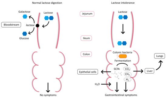
The digestion and metabolism of the disaccharide lactose, requires the hydrolysis of the glycosidic bond connecting its monosaccharides, galactose and glucose, by the enzyme lactase-phlorizin hydrolase, commonly known as lactase, which belongs to the beta-galactosidase family [17]. The lactase encoding gene (LCT) is localized on the long arm of chromosome 2 in position 21 (2q21). On the same chromosome, MCM6 encodes the minichromosome maintenance complex component 6, a regulatory element that controls the expression of LCT [5][18]. Initially, lactase is synthesized as pre-pro-lactase, which contains a signal sequence that is then cleaved in the endoplasmic reticulum to form pro-lactase. During intracellular transport, pro-lactase becomes N- and O-glycosylated in the endoplasmic reticulum and Golgi apparatus leading to the mature form of lactase, which is then exported and anchored to the apical brush border membrane of the intestinal epithelial cells [19].
The lactase enzyme is abundantly present in the proximal part of the jejunum, while its presence progressively declines towards the ileum [20]. After hydrolysis, galactose and glucose sugars are actively absorbed across the intestinal epithelial cells and transported into the bloodstream to be used as a source of energy (Figure 1). When lactase is absent or deficient, unhydrolyzed lactose is able to reach the terminal ileum and subsequently enters the colon. An excess of undigested and therefore non-absorbable lactose will draw water from the bloodstream into the intestinal lumen via an osmotic effect, causing loose stools or watery diarrhea [17][21]. Within the large intestine, the undigested lactose is then cleaved into monosaccharides by the colonic microbiota. This bacterial fermentation process forms short-chain fatty acids (SCFAs), such as acetate, propionate, and butyrate. SCFAs can either be used by the intestinal epithelial cells or excreted in the feces. In addition, gases are produced from the bacterial fermentation of lactose, primarily hydrogen (H2), carbon dioxide (CO2), and methane (CH4), which increase intracolonic pressure. All these factors lead to gastrointestinal symptoms including flatulence, bloating, abdominal pain, cramps, and nausea [22][23]. However, the severity of the symptoms after lactose ingestion depends on the amount of lactose ingested, intestinal transit time, lactase expression, variability of intestinal microbiota, individual sensitivity, and psychological factors [21][24].
Figure 1.
Normal lactose digestion and lactose intolerance.
Currently, the exact prevalence of LI remains unknown, but it is acknowledged that it varies considerably among different ethnic populations [21]. LP and LNP are determined by assessing genetic polymorphisms associated with LP. At least five single nucleotide polymorphisms (SNPs) occurring upstream of the LCT gene have been associated with LP into adulthood: LCT-13910C/T and LCT-22018G/A in populations of European descent, LCT-13915T/G in African and Middle Eastern populations, LCT-14010G/C and LCT-13907C/G in some African tribes [10][11][25][26][27]. For the most common SNPs associated with LP in the European population (LCT-13910C/T and LCT-22018G/A), homozygotes (TT and AA) and heterozygotes (CT and GA) are indicative of LP, whereas the wild type (CC and GG) results in LNP [28][29]. In consequence, homozygous carriers of LCT-13910C/C and LCT-22018G/G are typically found to develop LI. Interestingly, heterozygous carriers of LCT-13910C/T and LCT-22018G/A have shown intermediate enzymatic activity with the occurrence of LI symptoms during situations of stress or with intestinal infections [30][31]. The currently known SNPs only provide a partial description of the mechanisms involved in the development of LI and the exact process by which the lactase is downregulated in early childhood still remains unclear [25][28][32][33].

3. Types of Lactose Intolerance

The complete or partial inability to digest lactose often leads to the same clinical manifestations of LI, though the causes of lactose maldigestion differ. LI refers to a syndrome in which the lactose maldigestion causes an onset of gastrointestinal symptoms including diarrhea, bloating, flatulence, nausea, abdominal pain, and cramps [34] (Table 1). It should not be confused with milk or dairy allergies that are characterized by an abnormal immunologic response that can develop into severe life-threatening anaphylaxis [35]. LI is typically caused by lactase deficiency (LD) which implies a reduced or absent lactase enzyme activity in the small intestinal mucosa. There are three main forms of lactase deficiency: congenital, primary, and secondary. Congenital lactase deficiency (alactasia) is a rare autosomal recessive pediatric disorder associated with an absence of lactase expression in newborns. Primary lactase deficiency (adult-onset hypolactasia) is the condition resulting from the progressive and physiological decline of lactase enzyme activity that typically occurs during childhood. Conversely, secondary lactase deficiency (acquired LI) is induced by small intestine disease or injury such as gastroenteritis, celiac disease, inflammatory bowel disease, chemotherapy, and antibiotics treatment [21][34]. Of note, ethnicity has been shown to be a more important determinant of susceptibility to developing lactase deficiency in many patients with inflammatory bowel disease rather than disease markers (particularly for cases of ulcerative colitis and Crohn’s disease that does not involve the small intestine, distal obstruction, or bacterial overgrowth) [36][37]. Generally, the non-genetic aetiologies of LI can be reversed if the cause can be eliminated.

4. Diagnosis of Lactose Intolerance

The diagnosis of LI relies in part upon the development of gastrointestinal symptoms resulting from lactose ingestion, not all of which can be assessed objectively and many which overlap with other conditions, notably irritable bowel syndrome in which lactose maldigestion may be accompanied by sensitivity to other fermentable carbohydrates described as FODMAPs (fermentable oligosaccharide, disaccharide, monosaccharide, and polyols) [5][38][39][40]. In practice, the diagnosis of LI is usually made on the basis of clinical suspicion supported by the positive response to a dietary challenge such as a trial period of a lactose-free diet [41]. However, several clinical diagnostic tests are available including blood, breath, and genetic tests, which can confirm a diagnosis even in the absence of gastrointestinal symptoms.
Diagnosis of LI confirmed by a jejunal biopsy for the in vitro assessment of lactase activity [42], is regarded as the “gold standard” test for LI diagnosis and can be used to exclude other gastrointestinal conditions. False positives are rare for this method but false negatives may occur due to irregular dissemination of lactase in the intestine. However, this method is almost exclusively used in clinical research because of its high cost, invasiveness, and need for highly specialized equipment [5][21]. In contrast, the oral lactose tolerance test (LTT) is a minimally invasive metabolic test, which consists in the determination of blood glucose levels at various times, following the administration of an oral overload of lactose (25–50 g). As the digestion of lactose results in an elevation of serum glucose, lactose malabsorption is indicated by a failure in blood glucose level to rise ≥ 1.1 mmol/L above the basal value, 60–120 min after lactose ingestion [17][21]. However, due to individual variability in the gastrointestinal transit time and glucose metabolism, false-positive and false-negative test results are relatively frequent for the LTT, reducing its use in clinical practice [43]. Alternatively, the lactose hydrogen breath test (HBT) has been widely adopted for the detection of an increase in H2 in expired air at several time points, after an oral lactose challenge (25–50 g). H2 is produced due to bacterial fermentation of non-digested lactose in the colon and can be indirectly assessed in breath (Figure 1). A rise of exhaled H2 of ≥20 ppm from baseline within 90 min after lactose ingestion is indicative of lactose malabsorption [44][45]. Even though this test accuracy is influenced by the gut microbiome, relatively high sensitivity and specificity have been reported for HBT, making it the most common type of LI test used today [21][43][46][47].
The gaxilose test is a more recent and non-invasive diagnostic test based on oral administration of 4-galactosylxylose (gaxilose), a synthetic disaccharide and structural analogue of lactose. The intestinal lactase hydrolyzes the gaxilose compound into D-xylose, which is then absorbed into the blood and subsequently excreted in the urine. The D-xylose levels in urine or serum can be quantified by colorimetric methods [48]. Besides its high sensitivity and specificity, the gaxilose test is easy to use, does not require specialized equipment, and only induces minimal subject discomfort [49]. Genetic testing has also emerged as a less invasive tool for supporting the diagnosis of LI and tests based on the most common SNPs that are linked to LP in the Caucasian population (LCT-13910C/T and LCT-22018G/A) have been developed [29]. However, the use of these SNPs cannot be applied as a global diagnostic tool, as other polymorphisms that confer LP have been identified in several African and Arabian populations [5][10]. Moreover, this method may not detect all SNPs associated with LI that exist within multi-ethnic populations [10][33][50]. Currently genetic tests have a limited role in diagnosing LI in the clinical setting, as none of them achieve perfect sensitivity and specificity and the results do not always correlate with clinical symptoms [5].

5. Management of Lactose Intolerance

The main treatment options for LI consists in preventing gastrointestinal symptoms by reducing or eliminating the amount of lactose in the diet or by taking oral enzyme replacement therapy. In order to manage their symptoms, people with LI should avoid eating high-lactose foods, such as fresh milk or cream [51], while ensuring an adequate intake of nutrients from other foods [52]. It is also recommended that individuals with LI eat lactose-containing foods together with other foods and that they favor small repeated intakes of lactose over one single meal with a high amount of lactose [53]. Fermented dairy products like hard cheese, quark, or yogurt are suitable for the majority of individuals with LI [54]. In fact, most aged hard cheeses naturally contain very little, if any lactose [51]. Yogurt usually still contains an appreciable amount of lactose, but delivers lactic acid bacteria with beta-galactosidase activity known to improve lactose digestion [51][52][55].
It should also be noted that lactose is a common additive in many processed foods, such as frozen meals, sweets, cakes, and sauces [53]. This so-called “hidden lactose” is used for its texture and flavor enhancing properties. Moreover, lactose is commonly used in the pharmaceutical industry as an excipient for oral medications [56]. The dose of lactose in oral solid-dosage form is generally small as most pharmaceuticals provide less than 2 g of lactose per day. Nevertheless, alternative medications might be necessary for individuals suffering from severe LI [57]. Overall, individuals with LI should be aware of these hidden or added sources of lactose and their possible impact on LI symptoms when combined.
To support the management of LI, alternative foods that are naturally lactose-free, such as soy products (e.g., tofu, edamame) and plant-based drinks (e.g., soy, almond, and rice drink) are often recommended as an alternative for high-lactose foods [58]. In addition, the food industry has developed many “low-lactose” and “lactose-free” products using diverse processes to remove lactose from lactose-rich dairy foods [52]. Lactose can be physically removed from milk using ultrafiltration or chromatographic separation followed by subsequent hydrolysis of the remaining lactose [59]. Thus, the sensory properties of lactose-free milk produced are not affected, but this process may also remove some valuable minerals, like calcium [60]. Alternatively, lactose-free milk can be obtained by enzymatic hydrolysis of lactose to its monosaccharides, glucose and galactose, using microbial beta-galactosidase [59]. This process is known to generate extra sweetness and may also have an impact on the nutritional value of the hydrolyzed milk [52]. More recently, the use of “A2 milk” has been shown to reduce some of the gastrointestinal discomfort associated with drinking ordinary cow’s milk in individuals with LI [61]. “A2 milk” is a variety of cow’s milk containing mostly A2 beta-casein (like human milk, sheep and goat’s milk) that does not metabolize to the peptide beta-casomorphin-7 (BMC-7), which is implicated in adverse gastrointestinal effects, including inflammation [62]. Interestingly, the gastrointestinal symptoms resulting from the ingestion of cow’s milk in LI individuals are decreased when milk enriched in A2 beta-casein is consumed in place of regular milk (which contains A1 beta-casein) [61]. This indicates that the gastrointestinal symptoms due to LI might be confounded by the beta-casein variant present in milk.
Finally, in the case where food-based approaches to manage LI are insufficient or not feasible, lactase enzyme replacements may be taken prior to consuming a lactose-containing meal [63]. This therapeutic option uses exogenous lactase for lactose digestion, like the fungal beta-galactosidase tilactase, which may be helpful as an alternative to dietary restriction and thus avoid possible nutritional deficiencies [64][65].

References

  1. Kaput, J.; Kussmann, M.; Radonjic, M.; Virgili, F.; Perozzi, G. Human nutrition, environment, and health. Genes Nutr. 2015, 10, 489.
  2. Walther, B.; Lett, A.M.; Bordoni, A.; Tomás-Cobos, L.; Nieto, J.A.; Dupont, D.; Danesi, F.; Shahar, D.R.; Echaniz, A.; Re, R.; et al. GutSelf: Interindividual Variability in the Processing of Dietary Compounds by the Human Gastrointestinal Tract. Mol. Nutr. Food Res. 2019, 63, e1900677.
  3. Ordovas, J.M.; Ferguson, L.R.; Tai, E.S.; Mathers, J.C. Personalised nutrition and health. BMJ 2018, 361, bmj.k2173.
  4. Bush, C.L.; Blumberg, J.B.; El-Sohemy, A.; Minich, D.M.; Ordovás, J.M.; Reed, D.G.; Behm, V.A.Y. Toward the Definition of Personalized Nutrition: A Proposal by The American Nutrition Association. J. Am. Coll. Nutr. 2020, 39, 5–15.
  5. Mattar, R.; de Campos Mazo, D.F.; Carrilho, F.J. Lactose intolerance: Diagnosis, genetic, and clinical factors. Clin. Exp. Gastroenterol. 2012, 5, 113–121.
  6. Wiley, A.S. Lactose intolerance. Evol. Med. Public Health 2020, 2020, 47–48.
  7. Storhaug, C.L.; Fosse, S.K.; Fadnes, L.T. Country, regional, and global estimates for lactose malabsorption in adults: A systematic review and meta-analysis. Lancet Gastroenterol. Hepatol. 2017, 2, 738–746.
  8. Smith, G.D.; Lawlor, D.A.; Timpson, N.J.; Baban, J.; Kiessling, M.; Day, I.N.M.; Ebrahim, S. Lactase persistence-related genetic variant: Population substructure and health outcomes. Eur. J. Hum. Genet. 2009, 17, 357–367.
  9. Comerford, K.B.; Pasin, G. Gene-Dairy Food Interactions and Health Outcomes: A Review of Nutrigenetic Studies. Nutrients 2017, 9, 710.
  10. Itan, Y.; Jones, B.L.; Ingram, C.J.; Swallow, D.M.; Thomas, M.G. A worldwide correlation of lactase persistence phenotype and genotypes. BMC Evol. Biol. 2010, 10, 36.
  11. Tishkoff, S.A.; Reed, F.A.; Ranciaro, A.; Voight, B.F.; Babbitt, C.C.; Silverman, J.S.; Powell, K.; Mortensen, H.M.; Hirbo, J.B.; Osman, M.; et al. Convergent adaptation of human lactase persistence in Africa and Europe. Nat. Genet. 2007, 39, 31–40.
  12. Luca, F.; Perry, G.H.; Di Rienzo, A. Evolutionary adaptations to dietary changes. Annu. Rev. Nutr. 2010, 30, 291–314.
  13. Gerbault, P.; Liebert, A.; Itan, Y.; Powell, A.; Currat, M.; Burger, J.; Swallow, D.M.; Thomas, M.G. Evolution of lactase persistence: An example of human niche construction. Philos. Trans. R. Soc. Lond B Biol. Sci. 2011, 366, 863–877.
  14. Itan, Y.; Powell, A.; Beaumont, M.A.; Burger, J.; Thomas, M.G. The origins of lactase persistence in Europe. PLoS Comput. Biol. 2009, 5, e1000491.
  15. Segurel, L.; Guarino-Vignon, P.; Marchi, N.; Lafosse, S.; Laurent, R.; Bon, C.; Fabre, A.; Hegay, T.; Heyer, E. Why and when was lactase persistence selected for? Insights from Central Asian herders and ancient DNA. PLoS Biol. 2020, 18, e3000742.
  16. Ségurel, L.; Bon, C. On the Evolution of Lactase Persistence in Humans. Annu. Rev. Genom. Hum. Genet. 2017, 18, 297–319.
  17. Lomer, M.C.; Parkes, G.C.; Sanderson, J.D. Review article: Lactose intolerance in clinical practice-myths and realities. Aliment. Pharmacol. 2008, 27, 93–103.
  18. Ingram, C.J.E.; Mulcare, C.A.; Itan, Y.; Thomas, M.G.; Swallow, D.M. Lactose digestion and the evolutionary genetics of lactase persistence. Hum. Genet. 2009, 124, 579–591.
  19. Kuchay, R.A.H. New insights into the molecular basis of lactase non-persistence/persistence: A brief review. Drug Discov. Ther. 2020, 14, 1–7.
  20. Forsgård, R.A. Lactose digestion in humans: Intestinal lactase appears to be constitutive whereas the colonic microbiome is adaptable. Am. J. Clin. Nutr. 2019, 110, 273–279.
  21. Misselwitz, B.; Butter, M.; Verbeke, K.; Fox, M.R. Update on lactose malabsorption and intolerance: Pathogenesis, diagnosis and clinical management. Gut 2019, 68, 2080–2091.
  22. He, T.; Venema, K.; Priebe, M.G.; Welling, G.W.; Brummer, R.J.; Vonk, R.J. The role of colonic metabolism in lactose intolerance. Eur. J. Clin. Investig. 2008, 38, 541–547.
  23. Deng, Y.; Misselwitz, B.; Dai, N.; Fox, M. Lactose Intolerance in Adults: Biological Mechanism and Dietary Management. Nutrients 2015, 7, 8020–8035.
  24. Szilagyi, A. Adult lactose digestion status and effects on disease. Can. J. Gastroenterol. Hepatol. 2015, 29, 149–156.
  25. Swallow, D.M. Genetics of lactase persistence and lactose intolerance. Annu. Rev. Genet. 2003, 37, 197–219.
  26. Ingram, C.J.; Elamin, M.F.; Mulcare, C.A.; Weale, M.E.; Tarekegn, A.; Raga, T.O.; Bekele, E.; Elamin, F.M.; Thomas, M.G.; Bradman, N.; et al. A novel polymorphism associated with lactose tolerance in Africa: Multiple causes for lactase persistence? Hum. Genet. 2007, 120, 779–788.
  27. Enattah, N.S.; Sahi, T.; Savilahti, E.; Terwilliger, J.D.; Peltonen, L.; Järvelä, I. Identification of a variant associated with adult-type hypolactasia. Nat. Genet. 2002, 30, 233–237.
  28. Santonocito, C.; Scapaticci, M.; Guarino, D.; Annicchiarico, E.B.; Lisci, R.; Penitente, R.; Gasbarrini, A.; Zuppi, C.; Capoluongo, E. Lactose intolerance genetic testing: Is it useful as routine screening? Results on 1426 south-central Italy patients. Clin. Chim. Acta 2015, 439, 14–17.
  29. Tomczonek-Moruś, J.; Wojtasik, A.; Zeman, K.; Smolarz, B.; Bąk-Romaniszyn, L. 13910C>T and 22018G>A LCT gene polymorphisms in diagnosing hypolactasia in children. United Eur. Gastroenterol. J. 2019, 7, 210–216.
  30. Enattah, N.-S.; Kuokkanen, M.; Forsblom, C.; Natah, S.; Oksanen, A.; Jarvela, I.; Peltonen, L.; Savilahti, E. Correlation of intestinal disaccharidase activities with the C/T-13910 variant and age. World J. Gastroenterol. 2007, 13, 3508–3512.
  31. Gugatschka, M.; Dobnig, H.; Fahrleitner-Pammer, A.; Pietschmann, P.; Kudlacek, S.; Strele, A.; Obermayer-Pietsch, B. Molecularly-defined lactose malabsorption, milk consumption and anthropometric differences in adult males. Qjm 2005, 98, 857–863.
  32. Matthews, S.B.; Waud, J.P.; Roberts, A.G.; Campbell, A.K. Systemic lactose intolerance: A new perspective on an old problem. Postgrad Med. J. 2005, 81, 167–173.
  33. Troelsen, J.T. Adult-type hypolactasia and regulation of lactase expression. Biochim. Biophys. Acta 2005, 1723, 19–32.
  34. Szilagyi, A.; Ishayek, N. Lactose Intolerance, Dairy Avoidance, and Treatment Options. Nutrients 2018, 10, 1994.
  35. Walsh, J.; Meyer, R.; Shah, N.; Quekett, J.; Fox, A.T. Differentiating milk allergy (IgE and non-IgE mediated) from lactose intolerance: Understanding the underlying mechanisms and presentations. Br. J. Gen. Pr. 2016, 66, e609–e611.
  36. Mishkin, B.; Yalovsky, M.; Mishkin, S. Increased prevalence of lactose malabsorption in Crohn’s disease patients at low risk for lactose malabsorption based on ethnic origin. Am. J. Gastroenterol. 1997, 92, 1148–1153.
  37. Szilagyi, A.; Galiatsatos, P.; Xue, X. Systematic review and meta-analysis of lactose digestion, its impact on intolerance and nutritional effects of dairy food restriction in inflammatory bowel diseases. Nutr. J. 2016, 15, 67.
  38. Hasan, N.; Zainaldeen, M.; Almadhoob, F.; Yusuf, M.; Fredericks, S. Knowledge of lactose intolerance among clinicians. Gastroenterol. Insights 2018, 9.
  39. Martínez Vázquez, S.E.; Nogueira de Rojas, J.R.; Remes Troche, J.M.; Coss Adame, E.; Rivas Ruíz, R.; Uscanga Domínguez, L.F. The importance of lactose intolerance in individuals with gastrointestinal symptoms. Rev. Gastroenterol. México 2020, 85, 321–331.
  40. Lomer, M.C. Review article: The aetiology, diagnosis, mechanisms and clinical evidence for food intolerance. Aliment. Pharmacol. Ther. 2015, 41, 262–275.
  41. Lule, V.K.; Garg, S.; Tomar, S.K.; Khedkar, C.D.; Nalage, D.N. Food Intolerance: Lactose Intolerance. In Encyclopedia of Food and Health; Caballero, B., Finglas, P.M., Toldrá, F., Eds.; Academic Press: Oxford, UK, 2016; pp. 43–48.
  42. Di Rienzo, T.; D’Angelo, G.; D’Aversa, F.; Campanale, M.C.; Cesario, V.; Montalto, M.; Gasbarrini, A.; Ojetti, V. Lactose intolerance: From diagnosis to correct management. Eur. Rev. Med. Pharmacol. Sci. 2013, 17 (Suppl. 2), 18–25.
  43. Robles, L.; Priefer, R. Lactose Intolerance: What Your Breath Can Tell You. Diagnostics 2020, 10, 412.
  44. Law, D.; Conklin, J.; Pimentel, M. Lactose intolerance and the role of the lactose breath test. Am. J. Gastroenterol. 2010, 105, 1726–1728.
  45. Usai-Satta, P.; Scarpa, M.; Oppia, F.; Cabras, F. Lactose malabsorption and intolerance: What should be the best clinical management? World J. Gastrointest. Pharmacol. Ther. 2012, 3, 29–33.
  46. Furnari, M.; Bonfanti, D.; Parodi, A.; Franzè, J.; Savarino, E.; Bruzzone, L.; Moscatelli, A.; Di Mario, F.; Dulbecco, P.; Savarino, V. A comparison between lactose breath test and quick test on duodenal biopsies for diagnosing lactase deficiency in patients with self-reported lactose intolerance. J. Clin. Gastroenterol. 2013, 47, 148–152.
  47. Hovde, Ø.; Farup, P.G. A comparison of diagnostic tests for lactose malabsorption—Which one is the best? BMC Gastroenterol. 2009, 9, 82.
  48. Aragón, J.J.; Hermida, C.; Martínez-Costa, O.H.; Sánchez, V.; Martín, I.; Sánchez, J.J.; Codoceo, R.; Cano, J.M.; Cano, A.; Crespo, L.; et al. Noninvasive diagnosis of hypolactasia with 4-Galactosylxylose (Gaxilose): A multicentre, open-label, phase IIB-III nonrandomized trial. J. Clin. Gastroenterol. 2014, 48, 29–36.
  49. Domínguez Jiménez, J.L.; Fernández Suárez, A.; Muñoz Colmenero, A.; Fatela Cantillo, D.; López Pelayo, I. Primary hypolactasia diagnosis: Comparison between the gaxilose test, shortened lactose tolerance test, and clinical parameters corresponding to the C/T-13910 polymorphism. Clin. Nutr. 2017, 36, 471–476.
  50. Bayless, T.M.; Brown, E.; Paige, D.M. Lactase Non-persistence and Lactose Intolerance. Curr. Gastroenterol. Rep. 2017, 19, 23.
  51. Gille, D.; Walther, B.; Badertscher, R.; Bosshart, A.; Brügger, C.; Brühlhart, M.; Gauch, R.; Noth, P.; Vergères, G.; Egger, L. Detection of lactose in products with low lactose content. Int. Dairy J. 2018, 83, 17–19.
  52. Dekker, P.J.T.; Koenders, D.; Bruins, M.J. Lactose-Free Dairy Products: Market Developments, Production, Nutrition and Health Benefits. Nutrients 2019, 11, 551.
  53. Facioni, M.S.; Raspini, B.; Pivari, F.; Dogliotti, E.; Cena, H. Nutritional management of lactose intolerance: The importance of diet and food labelling. J. Transl. Med. 2020, 18, 260.
  54. McSweeney, P.L.H. Biochemistry of cheese ripening. Int. J. Dairy Technol. 2004, 57, 127–144.
  55. Savaiano, D.A. Lactose digestion from yogurt: Mechanism and relevance. Am. J. Clin. Nutr. 2014, 99, 1251s–1255s.
  56. Van der Merwe, J.; Steenekamp, J.; Steyn, D.; Hamman, J. The Role of Functional Excipients in Solid Oral Dosage Forms to Overcome Poor Drug Dissolution and Bioavailability. Pharmaceutics 2020, 12, 393.
  57. Mill, D.; Dawson, J.; Johnson, J.L. Managing acute pain in patients who report lactose intolerance: The safety of an old excipient re-examined. Adv. Drug Saf. 2018, 9, 227–235.
  58. Sethi, S.; Tyagi, S.K.; Anurag, R.K. Plant-based milk alternatives an emerging segment of functional beverages: A review. J. Food Sci. Technol. 2016, 53, 3408–3423.
  59. Harju, M.; Kallioinen, H.; Tossavainen, O. Lactose hydrolysis and other conversions in dairy products: Technological aspects. Int. Dairy J. Int. Dairy J. 2012, 22.
  60. Vyas, H.K.; Tong, P.S. Process for Calcium Retention During Skim Milk Ultrafiltration. J. Dairy Sci. 2003, 86, 2761–2766.
  61. Ramakrishnan, M.; Eaton, T.K.; Sermet, O.M.; Savaiano, D.A. Milk Containing A2 β-Casein ONLY, as a Single Meal, Causes Fewer Symptoms of Lactose Intolerance than Milk Containing A1 and A2 β-Caseins in Subjects with Lactose Maldigestion and Intolerance: A Randomized, Double-Blind, Crossover Trial. Nutrients 2020, 12, 3855.
  62. Jianqin, S.; Leiming, X.; Lu, X.; Yelland, G.W.; Ni, J.; Clarke, A.J. Effects of milk containing only A2 beta casein versus milk containing both A1 and A2 beta casein proteins on gastrointestinal physiology, symptoms of discomfort, and cognitive behavior of people with self-reported intolerance to traditional cows’ milk. Nutr. J. 2016, 15, 35.
  63. Suri, S.; Kumar, V.; Prasad, R.; Tanwar, B.; Goyal, A.; Kaur, S.; Gat, Y.; Kumar, A.; Kaur, J.; Singh, D. Considerations for development of lactose-free food. J. Nutr. Intermed. Metab. 2019, 15, 27–34.
  64. Montalto, M.; Nucera, G.; Santoro, L.; Curigliano, V.; Vastola, M.; Covino, M.; Cuoco, L.; Manna, R.; Gasbarrini, A.; Gasbarrini, G. Effect of exogenous beta-galactosidase in patients with lactose malabsorption and intolerance: A crossover double-blind placebo-controlled study. Eur. J. Clin. Nutr. 2005, 59, 489–493.
  65. Montalto, M.; Curigliano, V.; Santoro, L.; Vastola, M.; Cammarota, G.; Manna, R.; Gasbarrini, A.; Gasbarrini, G. Management and treatment of lactose malabsorption. World J. Gastroenterol. 2006, 12, 187–191.
More
Video Production Service