Bone Morphogenetic Protein 7: Comparison
Please note this is a comparison between Version 1 by Chandrakala Aluganti Narasimhulu and Version 2 by Vivi Li.

Bone morphogenetic protein-7 is (BMP-7) is a potent anti-inflammatory growth factor belonging to the Transforming Growth Factor Beta (TGF-β) superfamily. It plays an important role in various biological processes, including embryogenesis, hematopoiesis, neurogenesis and skeletal morphogenesis. BMP-7 stimulates the target cells by binding to specific membrane-bound receptor BMPR 2 and transduces signals through mothers against decapentaplegic (Smads) and mitogen activated protein kinase (MAPK) pathways. To date, rhBMP-7 has been used clinically to induce the differentiation of mesenchymal stem cells bordering the bone fracture site into chondrocytes, osteoclasts, the formation of new bone via calcium deposition and to stimulate the repair of bone fracture. However, its use in cardiovascular diseases, such as atherosclerosis, myocardial infarction, and diabetic cardiomyopathy is currently being explored. More importantly, these cardiovascular diseases are associated with inflammation and infiltrated monocytes where BMP-7 has been demonstrated to be a key player in the differentiation of pro-inflammatory monocytes, or M1 macrophages, into anti-inflammatory M2 macrophages, which reduces developed cardiac dysfunction. 

  • atherosclerosis
  • myocardial infarction
  • diabetic cardiomyopathy
  • inflammation

1. Introduction

In 1970, a physician named Marshall Urist coined the term bone morphogenetic protein (BMP) after demonstrating that these proteins play an important role in osteogenesis and bone formation. Thereafter, more than 20 BMPs have been identified and subdivided into the following four groups; (i) BMP-2/4, (ii) BMP-5/6/7/8a/8b, (iii) BMP-9/10, and (iv) BMP-12/13/14 based on their function and amino acid sequence similarity [1][2][3][4][1,2,3,4]. BMP signaling plays a crucial role in several developmental pathways. BMPs regulate erythropoiesis and neurogenesis during embryonic development by interacting with the BMP receptors (BMPR) I and II [5][6][5,6]. Accordingly, their function in embryogenesis has been extensively studied in several model organisms including frogs, mice, and zebrafish. After birth, they maintain bone mass by inducing the differentiation of mesenchymal stem cells (MSCs) into osteoblasts and regulating their differentiation potential [7][8][9][10][11][12][7,8,9,10,11,12]. Specifically, BMP-2/4/6/7/9/12/13 have the ability to induce MSC differentiation, whereas BMP-3 plays a role in inducing MSC proliferation [7][8][9][10][11][12][13][7,8,9,10,11,12,13]. In addition to MSCs, existing studies reveal that adipocyte, fibroblast, myoblast and neural cell differentiation and proliferation is also regulated by BMPs [14][15][16][17][18][14,15,16,17,18].
Evidence suggests that BMP-2, 4 and 10 deletion is embryonically lethal [19][20][21][19,20,21] whereas loss of BMP-7/11 leads to death immediately after birth [22][23][22,23]. Moreover, deletion of BMP receptors [24][25][26][24,25,26] and downstream transducers (Smad-1/4/5/7) are also embryonically lethal [27][28][29][30][27,28,29,30]. BMP-4 insufficiency prompted an imbalance in the hematopoietic stem cell (HSC) proliferation and differentiation, whereas the lack of BMP-4 has disrupted gastrulation and subsequent formation of the mesoderm, obstructing the generation of major tissues such as cardiac, skeletal, and vascular muscle cells that resulted in animal lethality [31]. BMP-2/10 play a key role in myocardial patterning, chamber formation and maturation [21][32][33][21,32,33]. The diverse biological activities of BMPs [3][25][26][3,25,26] along with their receptors are highlighted in Table 1 [34][35][36][34,35,36], and it is clear that BMP deficiency can result in numerous human pathophysiological diseases and death.
Table 1. Types of bone morphogenetic proteins (BMPs) and their functions.
Types Alternate Names Tissues that Express Functions Receptors
BMP-1 BMP-1 is a metalloproteinase major end organs (heart, lung, liver, pancreas, kidney, and brain), lymphoid organs (bone marrow, thymus, spleen and lymph nodes), exocrine glands (prostate and mammary gland) organ protectors (muscle and bone) Metalloprotease that cleaves COOH–propeptides of procollagens

I, II, and III/induces cartilage formation/cleaves BMP antagonist chordin
_____
BMP-2 BMP-2A, XBMP2, xBMP-2,

MGC114605
major end organs (lung, pancreas, and kidney), lymphoid organ (spleen) Induces bone and cartilage formation. Plays a role in skeletal repair and regeneration/heart formation ALK-2, 3, 6

BMPR-II; ActR-IIA, ActR-IIB
BMP-3a

& 3b
Osteogenin,

BMP-3A
major end organs (brain, heart, pancreas),

exocrine gland (prostate), organ protector (skeletal muscle), lymphoid organs (bone marrow, spleen and thymus), BMP-3b also expresses in spinal cord
Negative regulator of bone morphogenesis

Cell differentiation regulation; skeletal morphogenesis; Regulates cell growth and differentiation in both embryonic and adult tissues
ALK-4

ActR-IIA, ActR-IIB
BMP-4 BMP-2B, BMP2B1, ZYME, OFC11,

MCOPS6
major end organs (brain, heart, pancreas, liver, lung, kidney), exocrine gland (prostate), organ protector (skeletal muscle), lymphoid organs (bone marrow, spleen and thymus), spinal cord Skeletal repair and regeneration; kidney formation; Induces cartilage and bone formation; limb formation; tooth development. ALK-2,3,5,6

BMPR-II, ActR-IIA
BMP-5 MGC34244 major end organs (brain, heart, pancreas, liver, lung, kidney), exocrine gland (prostate), organ protector (skeletal muscle), lymphoid organs (bone marrow, spleen and thymus), spinal cord Limb development; induces bone and cartilage morphogenesis; connecting soft tissues ALK-3

BMPR-II; ActR-IIA, ActR-IIB
BMP-6 Vgr1, DVR-6 major end organs (brain, heart, pancreas, liver, lung, kidney); exocrine gland (prostate); organ protector (muscle and bone),

lymphoid organs (bone marrow, spleen and thymus); spinal cord
Cartilage hypertrophy; bone morphogenesis; nervous system development; Plays a role in early development ALK-2, 3, 6

BMPR-II; ActR-IIA, ActR-IIB
BMP-7 OP-1 major end organs (brain, heart, pancreas, liver, lung, kidney), exocrine gland (prostate) organ protector (skeletal muscle), lymphoid organs (bone marrow, spleen and thymus), spinal cord. Skeletal repair and regeneration; kidney and eye formation; nervous system development

plays a major role in calcium regulation and bone homeostasis
ALK 2, 3, 6

BMPR-II;
BMP-8a

& 8b
OP-2, FLJ14351, FLJ45264

OP-3, PC-8, MGC131757
major end organs (brain, heart, kidney, lung, liver, pancreas), exocrine gland (prostate), organ protector (skeletal muscle), lymphoid organs (spleen, thymus bone marrow) spinal cord Induces cartilage formation; Bone morphogenesis and spermatogenesis; calcium regulation and bone homeostasis. ALK 2; 3; 4; 6; 7

BMPR-II;

ALK3,6

BMPR-II; ActR-IIA, ActR-IIB
BMP-9 GDF-2 major end organ (liver) Bone morphogenesis; cholinergic neurons development; in glucose metabolism;

potent inhibitor of angiogenesis
ALK-1,2

BMPR-II; ActR-IIA, ActR-IIB
BMP-10 MGC126783 major end organs (brain, heart, kidney, lung, liver, pancreas), exocrine gland (prostate), organ protector (skeletal muscle), lymphoid organs (spleen, thymus, bone marrow) spinal cord. Heart morphogenesis maintains

the proliferative activity of embryonic cardiomyocytes by preventing premature activation of the negative cell cycle regulator;

inhibits endothelial cell migration and growth
ALK-1, 3, 6

ActR-IIA, ActR-IIB
BMP-11 GDF-11 major end organs (brain, pancreas), exocrine gland (prostate), lymphoid organs (spleen, thymus bone marrow) spinal cord. Pattering mesodermal and neural tissues, dentin formation ALK-3, 4, 5, 7

BMPR-II; ActR-IIA, ActR-IIB
BMP-12 GDF-7, CDMP-3 _____ Ligament and tendon development/sensory neuron development ALK-3, 6

BMPR-II; ActR-IIA
BMP-13 GDF-6, CDMP-2, KFS, KFSL, SGM1,

MGC158100, MGC158101
_____ Normal formation of bones and joins; skeletal morphogenesis and chondrogenesis

Plays a key role in establishing boundaries between skeletal elements during development
ALK-3, 6

BMPR-II; ActR-IIA, ActR-IIB
BMP-14 GDF-5, CDMP-1, OS5, LAP4,

SYNS2, MP52
sensory organs (eye, skin), major end organs (brain, heart; kidney, liver, lung), embryonic tissue, mixed connective tissue, pituitary gland, salivary gland; exocrine gland (prostate), reproductive system related (uterus), lymphoid organ (bone marrow) Bone and cartilage formation;

Skeletal repair and regeneration
ALK-3, 6

BMPR-II; ActR-IIA
BMP-15 GDF-9B, ODG2, POF4 _______ Oocyte and follicular development ALK-6
BMP-16 _____ embryonic tissue;

reproductive system (testis)
Skeletal repair and regeneration

Essential for mesoderm formation and axial

patterning during embryonic development
_____
BMP-17 _____ major end organ (brain, lung, liver, pancreas, spleen) lymphoid organ (lymph node); exocrine gland (mammary gland); sensory organ (skin); reproductive organ (testis); bladder; embryonic tissue; intestine; joints; Required for left-right axis determination as a regulator of LEFTY2 and NODAL _____
BMP-18 _____ major end organ (brain), embryonic tissue,

reproductive system (testis)
Required for left-right (L-R) asymmetry determination of organ systems in mammals. May play a role in endometrial bleeding _____
ALK: activin receptor-like kinase; Actr: activin receptor; BMPR: bone morphogenetic protein receptor.
In addition to BMP receptors, BMP-7 also exerts its biological effects through the type 1 and type 2 receptors of activin [35][36][35,36]. It has been reported that BMP-7 deletion leads to death and its deficiency induces different diseases such as osteoporosis. Therefore, BMP-7 was used for the treatment of osteoporosis [37][38][39][37,38,39], a widespread condition affecting several millions of people worldwide. This disease is characterized by the loss of bone mineral density, resulting in an increased susceptibility to osteoporosis induced bone fracture [40][41][42][40,41,42]. However, further studies are required to understand the role of BMP-7 in tissue-specific disease development and therapeutic applications. In recent years, the use of BMP-7 has been extended to several other inflammatory diseases, including cardiovascular diseases (CVD) and cellular plasticity to neurological disorders. Therefore, the focus of this review article was to provide an overall structure of BMP-7, mechanistic pathways and its potential therapeutic significance in CVD.

2. Structure of BMP-7

BMP-7 is expressed by several tissues, including, sensory organs (eye and skin), major end organs (heart, lung, liver, pancreas, kidney, and brain), lymphoid organs (bone marrow, thymus and lymph nodes), the reproductive system (testis, ovary, uterus and placenta), exocrine glands (prostate and mammary gland), and organ protectors (muscle and bone) [22][43][44][45][46][47][48][49][22,43,44,45,46,47,48,49]. It is synthesized in the cells as pro-protein form of 431 amino acid residues, including N-terminal signal peptide of 29 amino acid residues, a pro-peptide of 263 amino acids, and a mature peptide of 139 amino acid residues [50] (Figure 1). During processing, pro-BMP-7 is hydrolyzed in the cell by furin-like proteinase on its carboxy terminal, where it is converted into mature BMP-7 of 139 amino acid residues and secreted into the extracellular matrix [51]. BMP-7 is approximately a 35 kDa glycoprotein with three N-glycosylation sites and seven cysteine residues involved in three intramolecular disulfide bonds Cys38-104, Cys67-136 and Cys71-138 [52]. More importantly the intermolecular disulfide bond formed via the 103rd cysteine form dimers in two mature BMP-7 monomers with enhanced biological activity. BMP-7 has the ability to form homodimers as well as heterodimers to induce bone formation. It has been reported that BMP-7 can form heterogenous dimers with other BMPs, specifically, BMP-2 and BMP-4 [53][54][55][53,54,55]. However, heterodimers are more potent than homodimers in osteogenic differentiation assays [56][57][58][56,57,58]. Moreover, it has been demonstrated that the biological activity of these heterogenous dimers is almost 20 times higher than that of homodimers [39][58][59][39,58,59]. These heterodimers also showed enhanced activity in embryonic assays of Xenopus and Zebrafish [60][61][60,61]. According to these studies, co-injection of RNA encoding BMP-7 with BMP-2 or BMP-4 into embryonic blastomere enhanced embryo ventralization and patterning compared with individual injection. Additionally, combined injection of purified recombinant proteins of BMP4/7 or BMP2/7 increased BMP signaling (SMAD pathway) in Xenopus and Zebrafish, whereas varied concentrations of individual injections of homodimers did not have that level of BMP signaling alterations, suggesting that heterodimes are more potent in BMP cell signaling [55][61][62][55,61,62].
Figure 1. BMP-7 Structure: During processing, Pro-BMP-7 hydrolyzation by Furin on its carboxy terminal and converts into BMP-7 with three intra-chain disulfide bond forming cysteine residues and one inter-chain disulfide bond forming cysteine residue (highlighted).
Recently, to evaluate the heterodimer presence in vivo, Kim et al. generated knock in mice carrying a mutation (Bmp7R-GFlag) that prevents proteolytic activation of the dimerized BMP-7 precursor protein [63]. This mutation abolishes the ability of BMP-7 homo and heterodimer formation. Further, the presence of endogenous BMP4/7 heterodimer was confirmed with coimmunoprecipitation assays. These studies suggested that BMP-7 predominantly forms heterodimers with BMP-2 or BMP-4 and plays a major role during mammalian development. BMP-7 is a pleiotropic growth factor and plays a crucial role in the development of various tissues and organs as represented in Table 1. It maintains multiple physiological processes such as bone development, fracture healing, and differentiation of brown adipose tissue in the body. Reduction in BMP-7 expression is associated with various diseases including osteoporosis, CVD and diabetes. In 1980, the recombinant human BMP-7 (rhBMP-7) expressed in Chinese hamster ovary cells was approved to use as a therapeutic agent in the repair of bone fractures and has been successfully implemented in clinical trials [64][65][66][67][64,65,66,67]. Moreover, BMP-7containing osteogenic implants have been used widely for the treatment of long bone non-unions, spinal fusions, and acute fractures [68]. In addition, earlier reports from our laboratory have demonstrated the potential protective role of BMP-7 in inhibiting plaque formation, monocyte infiltration and in the inhibition of pro-inflammatory cytokine secretion [69][70][69,70]. Further, we also observed reduced circulatory BMP-7 levels as atherosclerosis progressed and that the exogenous supplementation of BMP-7 significantly attenuated disease progression [71]. Recent studies revealed that BMP-7 not only reduces body fat, but also strengthens insulin signaling, further improves glucose uptake and insulin resistance [72]. Considering the beneficial effects of BMP-7 in metabolism, this review focuses on the molecular aspects of BMP-7 and its regulation in inflammation in CVD. The current literature has suggested the therapeutic efficacy of BMP-7 mediated through canonical and non-canonical mechanistic pathways in various animal disease models of CVD, diabetes and obesity [65][66][65,66].

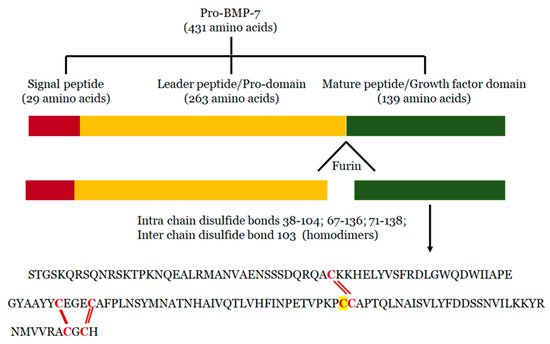
3. Mechanisms of BMP-7

BMP-7 binds to bone morphogenetic protein receptor 2 (BMPR2) on the surface of cells and activates two major signaling pathways: 1) Canonical/Smad dependent and 2) Non-canonical/Smad independent pathway [65][66][65,66] (Figure 2).
Figure 2. BMP signaling pathways. BMP-7 transduces signals in target cells by binding to a specific membrane bound receptor BMPR2 and phosphorylates BMPR1, which activates both canonical and non-canonical pathways. In the canonical pathway, activated BMPR2 leads to phosphorylation of Smad-1/5/8 which complexes with Smad-4 and translocate the signal. In the non-canonical pathway, p38 MAPK, JNK, ERK and NFKB were activated via the activation of XIAP, TAK1 and TAB1 whereas PI3K, Akt were activated by both BMPR2 and Smad-1/5/8. Altogether, this influences the different transcription factors and regulates the gene expression. BMP: Bone morphogenetic protein; BMPR: Bone morphogenetic protein receptor; XIAP: X-linked inhibitor of apoptosis protein; TAK1:TGF-beta activated kinase 1; TAB1: TAK1 binding protein; Runx2: Run-related transcription factor 2; MAPK: Mitogen-activated protein kinase; JNK: c-Jun-N terminal Kinase; ERK: Extracellular signal-regulated kinase; PI3K: Phosphotidylinositol 3 kinase; Akt: RAC-alpha serine/threonine-protein kinase; mTOR: mammalian target of rapamycin.
In the canonical or Smad dependent pathway (Figure 2), BMP-7 activates regulatory Smads (Smad-1, 5, and 8) for subsequent phosphorylation in the cytoplasm. Thereafter, phosphorylated regulatory Smad proteins form a complex with the co-stimulatory molecule Smad-4. This complex is then transduced to the nuclei to recruit cofactors and Run-related transcription factor 2 (Runx2) to regulate osteogenic gene expression and consequently influences osteoblast differentiation [65][73][74][65,73,74]. Mesenchymal stem cell differentiation into osteoblasts is a pre-requisite for embryonic skeletal formation, homeostatic skeletal remodeling and bone fracture repair. BMP-7 plays a major role in upregulating the transcription factor osterix (Osx) or SP7 which has the ability to stimulate differentiation of osteoblasts both in vitro and in vivo [65][75][76][65,75,76]. These studies suggested the involvement of canonical cell signaling pathway in osteoblast differentiation and embryo skeletal formation induced by BMP-7 [76][77][78][79][80][81][82][76,77,78,79,80,81,82]. BMP-7 induced activation of Smad-1/5 leads to the activation of osterix resulting in increased osteogenic markers alkaline phosphatase (ALP) activity and mineralization [83]. Lavery et al. demonstrated the BMP-7 mediated osteoblastic differentiation of primary human mesenchymal stem cells with strongly enriched established osteogenic marker genes including osteocalcin (OCN), osteopontin (OPN) and ALP along with several other osteogenic markers of unknown function [84]. It has been reported that BMP-7 differentiates murine C2C12 myoblasts into osteoblasts by suppressing myoblast determination protein 1 (MyoD) expression, and enhancing the ALP activity and the osteogenic specific gene expressions ALP, Runx2, and OCN via P38 mitogen-activated protein kinase (MAPK) dependent Smad-1/5/8 signaling pathways [85]. Alongside, a recent study from our laboratory demonstrated monocyte differentiation into anti-inflammatory M2 macrophages through the Smad-1/5/8 pathway [67]. On the other hand, in the non-canonical pathway (Figure 2), BMP-7 transduces the signal to the MAPK signaling via c-Jun-N terminal kinase (JNK)1/2/3, extracellular signal-regulated kinase (ERK)1/2, nuclear factor kappa-light-chain-enhancer of activated B (NFκB), and p38 to regulate different target gene expressions [86][87][86,87]. Activated BMPR1A receptor complex initiates these pathways through a series of protein interactions including bone morphogenetic protein receptor associated molecule 1 (BRAM1) or X-linked inhibitor of apoptosis protein (XIAP), and downstream signaling molecules TGF-beta activated kinase 1 (TAK1) and TAK1 binding protein (TAB1) [88]. TAK1 and TAB1 binding activates downstream NFkB, p38, and JNK pathways that induces cell death and differentiation [86][87][88][89][86,87,88,89]. In addition, BMP-7 activates ERK, Phosphoinositol 3-kinase (PI3K), Protein Kinase (PK) C, and D which play a role in cell survival, apoptosis, migration and differentiation [90][91][92][90,91,92]. Hu et al. showed that BMP-7 stimulates renal epithelial cell morphogenesis via p38 MAPK and that its action is counteracted by Smad-1. Further, these studies also revealed that responses to low doses of BMP-7 lead to increased cell proliferation, which are regulated by the p38 MAPK pathway while responses to high doses of BMP-7 suppress cell proliferation, and are controlled by the Smad pathway. In addition, suppression of the p38 MAPK activity by high doses of BMP-7 might integrate the dose-dependent cellular response to BMP-7 [93]. BMP-7 promotes proliferation of nephron progenitor cells through TAK1-mediated JNK activation as well as further activation of transcription factor Jun and activating transcription factor 2 (ATF2) [94]. BMP-7 also plays a major role in the induction of tissue factor in human mononuclear cells (MNCs) through NF-KB activity, leading to increased F3 (tissue factor gene) transcription [95] and resulting in an increased procoagulant activity. Additionally, it has been noticed that BMP-7 binding to its receptor BMPR-II can also activate the Smad dependent and independent PI3K pathways. In this process, activation of PI3K subunit p85 occurs either by Smad-1/5/8 or BMP-7 binding to BMPR II and its subsequent phosphorylation leads to down-stream phosphorylation of phosphotidylinositol biphosphate (PIP2) to phosphatidylinositol triphosphate (PIP3) [96][97][96,97] which, in turn, leads to the phosphorylation of RAC-alpha serine/threonine-protein kinase (Akt) and downstream activation of mammalian target of rapamycin (mTOR) [98]. In immune regulation, the PI3K pathway plays an important role in maintaining the anti-inflammatory environment [97]. Furthermore, studies from our laboratory demonstrated that the Smad-PI3K-Akt-mTOR pathway specifically inhibits pro-inflammatory cytokine secretion (TNF-α, IL-6 and MCP-1), enhances anti-inflammatory cytokines (IL-10 and IL-1ra) and plays a key role in M2 macrophage polarization [67][70][67,70].

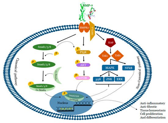
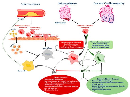
4. BMP-7 as an Anti-Inflammatory Agent in Atherosclerosis

Atherosclerosis is a serious cardiovascular condition that involves the constriction of the arterial wall leading to the development of myocardial infarction. Atherogenesis is regulated by cholesteryl ester (CE) accumulation, foam cell formation, smooth muscle cell migration, necrotic core formation, and increased calcification [66][99][100][101][66,130,131,132]. Moreover, the developed atherogenesis creates turbulence in blood flow leading to plaque rupture and thrombosis. Although these atherogenic factors are well-established, recent data suggests the involvement of modified LDL, extracellular components in the plaque activation and rupture [102][133]. Therefore, atherosclerosis was considered to be the product of lipoprotein accumulation, particularly LDL in the arterial wall [103][104][134,135]. Recently, it is speculated that atherosclerosis is a complex process that involves the participation of both immune systems, oxidative stress, various cell types, receptors, lipids, enzymes, signaling pathways, trace elements, and other products [105][106][107][136,137,138]. Inflammation and oxidative stress are considered to be major players in the progression of the disease [108][109][110][111][139,140,141,142]. Altered vessel wall structure and disturbed blood flow patterns include inflammation and varied stress levels in developed atherosclerosis [112][143]. Despite the abundance of research literature on the topic, the role of lipids, especially fatty acids and their oxidation products like peroxidized linoleic acid (HPODE), 4-hydroxynonenal (HNE), oxo-nonanoic acid (ONA), and their interaction with inflammatory molecules such as oxidized LDL, phospholipids, TNF-α, vascular cell adhesion molecule (VCAM1) in many of these processes are poorly understood. Monocytes, which are precursors of macrophages as well as dendritic cells (DCs) and migrate into the areas of “injury” as a result of a chemotactic stimuli such as monocyte chemotactic protein 1 and 3 (MCP-1&3). Migration of monocytes into the arterial wall has been considered as one of the initial events in atherogenesis which persists in different stages of disease progression [109][110][111][140,141,142]. In tissues, based on the environmental growth factors and pro-inflammatory cytokines, monocytes differentiate into either M1 macrophages or DCs. Monocyte adherence, their differentiation into pro-inflammatory macrophages/dendritic cells that release pro-inflammatory cytokines which are involved in the generation of complex pathophysiology of atherosclerosis [109][140] (Figure 3). Macrophages were initially viewed as a mere scavenger of altered lipoproteins. However, the presence of macrophages along with lymphocytes in atherosclerotic plaques showed enhanced inflammatory immune response and release of pro-inflammatory molecules. The specific roles of different stages of atherosclerosis and presence of these inflammatory macrophages, foam cells, lymphocytes, and vascular smooth muscle cells are not yet completely understood. For example, M2 macrophages, are known for high endocytic clearance capacity due to their higher expression of scavenger receptors (SR) during wound healing and repair processes [113][144]. Van Tits et al. demonstrated that M2 macrophages are susceptible in forming foam cells in presence of oxidized LDL and shift towards the M1 phenotype with enhanced secretion of the pro-inflammatory cytokines IL-6, IL-8 and MCP-1 [114][145]. Furthermore, this increased production of pro-inflammatory cytokines by polarized M1 macrophages from M2 macrophages which are residing in subendothelial space of the vessel wall might lead to the initiation of the inflammatory cascade that mediates disease progression [114][145]. Similarly, in human atherosclerotic lesions different macrophage phenotypes exist in different plaque locations. M2 (CD68+ CD206+) macrophages were located in plaque stable zones far from the lipid core, whereas M1 (CD68+ CCL2+) macrophages exhibited a distinct tissue localization pattern [115][146] suggesting that the tissue microenvironment decides the fate of macrophage polarization. Subsequent research studies confirmed this finding by demonstrating the presence of lipid droplets in CD68+ CD206+ macrophages in comparison with CD68+ CD206 macrophages [116][147]. This discovery suggests that despite the anti-inflammatory nature of M2 macrophages they tend to form foam cells, a significant contributor of atherogenesis.
Figure 3. BMP-7 in Heart Diseases: Schematic representation of how BMP-7 modulates inflammation in heart diseases by converting infiltrated monocytes into anti-inflammatory M2 macrophages. LDL: Low-density lipoprotein; Ox-LDL: oxidized lipoprotein; TNF-α: Tumor necrosis factor alpha; IFN gamma: Interferon gamma; GMCSF: granulocyte macrophage colony stimulating factor; MCSF: macrophage colony stimulating factor.
We demonstrated from our laboratory that rhBMP-7 is able to inhibit the atherosclerosis associated inflammation at both acute (Day-14) and mid-stage (Day-28) time points of atherosclerosis by promoting monocyte differentiation into the anti-inflammatory M2 phenotype via reducing phosphorylated kinases p-38 and JNK while increasing p-Smad and ERK pathways [69][71][69,71]. Additionally, a recent study from our laboratory demonstrated the significantly increased BMPR2 expression on monocytes following BMP-7 treatment, and further polarization into M2 macrophages [67]. BMP-7 treatment showed increased M2 macrophages [approximately 25% at Day-14 and 60% at Day-28] than M1 macrophages [15% at Day-14 and 30% at Day-28] leading to decrease in pro-inflammatory cytokines such as tumor necrosis factor alpha (TNF-α), IL-6 and MCP-1 associated with atherosclerotic lesion development and to an increase in anti-inflammatory cytokines such as IL-10 and IL-1ra levels. Further, BMP-7 improved blood flow in the artery after post ligation, reduced the inflammatory kinases, and completely slowed down disease progression (Figure 3). In addition, we also demonstrated that, upon macrophage depletion by liposomal clodronate, BMP-7 fails to significantly reduce plaque progression and inflammation suggesting the direct role of BMP-7 on macrophages [71]. The literature on BMP-7 in macrophage polarization is new and growing; however, there are certain unanswered questions such as whether BMP-7 can inhibit the formation of foam cells; and if BMP-7 can inhibit the conversion of M2 macrophage into foam cell formation in atherosclerosis?
Video Production Service