Isoliquiritigenin (ISL), a natural bioactive compound with a chalcone structure, demonstrates high antitumor efficacy.
- isoliquiritigenin
- cancer
Thanks so much for your check. The entry will be online only after submission. (Please remove this note when submitting, thanks.)
1. Indroduction
Figure 1. The previous studies demonstrated the six metabolites detected in phase I[1][2][3], including liquritigenin (M1), 2′,4,4′,5′-tetrahydroxychalcone (M2), sulfuretin (M3), butein (M4), davidigenin (M5), and
cis
Figure 1) [1][2][4]. Moreover, the previous study reported that the dominant metabolites of ISL are THC (2,4,2′,4′-tetrahydroxychalcone) and naringenin chalcone in lung cells [5] In vivo absorption of ISL occurs in the intestines, transported to the liver for phase II biotransformation[2]. In phase II metabolism, liquiritigenin, glucuronidated ISL, glucuronidated liquiritigenin, and glucuronidated ISL are produced. Only glucuronidated liquiritigenin is predominant[6]. Many studies have suggested that secondary metabolites are involved in different biological activities and pharmaceuticals[1][2][6][7]. Therefore, these metabolites may differ in various cell lines or organs; however, they all share a similar structure to that of chalcone, which contains two aromatic rings connected by an unsaturated carbon chain, resulting in interconnected biological activities.
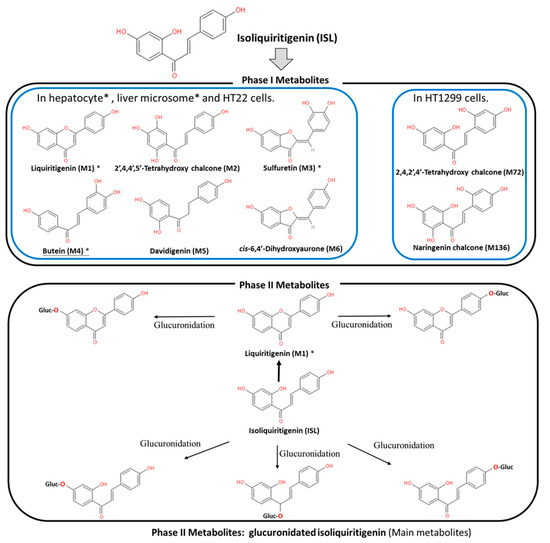
Figure 1.
cis-6,4′-dihydroxyaurone (M6). Phase II metabolites were glucuronide conjugated process. Note: Figure was modified from[1][2].
2. ISL Pharmacokinetics
1/2: 2–4.9 h)[6][8][9][10]. Moreover, the data showed similar trends among different analytic methods, including high-performance liquid chromatography (HPLC), HPLC–MS/MS, and fluorescence spectrometry (SFS)[6][8][9]. This means that the absorption of ISL is quickly and widely distributed throughout the body[6][8][9][10]. Concentrations of ISL may vary in different tissues, including the heart, liver, lungs, spleen, kidneys, brain, muscles, and fat. ISL distribution mainly relies on the blood circulation, with the brain showing the lowest level of ISL due to the blood–brain barrier (BBB). These results imply that ISL is able to penetrate the BBB and exhibits neuroprotective activity in a male middle cerebral artery occlusion (MCAO)-induced focal cerebral ischemia rat model and high fat diet (HFD)-induced ICR mice model[11][12]. Interestingly, only after oral administration does [ISL]
plasma exhibit a double-peak of ISL[10][13][14][15], the possible mechanism for which has been proposed as enterohepatic recycling. As a matter of fact, oral administration has become the most advanced application route.
3. ISL Nanoformulations and ISL Derivatives: Improved Efficacy
Generally speaking, poor bioavailability, rapid degradation, fast metabolism, and systemic elimination are the essential factors that lead to insufficient bioavailability. Insufficient bioavailability of ISL means that its efficacy is far less than 20%[6] [10]. The term insufficient bioavailability implies that patients show intolerance to bulk administration of ISL to reach the desired effect, thereby highlighting the need to improve its effectiveness. To improve solubility, enhancing its bioavailability and distribution, encapsulated ISL nanoparticles or nano-ISL have been developed. Below, we summarize various ISL nanoparticles applied in preclinical studies, for example, polymer nanoparticles, liposomes, micelles, solid lipid nanoparticles (SLNs), and polymer conjugates.
| (In Vitro) | Ref | |||
|---|---|---|---|---|
| Breast cancer | MCF-7 | Testing conc: 10 nM~10 µM (5 days; 10 nM is sufficient) |
|
[57] |
| MCF-7 MDA-MB-231 |
Effective conc: 25 µM and 50 µM (24 h) |
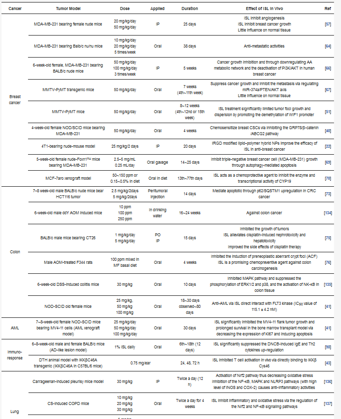
|
[41] | |
| MCF-7 MDA-MB-231 HUVEC |
Testing conc.: 0, 20, 40, 60, 80, 100 µM |
|
[58] | |
| Tumor cell line: MCF-7 IC50 estimated = ~33.39 µM MDA-MB-231 IC50 estimated = ~35.64 µM (48 h) |
||||
| HUVEC IC50 estimated = ~75.48 µM | ||||
| PMA-induced COX-2 in MCF-10A |
Effective conc: 0.1 µM and 10 µM (24 h; 1 µM is sufficient.) |
|
[59] | |
| BT549 MDA-MB-231 |
Effective conc.: 10, 20, 40 µM (12 h) |
|
[60] | |
| MDA-MB-231 Hs-578T |
Effective conc.: ~20 µM |
|
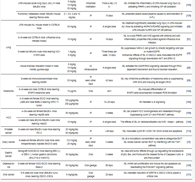
[61] | |
| Breast cancer | MCF-7 MDA-MB-231 |
Testing conc.: 0, 5, 10, 20 µM |
|
[62] |
| Tumor cell line: MCF-7 IC50 = 10.08 µM MDA-MB-231 IC50 = 5.5 µM (48 h) |
||||
| MCF-7 MDA-MB-231 |
Testing conc.: 0, 6.25, 12.5, 25, 50, 100 µM |
|
[63] | |
| Tumor cell line: MCF-7 IC50: 32.66 µM MDA-MB-231 IC50: 22.36 µM (24 h) |
||||
| MDA-MB-231 Hs-578T |
Effective conc.: 10 µM and 20 µM |
|
[64] | |
| MCF-7 MDA-MB-231 BT549 MCF-10 |
Testing conc.: 1, 5, 10 and 25 µM |
|
[36] | |
| Tumor cell lines: MCF-7 IC50 estimated: ~33.0 µM MDA-MB-231 IC50 estimated: ~21.2 µM BT549 IC50 estimated: ~18.1 µM (24 h) |
||||
| Normal cell line: MCF- 10A IC50 estimated: ~80.51 µM (24 h) |
||||
| Breast cancer | MCF-7 MDA-MB-231 H184B5F5/M10 |
Effective conc: 25 µM and 50 µM (48 h) Tumor cell lines: MCF-7 MDA-MB-231 |
|
[65] |
| Normal cell line: H184B5F5/M10 (ISL did not influence the viability) |
||||
| MCF-7 MCF-7/ADR MCF-10A |
Tumor cell lines: MCF-7 IC50 estimation: ~59.39 µM MCF-7/ADR IC50 estimation: ~38.86 µM (24 h) |
|
[53] | |
| Normal cell line: MCF-10A ISL (at 100 µM) had limited inhibitory effects on the proliferation |
||||
| MDA-MB-231 | Testing conc.: 0, 10, 25, 50 µM MDA-MB-231 IC50 estimated: ~24.23 µM (48 h) |
|
[66] | |
| MCF-7aro | Testing conc.: 0, 0.625, 1.25, 2.5, 5, 10 µM MCF-7aro IC50: 2.5 µM (24 h) |
|
[44] | |
| Colon cancer | HT29 | HT29 ED50: 11.1 µg/mL (42.32 µM) |
|
[67] |
| HT29 | Testing conc.: 0, 5,10, 20, 30, 40, 50 µM 40 µM was applied; (24 h) |
|
[68] | |
| HCT116 HT29 SW480 |
Testing conc.: 0,10, 20, 30, 40 µM HCT116 IC50 estimated = ~42.41 µM Working conc.: 30 or 40 µM; (24 h) |
|
[69] | |
| HCT116 | Testing Conc.: 0, 2.5,5, 10, 20, 40, 80, 160 µM HCT116 IC50 estimated: ~78.78 µM (48 h) HCT116 IC50 estimated: ~53.97 µM (72 h) HCT116 IC50 estimated: ~44.8 µM (96 h) |
|
[70] | |
| CT26 | Testing Conc.: 0, 10, 20, 40, 60, 80 µM CT26 IC50 estimated = ~54.48 µM |
|
[71] | |
| Colon26 RCN9 CoLo-320DM |
Testing Conc.: 0, 5, 25, 100 µM (24, 48 h) Colon26 IC50 estimated = ~17.55 µM (24 h) Colon26 IC50 estimated = ~12.59 µM (48 h) RCN9 IC50 estimated = ~41.73 µM (24 h) RCN9 IC50 estimated = ~18.21 µM (48 h) CoLo-320DM IC50 estimated = ~23.10 µM (24 h) CoLo-320DM IC50 estimated = ~10.82 µM (48 h) |
|
[72] | |
| Colon cancer | HCT116 | Applied 20 µM (48 h) |
|
[73] |
| Caco-2/TC-7 | Caco-2/TC-7 EC50: 42 μM |
|
[74] | |
| Ovary cancer | SKOV3 OVCAR5 ES2 |
Testing conc.: 2, 4, 8, 16, 32, 64, and 100 µM SKOV3 IC50: 83.2 µM (72 h) OVCAR5 IC50: 55.5 µM (72 h) ES2 IC50: 40.1 µM (72 h) Effective Conc.: 10 µM |
|
[75] |
| SKOV3 OVCAR5 |
Testing conc.: 0, 1, 5, 10, 20, 25, 50, 75, and 100 µM OVCAR5 IC50: 11 µM (48 h) ES2 IC50: 25 µM (48 h) |
|
[76] | |
| Antral follicle culture (female CD-1 mic) | Testing conc.: 0.6, 6, 36, and 100 μM |
|
[77] | |
| SKOV3 OVCAR3 | Testing conc.: 5~80 μM 30 μM applied |
|
[78] | |
| SKOV3 | N.A. |
|
[76][79] | |
| Lung cancer | H1299 H1975 A549 |
H1299 IC50 estimated: ~36.78~46.08 µM H1975 IC50: 48.14 µM A549 IC50: 75.08 µM (48 h) |
|
[9] |
| A549 | A549: applied 20 µM (24 h) |
|
[80][81] | |
| RAW 264.7 | Testing conc.: 5, 10, 20 µM for (Pretreated with 10mM of t-BHP for 18 h) RAW 264.7 (treated with t-BHP) EC50 = 10 µM (18 h) |
|
[82] | |
| Calu-3 | Calu-3 cells were infected with PR8/H1N1 virus; [EC50] = 24.7 μM |
|
[83] | |
| H1650 H1975 A549 |
H1650 IC50 estimated: ~26.88 µM (24 h) H1975 IC50 estimated: ~8.92 µM (24 h) A549 IC50 estimated: ~46.7 µM (24 h) |
|
[38] | |
| A549 | A549 IC50: 0.05 mg/mL (~191.21 µM ~117 µM) |
|
[84] | |
| Lung cancer | guinea-pig tracheal smooth muscle | N.A. |
|
[85] |
| A549 | A549 IC50: 27.14 µM |
|
[86] | |
| A549 | A549 IC50: 18.5 µM |
|
[87] | |
| AML (acute myeloid leukemia) |
HL-60 | HL-60 ED50: 5.5 µg/mL (~21.46 µM) 5.00 µg/mL = 19.5 µM (72 h) |
|
[61] |
| MV4-11 MOLM-13 OCI-LY10 |
MV4-11 IC50: 3.2 + 1.2 µM; MOLM-13 IC50: 4.9 + 2.1 µM OCI-LY10 IC50: 20.1 ± 6.7 µM (72 h) |
|
[31] | |
| LCLs | Testing conc.: 0, 20, 40, 60, 80, 100, 120, 140 µM LCLs IC50 estimated: 40~65 µM (24 h) Applied 50 µM for studies. |
|
[80] | |
| HL-60 | Testing conc.: 1~15 µg/mL (3.9 µM~58.54 µM) HL-60 IC50 estimated: ~40.42 µM (72 h) |
|
[81] | |
| RAW264.7 | Testing conc.: 20 and 50 μM |
|
[88] | |
| AML (acute myeloid leukemia) |
RAW264.7 | Testing conc.: 50 and 100 μM |
|
[89] |
| HL-60 | Testing conc.: 2.5~20 μg/mL (3.9 µM~78.05 µM) (Working conc.: 72 µM) |
|
[90] | |
| HL-60 | Testing conc.: 2.5~10 μg/mL (3.9 µM~39.0 µM) |
|
[91] | |
| Jurkat J-Jhan J16 HUT78 Karpas 45 |
Jurkat IC50: 0.49 ± 0.12 nM (72 h) J-Jhan IC50: 1.55 ± 1.12 nM (72 h) J16 IC50: 5.25 ± 1.12 µM (72 h) HUT78 IC50: 11 ±13.5 µM (72 h) Karpas 45 IC50: 6.61 ± 1.07 µM (72 h) |
|
[92] | |
| CCRF-CEM | CCRF-CEM IC50: 18.38 μM (24~72 h) |
|
[93] | |
| AML (acute myeloid leukemia) |
Human monocyte model THP-1 | N.A. |
|
[94] |
| Melanoma | A375 A2058 |
Testing Conc: 0, 10, 20, 40, 80 µM A375 IC50: 21.63 µM (24 h) A2058 IC50: 20.75 µM (24 h) |
|
[95] |
| B16F0 | N.A. |
|
[96] | |
| A375 | Testing Conc.: 0, 5, 10, 15 μg/mL (15 μg/mL = 58.53 µM) A375 IC50 estimated: ~48 µM |
|
[97] | |
| A375 | 40 μg/mL: 69.86% 60 μg/mL: 92.22% A375 IC50 estimated: ~73 µM (24 h) |
|
[98] | |
| Melanoma | B16F0 | Testing Conc.: 20, 40, 60 and 80 μg/mL B16F10 IC50 estimated: 35 μg/mL (~41.576 μM; 24 h) B16F10 IC50 estimated: 22 μg/mL (~86.77 μM; 48 h) |
|
[99] |
| B16F10 | Testing Conc.: 5, 10, 15, 20, and 25 μg/mL B16F10 IC50 estimated: ~19 μg/mL (~74.595 μM; 24 h) B16F10 IC50 estimated: ~10.5 μg/mL (~41.576 μM; 48 h) |
|
[100] | |
| ARH-77 U266 MPC-11 SP2/0 CZ-1 RPMI8226 |
ARH-77 IC50: ~13.54 µM MPC-11 IC50: ~4.45 µM SP2/0 IC50: ~22.91 µM CZ-1 IC50: ~13.93 µM U266 IC50: ~8.62 µM RPMI8226 IC50: ~9.09 µM IC50 of ISL was < 4 μg/mL (48 h) |
|
[101] | |
| SK-MEL-2 HaCaT |
Testing Conc.: 0, 1, 4, and 8 µM SK-MEL-2 cells and HaCaT cells (48 h) treated less than 8 µM showed no cytotoxic effects |
|
[102] | |
| Melanoma | B16 mouse melanoma 4A5 cells | Testing 150 and 200 µM (18 and 24 h) |
|
[103] |
| HCC/Hepato-ma | Hep3B | Hep3B IC50: 42.84 + 2.01 μM 50 μM applied (48 h) |
|
[104] |
| HepG2 Hep3B |
Testing conc.: 20, 40, 60, 80, and 100 μM (18 h) HepG2 IC50: 27.71 μM Hep3B IC50: 35.28 μM |
|
[105] | |
| HepG2 | Testing conc.: 1, 5, 10, 20 μg HepG2 IC50 estimated: ~88.46 μM (24 h) HepG2 IC50 estimated: ~31.07 μM (48 h) |
|
[106] | |
| HepG2 | HepG2 IC50: 10.51 μg/mL (~39 μM; 48 h) |
|
[107] | |
| HCC/Hepato-ma | SNU475 | SNU475 IC50: 0.243 + 0.21 mM |
|
[58] |
| Hepa 1c1c7 | Hepa 1c1c7 IC50: 36.3 μM |
|
[108] | |
| Hep3B | Hep3B IC50: 50.8 μM |
|
[43] | |
| SK-Hep-1 | SK-Hep-1 IC50: 19.08 μM |
|
[109] | |
| PC-3 22RV1 |
Testing conc: 0, 1, 10, 25, 50, and 100 μM) PC-3 IC50: 19.6 μM (48 h) 22RV1 IC50: 36.6 μM (48 h) |
|
[110] | |
| Prostate cancer | C4-2 LNCaP IEC-6 |
10~100 μM (24 h) C4-2 IC50: 87.0 μM |
|
[59] |
| DU145 | Applied conc.: 5~20 μM |
|
[111] | |
| DU145 | Applied conc.: 0~20 μM |
|
[112] | |
| DU145 | Applied conc.: 0~20 μM |
|
[113] | |
| Prostate cancer | MAT-LyLu DU145 |
Applied conc.: 0~20 μM MAT-LyLuIC50 estimated: ~13.74/5.67/5.01 µM DU145 IC50 estimated: ~56.87/31.49/17.60 µM (24 h/48 h/72 h) |
|
[114] |
| DU145 LNCaP | Testing conc.: 0, 5, 10, 15, and 20 μM DU145 IC50 estimated: ~10.561 µM (48 h) LNCaP IC50 estimated: ~10.775 µM (48 h) |
|
[115] | |
| Cervical cancer | Ca Ski SiHa HeLa C-33A |
Testing conc: 10, 20, 40, and 80 µM Ca Ski IC50 estimated: 39.09 μM (72 h) SiHa IC50 estimated: 53.76 μM (72 h) HeLa IC50 estimated: 58.10 μM (72 h) C-33A IC50 estimated: 32.83 μM (72 h) |
|
[116] |
| HeLa | Testing conc: 2, 5, 10, 30, 40, and 60 μg/mL HeLa IC50 estimated: ~21.24 μM (24 h) |
|
[117] | |
| HeLa | HeLa IC50: 9.8 μM (48 h) |
|
[118] | |
| Gastric cancer | MKN28 | MKN28 IC50: ~20.84 µM (48 h) |
|
[119] |
| MKN-45 | 5 µM applied |
|
[120] | |
| MGC-803 | 0.11 g/L applied (24 h) |
|
[121] | |
| SGC-7901 BGC-823 | BGc-823 IC50: 23.18 µM (48 h) SGC-7901 IC50: 12.91 µM (48 h) |
|
[34] | |
| Uterine leiomyoma | Leiomyma Myomentrium |
Testing conc: 0, 10, 20, 50 µM Leiomyma IC50 estimated = ~39.33 µM Myomentrium IC50 estimated = ~698.8 µM (48 h) |
|
[122] |
| Osteosarcoma | U2OS | Testing conc: 5, 10, and 20 µM 20 μM applied |
|
[123][128] |
| Saos‑2 MC3T3-E1 |
Saos‑2 IC50 estimated = ~24.23 μM 30 μM applied |
|||
| Glioma | SK-N-BE(2) IMR-32 | Effective conc. > 5 µM |
|
[125] |
| U87 | U87 IC50: 6.3 µM |
|
[124] | |
| PC12 | PC12 IC50: 17.8 ± 1.8 μM |
|
[126] | |
| Bladder cancer | T24 | Effective conc.: 30 and 70 µg/mL (24 h) |
|
[127] |
| Oral squamous cell carcinomas (OSCC) |
SG SAS-CSCs OECM-1 |
SG cells IC50: 386.3 ± 29.7 μM SAS-CSCs IC50: 144.9 ± 25.7 μM OECM-1-CSCs IC50: 104.5 ± 26.2 μM |
|
[54] |
Nanosuspension: ISL is milled with HPC (hydroxypropyl cellulose) SSL and PVP (polyvinylpyrrolidone) K30 to form a lamelliform or ellipse shape of the nanosuspension. HPC SSL and PVP K30 act as stabilizer. These two nanosuspension particles (size: 238.1 ± 4.9 nm with SSL; 354.1 ± 9.1 nm with K30) do not only improve the solubility issue, but also enhance the cytotoxicity a 7.5–10-fold[16].
Nanosuspension: ISL is milled with HPC (hydroxypropyl cellulose) SSL and PVP (polyvinylpyrrolidone) K30 to form a lamelliform or ellipse shape of the nanosuspension. HPC SSL and PVP K30 act as stabilizer. These two nanosuspension particles (size: 238.1 ± 4.9 nm with SSL; 354.1 ± 9.1 nm with K30) do not only improve the solubility issue, but also enhance the cytotoxicity a 7.5–10-fold [20].
Nanoencapsulation: Mesoporous silica nanoparticles (MSNs) are a solid material, acting as a biodegradable nanoscale drug carrier. When MSNs are encapsulated with ISL, they improve the efficacy of ISL in vitro and in vivo[17].
Nanoencapsulation: Mesoporous silica nanoparticles (MSNs) are a solid material, acting as a biodegradable nanoscale drug carrier. When MSNs are encapsulated with ISL, they improve the efficacy of ISL in vitro and in vivo [21].
- Lipid–polymer hybrid nanoparticle system:
- 3.1.
- ]
- .
- 3.1.
-
iRGD hybrid NPs: The composition of lipid–polymer hybrid nanoparticles (NPs) include lactic-co-glycolic acid (PLGA), lecithin, and a hydrophilic poly-ethylene-glycol (PEG). ISL-loaded hybrid NPs are composed of an inner PLGA core with an outer lipid layer (PEG, lecithin, and iRGD peptides). iRGD peptides (CRGDK/RGPD/EC, a tumor-homing peptides), can deliver drugs to a tumor. In vitro, ISL–iRGD NPs show stronger inhibition effects and induce apoptosis effects. In vivo, ISL–iRGD NPs show stronger effects in the viability of tumor cells. Herein, iRGD-modified lipid–polymer NPs showed better solubility, bioavailability, and targeting distribution [22].
- 3.2.
-
Hydrophilic polyanion solid lipid nanoparticles (SLNs): SLNs are composed of natural lipids such as lecithin or triglycerides that remain solid at 37 °C. SLNs can protect labile compounds from chemical degradation and can improve bioavailability. Low-molecular-weight heparins (LMWHs) are fragments of heparin showing hydrophilic polyanions that can improve the efficacy of ISL [23].
- iRGD hybrid NPs: The composition of lipid–polymer hybrid nanoparticles (NPs) include lactic-co-glycolic acid (PLGA), lecithin, and a hydrophilic poly-ethylene-glycol (PEG). ISL-loaded hybrid NPs are composed of an inner PLGA core with an outer lipid layer (PEG, lecithin, and iRGD peptides). iRGD peptides (CRGDK/RGPD/EC, a tumor-homing peptides), can deliver drugs to a tumor. In vitro, ISL–iRGD NPs show stronger inhibition effects and induce apoptosis effects. In vivo, ISL–iRGD NPs show stronger effects in the viability of tumor cells. Herein, iRGD-modified lipid–polymer NPs showed better solubility, bioavailability, and targeting distribution[18].
- 3.2.
-
Hydrophilic polyanion solid lipid nanoparticles (SLNs): SLNs are composed of natural lipids such as lecithin or triglycerides that remain solid at 37 °C. SLNs can protect labile compounds from chemical degradation and can improve bioavailability. Low-molecular-weight heparins (LMWHs) are fragments of heparin showing hydrophilic polyanions that can improve the efficacy of ISL[19
Microemulsion: The self-microemulsifying drug delivery system (SEMDDS) was designed for improving the solubility, absorption, and bioavailability of lipophilic drugs. The SMEDDS comprises ethyl oleate (EO; oil phase), Tween 80 (surfactant), and PEG 400 (co-surfactant). ISL-loaded SMEDDS has been proven to improve the solubility and oral in vivo availability
[
13].
Microemulsion: The self-microemulsifying drug delivery system (SEMDDS) was designed for improving the solubility, absorption, and bioavailability of lipophilic drugs. The SMEDDS comprises ethyl oleate (EO; oil phase), Tween 80 (surfactant), and PEG 400 (co-surfactant). ISL-loaded SMEDDS has been proven to improve the solubility and oral in vivo availability [17].
ISL-loaded nanostructured lipid carriers (ISL-NLCs): NLCs mix solid lipids with spatially incompatible liquid lipids, which leads to a special nanostructure with improved properties for drug loading. ISL-loaded NLCs are constructed by glycerol monostearate (MS) and Mi-glyol-812 as the solid and liquid lipid materials to carry the ISL
[
]. In pharmacokinetic studies, less than 10% of the NLCs remains in the stomach after oral administration, mainly absorbed in the colon[19]. Moreover, the antitumor effect of ISL-loaded NLCs has been evaluated in sarcoma 180 (S180)-bearing and murine hepatoma (H22)-bearing mice models via IP administration[20]. A biodistribution study showed that the ISL concentration of ISL-loaded NLCs in the tumor is higher 2.5-fold than free ISL. In a skin permeability study, the previous study suggested NLCs as a promising carrier to deliver the ISL[21].
ISL-loaded nanostructured lipid carriers (ISL-NLCs): NLCs mix solid lipids with spatially incompatible liquid lipids, which leads to a special nanostructure with improved properties for drug loading. ISL-loaded NLCs are constructed by glycerol monostearate (MS) and Mi-glyol-812 as the solid and liquid lipid materials to carry the ISL [24]. In pharmacokinetic studies, less than 10% of the NLCs remains in the stomach after oral administration, mainly absorbed in the colon [19]. Moreover, the antitumor effect of ISL-loaded NLCs has been evaluated in sarcoma 180 (S180)-bearing and murine hepatoma (H22)-bearing mice models via IP administration [24]. A biodistribution study showed that the ISL concentration of ISL-loaded NLCs in the tumor is higher 2.5-fold than free ISL. In a skin permeability study, the previous study suggested NLCs as a promising carrier to deliver the ISL [25].
TPGS-modified proliposomes: D-α-tocopheryl polyethylene glycol 1000 succinate (TPGS) has been selected as an excipient for ISL-loaded TPGS-modified proliposomes (ISL-TPGS-PLP), prepared using the film dispersion method with ISL-loaded proliposomes (ISL–PLP). ISL-TPGS-PLP can enhance the solubility, bioavailability and liver-targeting ability of ISL
[
]
.
TPGS-modified proliposomes: D-α-tocopheryl polyethylene glycol 1000 succinate (TPGS) has been selected as an excipient for ISL-loaded TPGS-modified proliposomes (ISL-TPGS-PLP), prepared using the film dispersion method with ISL-loaded proliposomes (ISL–PLP). ISL-TPGS-PLP can enhance the solubility, bioavailability and liver-targeting ability of ISL [18].
Polymeric micelles: PEO (polyethylene oxide)–PPO (polypropylene oxide)–PEO (polyethylene oxide) triblock copolymers are highly biocompatible and act as surface-active agents. P123 (PEO20–PPO65–PEO20) can remarkably enhance the retention of poorly soluble drugs in the blood circulation. Another important derivative of Pluronic, F127 (PEO100–PPO69–PEO100), possesses high biocompatibility. Therefore, mixed F127/P123 polymeric micelles have been developed, which have remarkably enhanced bioavailability with high encapsulation efficiency and low particle size. ISL-loaded F127/P123 polymeric micelles (ISL-FPM) improve the solubility as well as enhance the bioavailability and antioxidant activity of ISL
[
]
.
- Polymeric micelles: PEO (polyethylene oxide)–PPO (polypropylene oxide)–PEO (polyethylene oxide) triblock copolymers are highly biocompatible and act as surface-active agents. P123 (PEO20–PPO65–PEO20) can remarkably enhance the retention of poorly soluble drugs in the blood circulation. Another important derivative of Pluronic, F127 (PEO100–PPO69–PEO100), possesses high biocompatibility. Therefore, mixed F127/P123 polymeric micelles have been developed, which have remarkably enhanced bioavailability with high encapsulation efficiency and low particle size. ISL-loaded F127/P123 polymeric micelles (ISL-FPM) improve the solubility as well as enhance the bioavailability and antioxidant activity of ISL [
- ].
Nanoliposomes (NLs): Drug-loaded PEGylated nanomaterials have shown effective cancer cell-killing ability, PEG2000-DPSE-QUE-NLs (polyethyleneglycol-2000-distearoyl phosphatidyl ethanolamine loaded with querce-tin (QUE)) can efficiently disperse in aqueous media compared to controls, and PEGylated (PEG2000-DPSE) NLs have been found to be effective drug delivery vehicles when simply loaded with ISL. ISL-NLs as tumor-targeted drug carriers are more effective in regulating glycolysis in colon cancer cell lines (CRC: HCT116)
[
]
.
- Nanoliposomes (NLs): Drug-loaded PEGylated nanomaterials have shown effective cancer cell-killing ability, PEG2000-DPSE-QUE-NLs (polyethyleneglycol-2000-distearoyl phosphatidyl ethanolamine loaded with querce-tin (QUE)) can efficiently disperse in aqueous media compared to controls, and PEGylated (PEG2000-DPSE) NLs have been found to be effective drug delivery vehicles when simply loaded with ISL. ISL-NLs as tumor-targeted drug carriers are more effective in regulating glycolysis in colon cancer cell lines (CRC: HCT116) [
- ].
Hydrogel: Hydrogels are composed of hyaluronic acid (HA) and hydroxyethyl cellulose (HEC), and they can improve the skin permeation of ISL
[
]
.
- Hydrogel: Hydrogels are composed of hyaluronic acid (HA) and hydroxyethyl cellulose (HEC), and they can improve the skin permeation of ISL [
- ].
Table 1), demonstrating that ISL nanoformulations improve the bioavailability by 2–10-fold[13][20][22].
Table 1. Nano-formulation of ISL.
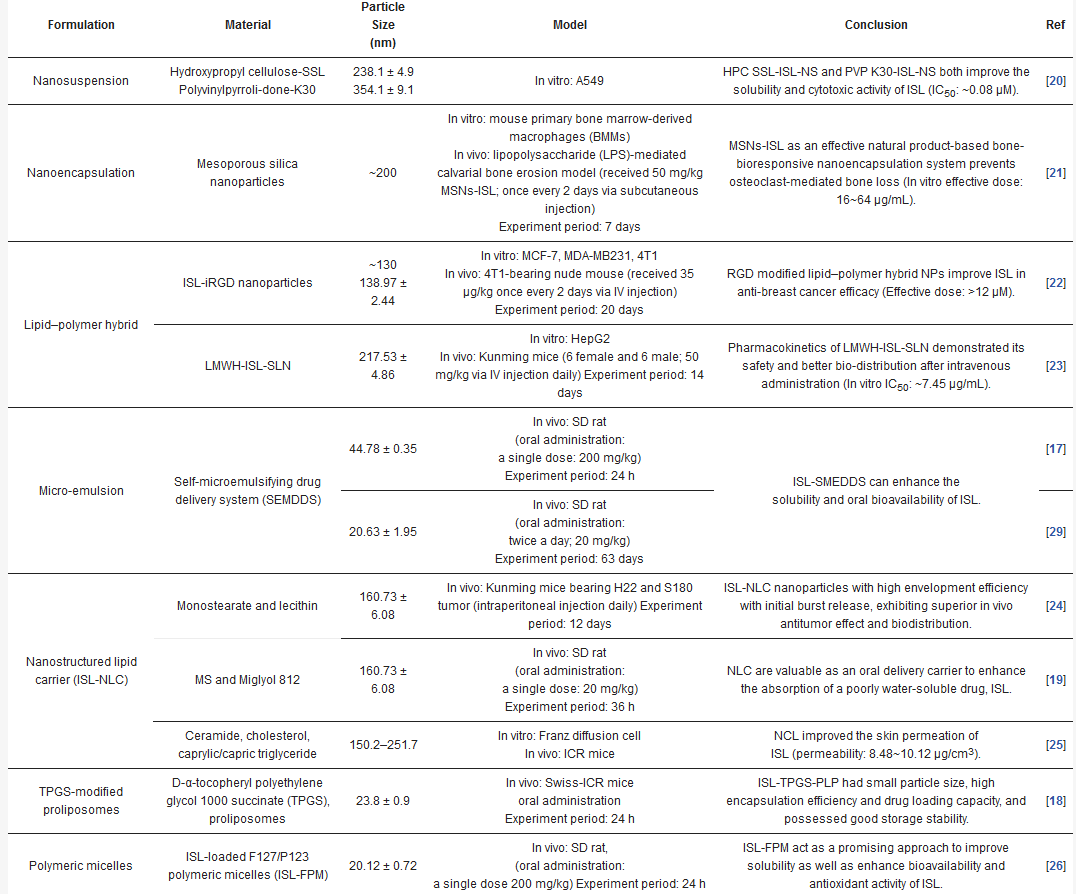
| Formulation | Material | Particle Size (nm) |
Model | Conclusion | Ref |
|---|---|---|---|---|---|
| Nanosuspension | Hydroxypropyl cellulose-SSL Polyvinylpyrroli-done-K30 |
238.1 ± 4.9 354.1 ± 9.1 |
In vitro: A549 | HPC SSL‑ISL‑NS and PVP K30-ISL‑NS both improve the solubility and cytotoxic activity of ISL (IC50: ~0.08 µM). | [16] |
| ] | |||||
| Polymeric micelles | |||||
| ISL-loaded F127/P123 polymeric micelles (ISL-FPM) | 20.12 ± 0.72 | In vivo: SD rat, (oral administration: a single dose 200 mg/kg) Experiment period: 24 h |
ISL-FPM act as a promising approach to improve solubility as well as enhance bioavailability and antioxidant activity of ISL. | [22] | |
| Liposome | Phospholipid and cholesterol | 233.1 | In vitro: HeLa and SiHa | ISL liposome can significantly inhibit the proliferation of human cervical cancer cells in vitro. | [26] |
| Nanoliposome | Sodium cholate, cholesterol and IPM were melted with a ratio of 5:1:4 (w/w/w) | 82.3 ± 35.6 | In vitro: HCT116 and HT29 | ISL involved in the glucose metabolism in colon cancer. | [23] |
| Hydrogel systems | HA-HEC hydrogels | N.A. | In vitro: skin permeation study Franz diffusion cells | HA-HEC hydrogel showing the stable viscoelastic be haviour and the optimal adhesiveness has potential to enhance skin permeation of IS (permeability: 20 μg/cm3). | [24] |
Figure 2.
-
4-C-β-D-glucosylated ISL (Figure 2a): Glucosylation of low molecular weight compounds have improve water solubility and bioavailability with a good inhibition of aldose reductase (AR) [37].
-
Synthetic isoliquiritigenin derivatives (BS5 and BS11 in Figure 2b,c): The compounds BS5 and BS11 with m-, p-dimethoxy, o-bromo phenyl group shows neuroprotective effects at 3 μM to 6 μM with higher viability (~80–100%) [36].
-
Robtein (ISL-derivative #10; Figure 2d): Robtein exhibited osteoclast differentiation and activation without any significant changes of viability or cytotoxicity [34].
-
3′,4′,5′,4″-tetramethoxychalcone (TMC; Figure 2f): Introducing methylation of hydroxy groups significant increase cytotoxic activity in breast cancer [31], especially targeting on triple-negative breast cancer (TNBC) [33].
-
ISL-17 (Figure 2g): A fluorine atom was introduced to the structure of ISL named ISL-17 showed the anti-tumor activities in gastric cancer [32].
| Nanoencapsulation | |||||
| Mesoporous silica nanoparticles | ~200 | In vitro: mouse primary bone marrow-derived macrophages (BMMs) In vivo: lipopolysaccharide (LPS)-mediated calvarial bone erosion model (received 50 mg/kg MSNs-ISL; once every 2 days via subcutaneous injection) Experiment period: 7 days |
MSNs-ISL as an effective natural product-based bone-bioresponsive nanoencapsulation system prevents osteoclast-mediated bone loss (In vitro effective dose: 16~64 µg/mL). | [17] | |
| Lipid–polymer hybrid | ISL-iRGD nanoparticles | ~130 138.97 ± 2.44 |
In vitro: MCF-7, MDA-MB231, 4T1 In vivo: 4T1-bearing nude mouse (received 35 µg/kg once every 2 days via IV injection) Experiment period: 20 days |
RGD modified lipid–polymer hybrid NPs improve ISL in anti-breast cancer efficacy (Effective dose: >12 µM). | [18] |
| LMWH-ISL-SLN | 217.53 ± 4.86 | In vitro: HepG2 In vivo: Kunming mice (6 female and 6 male; 50 mg/kg via IV injection daily) Experiment period: 14 days |
Pharmacokinetics of LMWH-ISL-SLN demonstrated its safety and better bio-distribution after intravenous administration (In vitro IC50: ~7.45 µg/mL). | [19] | |
| Micro-emulsion | Self-microemulsifying drug delivery system (SEMDDS) | 44.78 ± 0.35 | In vivo: SD rat (oral administration: a single dose: 200 mg/kg) Experiment period: 24 h |
ISL-SMEDDS can enhance the solubility and oral bioavailability of ISL. |
[13] |
| 20.63 ± 1.95 | In vivo: SD rat (oral administration: twice a day; 20 mg/kg) Experiment period: 63 days |
[25] | |||
| Nanostructured lipid carrier (ISL-NLC) | Monostearate and lecithin | 160.73 ± 6.08 | In vivo: Kunming mice bearing H22 and S180 tumor (intraperitoneal injection daily) Experiment period: 12 days | ISL-NLC nanoparticles with high envelopment efficiency with initial burst release, exhibiting superior in vivo antitumor effect and biodistribution. | [20] |
| MS and Miglyol 812 | 160.73 ± 6.08 | In vivo: SD rat (oral administration: a single dose: 20 mg/kg) Experiment period: 36 h |
NLC are valuable as an oral delivery carrier to enhance the absorption of a poorly water-soluble drug, ISL. | [15] | |
| Ceramide, cholesterol, caprylic/capric triglyceride | 150.2–251.7 | In vitro: Franz diffusion cell In vivo: ICR mice |
NCL improved the skin permeation of ISL (permeability: 8.48~10.12 μg/cm3). |
[21] | |
| TPGS-modified proliposomes | D-α-tocopheryl polyethylene glycol 1000 succinate (TPGS), proliposomes |
23.8 ± 0.9 | In vivo: Swiss-ICR mice oral administration Experiment period: 24 h |
ISL-TPGS-PLP had small particle size, high encapsulation efficiency and drug loading capacity, and possessed good storage stability. | [14 |
-
4-C-β-D-glucosylated ISL (Figure 2a): Glucosylation of low molecular weight compounds have improve water solubility and bioavailability with a good inhibition of aldose reductase (AR)[33].
-
Synthetic isoliquiritigenin derivatives (BS5 and BS11 in Figure 2b,c): The compounds BS5 and BS11 with m-, p-dimethoxy, o-bromo phenyl group shows neuroprotective effects at 3 μM to 6 μM with higher viability (~80–100%)[32].
-
Robtein (ISL-derivative #10; Figure 2d): Robtein exhibited osteoclast differentiation and activation without any significant changes of viability or cytotoxicity[28].
-
2′,4′-dimethoxy-4-hydroxychalcone (Figure 2e): shows in vivo antidiabetic activity[31].
-
3′,4′,5′,4″-tetramethoxychalcone (TMC; Figure 2f): Introducing methylation of hydroxy groups significant increase cytotoxic activity in breast cancer[27], especially targeting on triple-negative breast cancer (TNBC)[29].
-
ISL-17 (Figure 2g): A fluorine atom was introduced to the structure of ISL named ISL-17 showed the anti-tumor activities in gastric cancer[28].
However, the poor bioavailability and water-solubility issues remain in clinical applications. Future studies are still needed to elucidate the ISL formulations that would be more suitable for human clinical trials.
4. ISL Docking Model
Figure 3), such as SIRT1 [34]VEGF2 receptor[35], GRP78[36], FLT3[37], EGFR[38], IKKβ[39], Toll-like receptors (TLRs)[40], CK-2 (IC
50: 17.3 µM)[41], H2R[42], COX-2[43], aromatase (Ki: 2.8 µM)[44][45], topoisomerase I [46] and DNMT1[47]. These docking results imply that the binding pocket is composed of hydrophobic regions and is stabilized by a hydrogen bond with its neighboring carbonyl group. The hydrogen bond interactions and π–π stacking contribute to a tight interaction with the binding site. These docking results provide valuable information about the binding interactions of ISL and the active site, although more studies are required to approve them. Using a bioassay-guided purification method, suggested that isolated ISL acts as a xanthine oxidase inhibitor (IC
50: 55.8 µM; Ki: 17.4 µM) to avoid transplantation rejection and ischemia reperfusion damage [48]. In brief, multiple docking candidates indicate that ISL exhibits multiple biological properties and serves as a potential lead compound for developing new therapy in cancer treatment.
Figure 3.
a
b
c
d
e
f
g
h
i
j
k
5. ISL Biology Effects
2.5. ISL Biology Effects
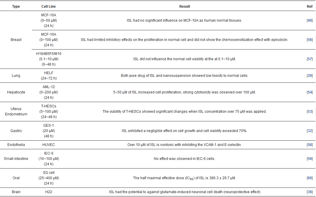
Table 3), and the effective dose in tumor cell lines shows very little cytotoxic effect on normal cells. Most studies have claimed that ISL significantly inhibits the viability of cancer cell but has little toxicity on normal cells. For example, Wu et al. (2017) compared the human endometrial stromal cells (T-HESCs; as a control) and human endometrial cancer cell lines (Ishikawa, HEC-1A, and RL95-2 cells). Their results indicated that ISL inhibits the growth of cancer cells at concentrations below 27 μM, but has little effect on normal cells[49]. Na et al. (2018) claimed that ISL shows little toxicity on normal hepatocyte cell lines (AML-12); only when applied in concentrations of over 100 μM is ISL harmful to normal hepatocytes [50]. Most studies have focused on the cytotoxicity between tumor and normal cells, and the effects of ISL on normal cells remain unknown. As Peng et al. (2015) mentioned, further research on the target organ toxicity or side effects of ISL is needed. The safety of ISL is always one of the most important concerns that must be evaluated.
Figure 4.
Figure 5. ISL-mediated regulation of molecular targets underlying anti-tumor effects, including tumor proliferation suppression, apoptosis induction, EMT/metastasis, epigenetic responses and sensitization to chemotherapy. Downward arrows (↓) represent downregulation while upward arrows (↑) represent upregulation. This figure was modified from[51].
Table 2.
| Type | Cell Line | Result | Ref |
|---|---|---|---|
| Breast | MCF-10A (0~50 µM) (24 h) |
ISL had no significant influence on MCF-10A as human normal tissues. | [36] |
| MCF-10A (0~100 µM) (24 h) |
ISL had limited inhibitory effects on the proliferation in normal cell and did not show the chemosensitization effect with epirubicin. | [52] | |
| H184B5F5/M10 (0.1~10 µM) (6~48 h) |
ISL did not influence the normal cell viability at the at 0.1~10 µM. | [53] | |
| Lung | HELF (24~72 h) |
Both pure drug of ISL and nanosuspension showed low toxicity to normal cells. | [16] |
| Hepatocyte | AML-12 (0~200 µM) (24 h) |
5~50 μM of ISL increased cell proliferation, strong cytotoxicity was observed over 100 μM. | [50] |
| Uterus Endometrium |
T-HESCs (5~100 µM) (24~48 h) |
The viability of T-HESCs showed significant changes when ISL concentration over 75 μM was applied. | [49] |
| Gastric | GES-1 (20 µM) (48 h) |
ISL exhibited a negligible effect on cell growth and cell viability exceeded 70%. | [28] |
| Endothelia | HUVEC | Over 10 µM of ISL is nontoxic with inhibiting the VCAM-1 and E-selectin. | [54] |
| Small intestine | IEC-6 (10~100 µM) (24 h) |
No effect was observed in IEC-6 cells. | [55] |
| Oral | SG cell (25~400 μM) (24 h) |
The half maximal effective dose (IC50) of ISL is 386.3 ± 29.7 μM. | [56] |
| Brain | H22 | ISL had the potential to against glutamate-induced neuronal cell death (neuroprotective effect) | [32] |
Different pathways of various cancers regulated by ISL.
| Type of Cancer | Cell | Testing Range/IC50 | Signaling Pathways Effect of ISL |
|---|
ote: The ‘’IC50 estimated’’ indicated Data extracted from published figures using Web Plot Digitizer (https://automeris.io/WebPlotDigitizer), then analyzed IC50 by “Quest Graph™ IC50 Calculator.” AAT Bioquest Inc, 27 October 2020, https://www.aatbio.com/tools/ic50-calculator [133].