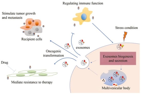
Exosomes distributed by extracellular vesicles carry various information highly consistent with cells, becoming a new type of biomarker for tumor screening. However, although conventional characterization technologies can quantify size and morphology for exosomes, they are limited in related fields such as function tracing, protein quantification at unit point, and microstructural information.细胞外囊泡分布的外泌体携带与细胞高度一致的各种信息,成为肿瘤筛查的新型生物标志物。然而,虽然传统的表征技术可以量化外泌体的大小和形态,但在功能追踪、单位点蛋白质定量和微观结构信息等相关领域受到限制。
- exosome
- tumor diagnosis
- optical analysis technology
- super-resolution microscope
1. Introduction引言
2. Conventional Characterization Technologies常规表征技术
Due to the unique biological function of exosomes, an increasing amount of basic research is being concentrated on it [37][38][39][40][41]. Characterization technologies play important roles in the study of exosomes [30]. Generally speaking, various approaches for analysis are categorized into two primary types: biochemical analysis and physical analysis. Biochemical analysis mainly determines the source and composition of exosomes, including Western blot and enzyme-linked immunosorbent assay (ELISA), in which the specific binding of antibody antigens decides the effect qualitatively or quantitatively [42]. However, the disadvantage is that the morphological characteristics and concentration of exosomes cannot be obtained.2.1. 可调电阻脉冲检测
2.1. Tunable Resistive Pulse Sensing
Tunable可调电阻脉冲检测 Resistive Pulse Sensing ((TRPS) is based on Coulter’s principle. The suspension was mixed in the electrolyte, which could go through the nanopore chip with a specific aperture. The resistance between the two electrodes inside and outside changes instantaneously at the moment of passing through the nanopore, the result of which is a pulse signal as shown in Figur) 基于库尔特原理。将悬浮液混合在电解液中,电解液可以通过具有特定孔径的纳米孔芯片。两个电极内外之间的电阻在通过纳米孔的那一刻瞬时发生变化,其结果是脉冲信号,如图2所示。信号的强度和频率与外泌体的大小和数量有关。通过脉冲信号计数外泌体的表达。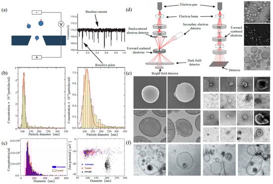
2.2. 电子显微镜
2.2. Electron Microscope
Electron Microscope (EM) is the most direct method to measure the size and morphology of a single EV [42]. It is divided into scanning electron microscope (SEM), transmission electron microscope (TEM), and cryo-transmission electron microscope (cryo-TEM). It is noted that EM can characterize the particle morphology and size of a single EV [53][54].
2.3. 动态光散射
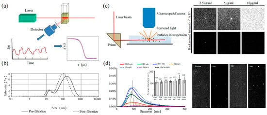
动态光散射(DLS)是一种测量亚微米颗粒尺寸和分布的光学分析方法,其基本原理如图3所示。粒子的布朗运动引起光散射信号的变化,该信号由数字自相关器监测,以计算粒子的扩散速度和粒子分布[62]。小颗粒的运动速率更高,其强度波动更大。结果,相关曲线迅速而明显地下降。DLSEM, TEM has a higher resolution and can realize the imaging analysis of a single E的灵敏度和FOV at the nanoscale [58]. 均优于上述论文,可实现大尺寸外泌体纳米颗粒的基本表征。对于较小的颗粒,自聚合行为会干扰光强信号。因此,无法实现高浓度样品的精密分析和检测。2009年,Liu et al. successfully characterized exosomes extracted from breast tumor cells including their morphology and distribution [59]. Park et al. found the morphology of EVs derived from prostate cancer cells (PCa) was round and that the number of these EVs was significantly higher than that of normal cells [60]. It is worth noting that sample protocol is complicated and that the sample is in a vacuum during the process of imaging. In order to reduce the complexity of the protocol, the researchers further developed cryo-Tie等人使用DLS表征了源自红细胞的EM to maintain the integrity and authenticity of the sample to the greatest extent [52]. This element changes the structure of the exosome from a round to a cup-like shape as shown in the right of Figure 2d. Conde-Vancells et al. realized the morphological characterization of exosomes derived from hepatocytes [50]. Kurtjak et al. identified the single, double, and multi-membrane morphology of C的大小分布[63]。DLSF extracellular vesicles by combining cryo-TEM with atomic force microscopy (AFM) [61]. This proves that the cryo-TEM technique can not only restore the EV shape but also allow the analysis of the EV interior.2.3. Dynamic Light Scattering
Dynamic light scattering (DLS) is an optical analysis method for measuring the size and distribution of sub-micron particles, and its basic principle is shown in Figure 3a. The Brownian motion of the particles causes the change in the light scattering signal, which is monitored by a digital autocorrelator to calculate the diffusion velocity and particle distribution of the particles [62]. The motion rate of small particles is higher, and its intensity fluctuation is larger. As a result, there is a swift and pronounced decline in the correlation curve. The sensitivity and FOV of DLS are better than the above papers, which can realize the basic characterization of large-size exosome nanoparticles. For smaller particles, the behavior of self-polymerization interferes with the light-intensity signal. Thus, it is impossible to realize the precision analysis and detection of high-concentration samples. In 2009, Lawrie et al. used DLS to characterize the size distribution of EV derived from red blood cells [63]. DLS analyzes all particles in a sample simultaneously, and therefore the information on the number or concentration of a certain category of particle cannot be provided [64]. For example, DLS provides a clear range of the diameter of EVs derived from ovarian cancer cells, but the concentration is difficult to analyze [65]. Therefore, DLS technology is often combined with other technologies to complete the characterization of EVs. For example, Tajik T combined DLS technology with electron microscopy, showed cannabis-derived EVs (CDEVs) can be considered exosome-like nanovesicles, and highlighted that CDEVs can be an ideal natural vehicle for bioactive phytocannabinoids, promoting the research of EVs in cancer diagnosis [66].

2.4. Nanoparticle Tracking Analysis
2.4. 纳米粒子跟踪分析
2.5. Flow Cytometry
Size and morphology are the basic parameters for the characterization of exosomes. The characterization of functional parameters such as surface protein quantitative expression and signal transduction mode is of paramount importance. Flow Cytometry (FCM) realizes the rapid multi-parameter quantitative analysis of cells or submicron particles based on light scattering changes, and its basic principle is shown in Figure 4a. Scatters of light from particles suspended in a sheath stream reflect the size and density of the cells or particles, and they were acquired by a detector array. At the same time, the specific gene expression, protein expression, enzyme activity, ion concentration, and other biomolecular substances labeled by fluorescent dyes were specifically measured by different channels. The sensitivity of traditional flow cytometry is limited to 300 to 500 nm [73][76], so it is obviously difficult to measure exosomes. Yan Xiaomei’s team developed nFCM by combining Rayleigh scattering with sheath flow single-molecule fluorescence detection technology, which enables the high-throughput analysis of exosomes with a size of 40 nm, as shown in Figure 4 [74][75][77,78]. Compared with traditional flow cytometry, the scattered light detection sensitivity of nFCM is improved by four to six orders.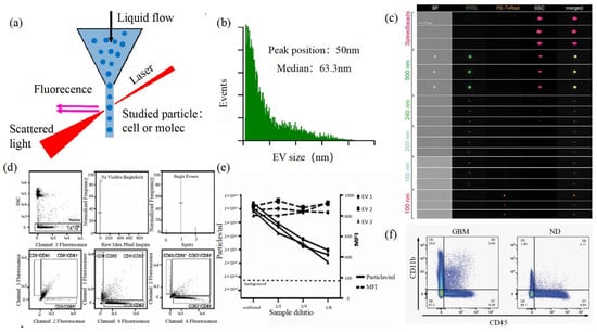
3. Super-Resolution Imaging Technology
3.1. Single Molecule Localization Imaging Technology
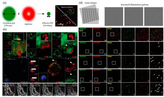
The basic principle of SMLM is based on the flicker of a single fluorescent molecule to locate a single molecule and then reconstruct super-resolution images. Compared with other technologies, SMLM has the advantages of low phototoxicity and low cell damage. It is more suitable for living cells, thus becoming a new super-resolution analysis method for exosomes in vivo observation. SMLM opens a new observation perspective for exosome-related studies.3.1.1. Stochastic Optical Reconstruction Microscopy and Photoactivated Localization Microscopy Technology
PALM and STORM technologies are classical technologies in SMLM. In 2006, Eric Betzig et al. proposed the PALM technology [78][82], and Xiaowei Zhuang et al. proposed the STORM technology [79][83] at the same time. Both of them are based on single-molecule localization technology to achieve the super-resolution imaging of subcellular structure molecules. One of the key elements is the switched fluorophores. For example, PALM uses photoactivated green fluorescent protein (PA-GFP) to label the protein and irradiate the cell surface with different lasers so as to cause the fluorescence molecule cycle to complete the excitation localization process. One of the key points is the spatial and temporal resolution for SMLM. That requires more than 10,000 frames of images during the process of reconstruction, which needs much more time. The rapid development of EMCCD cameras has greatly improved imaging speed. In 2011, Zhuang Xiaowei’s group pictured extracellular vesicles with a high-speed EMCCD. The temporal resolution was improved to 0.5 s, which means that STORM has the potential to monitor live cell imaging in real-time [80][85]. In 2011, Zhuang’s team used the stage-specific neurite-associated protein (SNAP) label to label the Alexa Flour 467 optical switching probe to clathrin in living BS-C-1 cells. STORM technology was successfully used to obtain a 30 nm horizontal resolution and a 50 nm vertical resolution [80][85]. In 2012, Shim et al. determined the STORM membrane probe for live cell imaging through a large number of experiments and performed the super-resolution imaging of organelle membranes in live cells, reaching a spatial resolution of 20~60 nm [81][92]. In 2018, Zong Shenfei et al. discovered that silicon quantum dots (Si QD) have fluorescent scintillation behavior and applied them as SMLM imaging nanoprobes to stain CD63 of breast cancer cell (SKBR3)-derived EVs using CD63 aptamers fused with Si QD, achieving an imaging accuracy of about 30 nm. They demonstrated that Si QD can be used for the SMLM imaging of small objects such as exosomes. Moreover, Si QD has the characteristics of high biocompatibility and low cytotoxicity, which makes it a better choice of fluorophores for SMLM live cell imaging [82][93].3.1.2. DNA-PAINT Technology
Similar to the STORM/PALM technology, DNA-PAINT also achieves super-resolution imaging by controlling the flicker of individual fluorophores. In 2014, Ralf Jungmann et al. proposed the DNA-PAINT technology, which uses reversible binding between complementary DNA sequences to produce an effect similar to the “flicker” of fluorescent molecules [83][101]. In double-strand DNA, one strand is connected to the fluorophores, called the imager strand, and the other is connected to the target molecule, called the docking strand. Due to the highly specific binding of the double-strand DNA, the imaging strand and the docking strand are bound spontaneously, producing single-molecule fluorescence in the focal plane [84][102] as shown in Figure 56. In addition, multi-channel fluorescence imaging can be realized by different proteins labeled with various docking strands.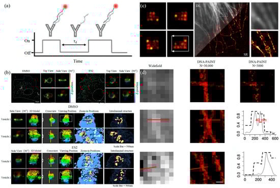
3.2. Stimulated Emission Depletion Technology