Rheumatoid arthritis (RA) is an autoimmune disease that causes inflammation, pain, and ultimately, bone erosion of the joints. The causes of this disease are multifactorial, including genetic factors, such as the presence of the human leukocyte antigen (HLA)-DRB1*04 variant, alterations in the microbiota, or immune factors including increased cytotoxic T lymphocytes (CTLs), neutrophils, or elevated M1 macrophages which, taken together, produce high levels of pro-inflammatory cytokines. TIn this review, we focused on the function exerted by osteoclasts on osteoblasts and other osteoclasts by means of the release of exosomal microRNAs (miRNAs) was focused on. Based on a thorough revision, researcherswe classified these molecules into three categories according to their function: osteoclast inhibitors (miR-23a, miR-29b, and miR-214), osteoblast inhibitors (miR-22-3p, miR-26a, miR-27a, miR-29a, miR-125b, and miR-146a), and osteoblast enhancers (miR-20a, miR-34a, miR-96, miR-106a, miR-142, miR-199a, miR-324, and miR-486b). Finally, we analyzed potential therapeutic targets of these exosomal miRNAs, such as the use of antagomiRs, blockmiRs, agomiRs and competitive endogenous RNAs (ceRNAs), which are already being tested in murine and ex vivo models of RA. These strategies might have an important role in reestablishing the regulation of osteoclast and osteoblast differentiation making progress in the development of personalized medicine.
1. Introduction
The study of miRNAs in RA has focused on their potential utility as diagnostic markers (including let-7d-5p, miR-24-3p, miR-126-3p, miR-130a-3p, miR-221-3p, and miR-431-3p), indicators of disease progression (such as miR-22, miR-486-3p, and miR-382), or markers associated with bone erosion (like miR-99b-5p). This analysis typically involves assessing these miRNAs in synovial fluid or serum
[1][2][3][109,110,111]. However, few articles have delved into the specific function of osteoclasts in bone erosion through the release of exosomal miRNAs. For this reason, researchers have reviewed the miRNAs contained in exosomes released by osteoclasts and grouped them according to their ability to promote or inhibit osteoblast and osteoclast differentiation (
Figure 1).
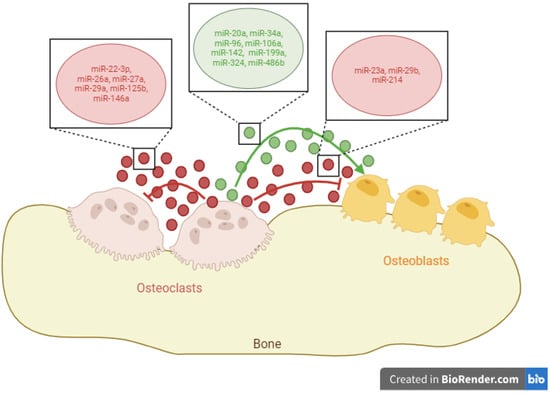
Figure 1. Exosomal microRNAs (miRNAs) released by osteoclasts and their functions over osteoblasts and other osteoclasts. Green circles/lines indicate exosomal miRNAs that promote cellular differentiation, while red circles/lines show exosomal miRNAs that inhibit cellular differentiation. Created with
BioRender.com.
2. Exosomal miRNAs with the Ability to Promote Osteoblast Differentiation
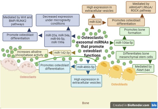
Exosomes isolated from osteoclasts derived from bone marrow macrophages of C57 black 6 (C57BL/6) mice contain miR-18a-5p, miR-185-5p, miR-142-3p, miR-106a-5p, and miR-132-3p, but only miR-106a-5p has the ability to promote bone formation and differentiate bone mesenchymal stem cells through its action on family with sequence similarity 134, member A (
FAM134A), a gene related to tumor proliferation, metastasis, etc. (
Figure 2)
[4][5][112,113]. Additionally, miR-142-3p increases alkaline phosphatase enzymatic activity in bone mesenchymal stem cells (
Figure 2)
[5][113], an enzyme produced by osteoblasts and used as a marker of their activity
[6][7][114,115]. Furthermore, a study conducted under microgravity conditions with the murine macrophage cell line Ralph, Raschke, Watson (RAW) 264.7, differentiated into osteoclasts with RANKL, concluded that miR-142a-3p decreased its expression in the extracellular vesicles of osteoclasts
[8][116]. In the same study, a decrease in the expression of miR-34a, miR-96-5p, miR-199a, and miR-20a was observed, while miR-486b-5p showed higher expression in extracellular vesicles released by osteoclasts (
Figure 2)
[8][116]. In all cases, these miRNAs have been associated with the ability to promote osteoblast differentiation, with miR-96 acting through the Wnt pathway and miR-20a acting through the bone morphogenetic protein (BMP)/runt-related transcription factor 2 (RUNX2) pathway
[9][10][11][12][13][14][117,118,119,120,121,122]. Another miRNA capable of promoting osteoblast differentiation is miR-324 (
Figure 2). Osteoclasts differentiated from the bone marrow macrophages of mice released extracellular vesicles with high miR-324 expression, which exhibited osteogenic capacity through the regulation of the Ras homology GTPase-activating protein 1 (ARHGAP1)/Ras homolog family member A (RhoA)/Rho-associated coiled-coil containing protein kinase (ROCK) signaling pathway
[15][123]. This led to increased mineralization, regeneration, and bone density.
Figure 2. Diagram of exosomal miRNAs produced by osteoclasts (miR-20a, miR-34a, miR-96-5p, miR-106a, miR-142-3p, miR-199a, miR-324, and miR-486b-5p) and their function as enhancers of osteoblast functions. Green circles/lines indicate exosomal miRNAs that promote cellular differentiation. Wingless-related integration site (Wnt); bone morphogenetic protein (BMP)/runt-related transcription factor 2 (RUNX2); Ras homology GTPase-activating protein 1 (ARHGAP1)/Ras homolog family member A (RhoA)/Rho-associated coiled-coil containing protein kinase (ROCK); family with sequence similarity 134, member A (FAM134A). Created with
BioRender.com.
3. Exosomal miRNAs with the Ability to Inhibit Osteoblast Differentiation
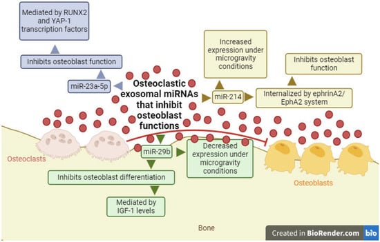
The group led by Yang analyzed exosomes released by the RAW 264.7 cell line after differentiation into osteoclasts. The results of their research showed that exosomes from osteoclasts contain miR-23a-5p, with an inhibitory function on osteoblasts (
Figure 3). Furthermore, using bioinformatics techniques, these authors demonstrated that the osteogenic effect of miR-23a-5p could be due to the inhibition of RUNX2 transcription factor. As suggested in the article, this inhibition could be mediated by the yes-associated protein 1 (YAP-1) transcription factor, which interacts with RUNX2 and inhibits metallothionein 1D, pseudogene (MT1DP)
[16][124], a long non-coding RNA (lncRNA) that promotes cytotoxic function in cells
[17][125].
Figure 3. Diagram of exosomal miRNAs produced by osteoclasts (miR-23a-5p, miR29b, and miR-214) and their function as inhibitors of osteoblast functions. Red circles/lines show exosomal miRNAs that inhibit cellular differentiation. Yes-associated protein 1 (YAP-1); ephrin A2 receptor (EphA2); insulin-like growth factor 1 (IGF-1). Created with
BioRender.com.
In another study, the same cell line used previously was differentiated into osteoclasts with RANKL and, after analyzing the released exosomes, high concentrations of miR-214 were observed (
Figure 3)
[18][19][126,127]. Moreover, these exosomes could be internalized by osteoblasts through the ephrin A2/ephrin A2 receptor (EphA2) system, inhibiting the function of these cells
[19][127]. The expression of this miRNA was also observed to be increased in extracellular vesicles released by osteoclasts under microgravity conditions, while miR-29b levels were decreased (
Figure 3)
[8][116]. Both miRNAs act as inhibitors of osteoblast differentiation, and in the case of miR-29b, it exerts its function by reducing insulin-like growth factor 1 (IGF-1) levels, as observed in an ex vivo model with murine osteocyte cells stimulated by mechanical stretching
[20][21][128,129].
4. Exosomal miRNAs with the Ability to Inhibit Osteoclast Differentiation
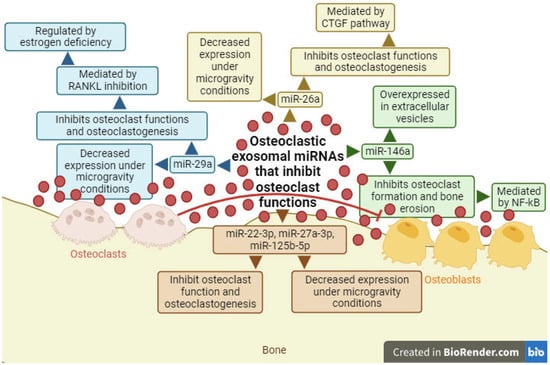
In other studies conducted with osteoclasts differentiated from mouse bone marrow precursors, miR-146a was found to be overexpressed in the extracellular vesicles of osteoclasts (
Figure 4)
[18][22][126,130]. Studies carried out with osteoclasts differentiated from monocytes from the peripheral blood mononuclear cells of patients with Paget’s syndrome showed decreased levels of miR-146a, leading to inhibition of osteoclasts and bone resorption, mediated by the nuclear factor—κB (NF-κB) transcription factor
[23][131].
Figure 4. Diagram of exosomal miRNAs produced by osteoclasts (miR-22-3p, miR-26a, miR-27a-3p, miR-29a, miR-125b-5p, and miR-146a) and their function as inhibitors of others osteoclast functions. Red circles/lines show exosomal miRNAs that inhibit cellular differentiation. Receptor activator for nuclear factor κ B ligand (RANKL); connective tissue growth factor (CTGF); nuclear factor—κB (NF-κB). Created with
BioRender.com.
The results of miRNA expression contained in extracellular vesicles released by osteoclasts under microgravity conditions concluded that miR-22-3p, miR-26a-5p, miR-27a-3p, miR-29a-3p, and miR-125b-5p showed decreased levels (
Figure 4)
[8][116]. All these miRNAs play a crucial role as inhibitors of both osteoclasts and osteoclastogenesis, with miR-29a, for instance, acting as an inhibitor of RANKL, which can be regulated due to estrogen deficiency, and miR-26a acting through the connective tissue growth factor (CTGF) pathway
[24][25][26][27][28][132,133,134,135,136].