The field of biosensors is filled with reports and designs of various sensors, with the vast majority focusing on glucose sensing. However, in addition to glucose, there are many other important analytes that are worth investigating as well. In particular, L-amino acids appear as important diagnostic markers for a number of conditions. The need to determine L-amino acids from clinical samples has risen. More clinical data appear to demonstrate that abnormal concentrations of L-amino acids are related to various clinical conditions such as inherited metabolic disorders, dyslipidemia, type 2 diabetes, muscle damage, etc.
- amino acids
- detection
- biosensors
- diagnostics
- clinical condition
- disease
1. Typical Concentrations of Total L-Amino Acids in Human Blood
2. Overview of L-Amino Acids in Clinical Conditions
The importance of L-amino acids for clinical diagnostics is not sufficiently clear and is often overlooked. Several factors are contributing to this issue, such as the relationship of L-amino acids to many biochemical processes, and, in turn, to many various conditions from metabolic disorders to cancers, giving them lower specificity. Additionally, to this date, the measurement methods and/or devices for L-amino acids are quite expensive and usually they are not routinely applied for clinical sample analysis. The most common clinical conditions related to L-amino acids are discussed in detail and summarized in Figure 1.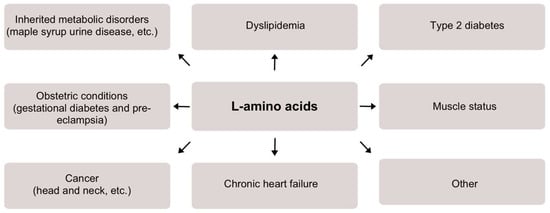
2.1. Inherited Metabolic Disorders
Inherited metabolic disorders, such as maple syrup urine disease, are closely related to serum L-amino acids both for diagnostic and therapeutic purposes [3][4][4,5]. Maple syrup urine disease has been shown to result in an increase in branched-chain L-amino acids (BCAAs) in plasma [5][6]. Long-term treatment requires restriction of dietary BCAAs and supplementation with a BCAA-free mixture. Frequent monitoring of the plasma concentration of L-amino acids could help identify deficiencies in essential L-amino acids secondary to a protein-restricted diet, and thus guide treatment.2.2. Dyslipidemia
BCAA, tyrosine, tryptophan, glutamate, and L-homocysteine concentrations are reported to be considerably lower in healthy diet individuals [6][7]. In one study, forty healthy men and women with a body mass index of 25.6 ± 0.6 were overfed by 1250 kCal per day for 28 days and, as a consequence, their serum BCAAs increased from 397 ± 10 µmol/L to 428 ± 9 µmol/L. This highlights the role of food overconsumption in increased BCAA concentrations [7][8]. Moreover, BCAAs and total L-amino acid levels are increased in obese individuals. Higher concentrations of all BCAAs, phenylalanine, tyrosine, and alanine are associated with a higher fat mass index, and some plasma-free L-amino acids are correlated with parameters used to measure central obesity (e.g., waist circumference, waist-to-hip ratio, BMI) [8][9][9,10]. Interestingly, medical studies report that serum amino acid profiles and increased levels of leucine, arginine, valine, proline, phenylalanine, isoleucine, and lysine could predict the development of hypertriglyceridemia later in life in healthy individuals even before they become obese [10][11]. For example, serum L-amino acid concentrations of 1125 individuals were analyzed with the follow-up data available after 7 years for comparison and a relationship was found between L-amino acids and an increased risk of developing hypertriglyceridemia [11][12]. In summary, measurements of L-amino acid concentrations can serve as early biomarkers to identify people at high risk of developing dyslipidemia and diseases related to dyslipidemia.2.3. Type 2 Diabetes
Increased levels of L-amino acids have been reported to be an early manifestation of insulin resistance; i.e., changes in blood concentrations of particular L-amino acids (e.g., sulfur amino acids, tyrosine, phenylalanine) are apparent with obesity and insulin resistance, often before the onset of clinical diagnosis [12][13][13,14]. Furthermore, BCAAs and aromatic L-amino acids predict the risk of developing type 2 diabetes. For example, during a study conducted by Wang et al., 2422 normoglycemic individuals were profiled for metabolites. After 12 years, 201 individuals were diagnosed with type 2 diabetes. A significant correlation between the disease and several amino acids, i.e., isoleucine, leucine, valine, tyrosine, and phenylalanine, was established. The risk of future diabetes was concluded to be at least fourfold greater in those with high plasma amino acid concentrations in both the discovery and replication samples [14][15]. Similar results were obtained from the Southall And Brent REvisited (SABRE) study. Asian-American individuals have been reported to have an increased risk of diabetes compared to Europeans that is not related to obesity or established traditional metabolic measures [15][16]. In total, 643 Asian and 801 European non-diabetic males were tested for baseline levels of nine L-amino acids in serum. Serum concentrations of isoleucine, phenylalanine, tyrosine, and alanine were significantly higher in the Asian men, while glycemia was similar in the two groups. Diabetes developed in 35% of the Asian and 14% of the European males. Strong associations between L-amino acids and diabetes were also observed in other studies [16][17][18][17,18,19]. In summary, L-amino acids could serve as a predictor of early-onset type 2 diabetes in healthy individuals.2.4. Obstetric Conditions
There are data showing that L-amino acids are related to some obstetric conditions, such as gestational diabetes and pre-eclampsia. Significant differences in serum levels of particular amino acids (e.g., L-arginine, L-glycine, and 3-hydroxy-isovalerylcarnitine) are observed between females who develop gestational diabetes and controls [19][20]. According to that article, these differences exist already in the first trimester of pregnancy and may be useful for early screening. Additionally, increased concentrations of L-amino acids were shown to be more prevalent in women with pre-eclampsia [20][21]. In the reviewed article, maternal and cord blood L-amino acid levels were reported to be significantly higher in women with pre-eclampsia compared to pregnant women without this condition. Most likely, pre-eclampsia is associated with increased placental L-amino acid transport or reduced utilization of fetal L-amino acids.2.5. Muscle Status
A strong relationship between L-amino acids and muscle status has been reported. For example, L-amino acid imbalance could explain fatigue after long-term exercise [21][22]. During overtraining, brain tryptophan uptake and 5-hydroxytryptamine synthesis are increased. In a 1993 study, the serum L-amino acid profile was determined in nine athletes before and after competing in the Colmar ultratriathlon. Serum concentrations of 25 L-amino acids (22 proteinogenic L-amino acids and citrulline, α-Aminobutyric acid, and taurine) decreased by 18% with a decrease in 18 individual L-amino acids by 9–56% and an increase in tyrosine, phenylalanine, methionine, cystine, and free tryptophan. In addition, a decrease in body mass (approximately 3.3 kg) and an increase in plasma volume (approximately 7.6%) were observed. The decrease in intracellular water may be seen as a protein catabolic signal. Interestingly, it was also reported that L-amino acid supplementation could be used to prevent muscle damage. For example, during prolonged exercise, L-branched-chain amino acid (BCAA) supplementation decreases the serum concentration of intramuscular enzymes (creatine kinase and lactate dehydrogenase) and thus may reduce muscle damage [22][23]. L-amino acids were also reported to be important for patients who have diseases that induce muscle mass decrease. It was reported that the supplementation of BCAAs may reduce the muscle damage for patients with rheumatic diseases. It was demonstrated that plasma BCAAs, aspartic acid, and glutamate concentrations correlate positively with the rate of improvement in biceps femoris muscle atrophy, suggesting that these amino acids are associated with the BCAA-induced increase in muscle mass [23][24]. It was also demonstrated that, for patients treated in intensive care units, the supplementation of L-amino acids can protect skeletal muscle mass and function; for severely ill patients, a higher provision of protein and L-amino acids has been associated with a lower mortality [24][25]. Another recent report also indicated that significant amounts of L-amino acids are lost during continuous renal replacement therapy and may indicate the need for clinical intervention for clinical care patients [25][26]. It was demonstrated that the amount of L-amino acids lost during continuous renal replacement therapy is increased not only by the set-up specifics, but also by individual considerations of the patients (e.g., by a higher systemic concentration of amino acids and by a higher fat-free mass index (mostly muscles)). To summarize, L-amino acids are strongly associated with body muscle status, and they could show muscle damage and help to prevent it.2.6. Cancer
L-amino acids could be an important parameter for cancer monitoring, diagnosis, and prognosis. It was reported that L-amino acids could be predictors of cancer cachexia and sarcopenia [26][27]. Cancer cachexia and sarcopenia cause ongoing muscle loss and a higher serum essential/total L-amino acid ratio is associated with sarcopenia in patients with advanced gastrointestinal cancers. It is reported that the levels of L-amino acids are also associated with the stage of disease and prognosis in patients with head and neck cancer [27][28]. Increased levels of alpha-aminobutyric acid, aminoadipic acid, L-histidine, L-proline, and L-tryptophan are associated with a reduced risk of advanced stage head and neck cancer. Increased levels of beta-alanine, beta-aminobutyric acid, ethanolamine, glycine, isoleucine, 4-hydroxyproline, and phenylalanine are associated with an increased risk of advanced stage head and neck cancer. Increased levels of alpha-aminobutyric acid are associated with increased overall survival, while increased levels of arginine, ethanolamine, glycine, histidine, isoleucine, 4-hydroxyproline, leucine, lysine, 3-methylhistidine, phenylalanine, and serine are associated with decreased overall survival. Other papers also report a relationship between cancers and L-amino acids. For example, in a recent review by Kang, the importance of L-amino acid restriction for cancer therapy was highlighted [28][29]. It was stated that L-amino acid restriction, in particular L-leucine, could be a simple metabolic intervention for cancer therapy.2.7. Chronic Heart Failure
Plasma L-amino acids can predict chronic heart failure. It was demonstrated that 17 L-amino acids and two concentration ratios between specific L-amino acids were significantly different in a heart failure group compared with those in the control [29][30]. The heart failure group had five specific L-amino acids, i.e., L-monoethanolamine, L-methionine, L-tyrosine, L-methylhistidine, and L-histidine, in correlation with cardiac function indicators such as B-type natriuretic peptide, left ventricle ejection fraction, and others. Moreover, in another study, it was shown that concentrations of essential L-amino acids and L-BCAAs are significantly lower for patients with severe chronic heart failure, which is associated with low nutritional status along with the loss of skeletal muscle [30][31]. To summarize, research papers indicate that chronic heart failure is related to changes in L-amino acid concentrations.2.8. Other
A relationship between L-amino acids and other conditions also were reported. Several papers have highlighted the importance of L-amino acids, especially L-BCAAs, for the prediction of septic shock resolution and survival [31][32][32,33]. A significant number of papers report that L-amino acids play important roles in brain functioning and could be related to various mental and neurological conditions, such as autism spectrum disorders, cerebral palsy, and Parkinson’s disease [33][34][35][36][34,35,36,37]. Papers have been published indicating a relationship between L-amino acids and liver cirrhosis [37][38][38,39] and chronic kidney disease [39][40]. Some papers also reported that L-amino acids may be related to a patient’s frailty and longevity in general [40][41][41,42]. For example, Hamed et al. reported that L-amino acids could be important in age-related diseases and that protein and/or L-amino acid restriction could be related with reduced occurrence in cancer, diabetes, and overall mortality [41][42]. Another paper also indicated that L-amino acids are highly important in aging [42][43].3. Biosensors for Measurement of Total L-Amino Acid Concentration
3.1. General Overview of Enzymatic Biosensors
Advantages and disadvantages of all generations of enzymatic electrodes are summarized in Table 1.|
Generation |
Key Advantages |
Key Disadvantages |
|---|---|---|
|
First (electrons are being transferred via native mediator, e.g., H2O2) |
Simple to design Native mediator is constantly being produced by the enzyme, thus cannot be depleted The sensing layer can be separated from the electrode surface The sensing layer can be made using excessive amount of enzyme to thus increase stability |
Depended on the concentration of the dissolved oxygen in a solution Lack of sensitivity High operational potential (e.g., +0.6 V vs. Ag/AgCl) |
|
Second (electrons are being transferred via synthetic mediator, e.g., tetrathiafulvalene) |
Usually do not depend on the concentration of the dissolved oxygen in a solution Have better sensitivities in comparisonto first generation The sensing layer can be made using excessive amount of enzyme to thus increase stability Low operational potential (e.g., +0.2 V vs. Ag/AgCl) |
Complex design Synthetic mediator needs to be constantly added to the solution Desorption of mediator from the surfacecan occur |
|
Third (electrons are being transferred directly) |
Usually do not depend on the concentration of the dissolved oxygen in a solution Highest sensitivities in comparison to the first and second generations Lowest operational potentials (e.g., 0 V vs. Ag/AgCl) No mediator (native or synthetic) is needed for the electrode operation |
Most complex design Can be implemented for a limited number of selective enzymes Low operational stability in comparison to other generations |
3.2. Overview of Biosensors for L-Amino Acid Detection
Table 2 summarizes most significant biosensors for L-amino acid detection.
Table 2.
A comparative table of most significant biosensors for L-amino acid detection (majority for total L-amino acids, some for specific).
|
Biosensor |
Enzyme, Nanomaterials Used |
Analysis Method |
Analytical Parameters (as Reported) |
Real Sample |
|---|---|---|---|---|
|
Enzyme: L-amino acid oxidase (Crotalus adamanteus) Nanomaterials: N/A |
Chronoamperometry |
Sensitivity: N/A LOD: 10−6–10−5 M Linear range: N/A Stability: stable for at least 4 months |
N/A |
|
|
Enzyme: L-amino acid oxidase (Crotalus adamanteus), D-amino acid oxidase (porcine kidney) Nanomaterials: N/A |
Flow-through amperometry |
Sensitivity: N/A LOD: N/A Linear range: 0.1–3 mM Stability: 900–1000 measurements |
Brewing process samples (ginger and brown beer) |
|
|
Enzyme: L-amino acid oxidase (Crotalus adamanteus), D-amino acid oxidase (porcine kidney) Nanomaterials: N/A |
Chronoamperometry |
Sensitivity: N/A LOD: 0.15–0.47 mM Linear range: 0.47–2.5 mM Stability: 40% activity loss after 56 days |
Milk, fruit juice, urine |
|
|
Enzyme: L-amino acid oxidase (goat kidney) Nanomaterials: MWCNT |
Linear square voltammetry |
Sensitivity: N/A LOD: 0.5 µM Linear range: 0.5 µM–100 mM Stability: 70% left after 140 days |
Fruit juices, alcoholic beverages |
|
|
Enzyme: L-amino acid oxidase (Gratelis Adamate) *, D-amino acid oxidase (porcine kidney) Nanomaterials: N/A |
Chronoamperometry |
Sensitivity: 10–480 µA M−1 LOD: 1.1–160 µM Linear range: 10−6–10−4 M Stability: around 10 days with no need to regenerate the electrode surface |
Grapes |
|
|
Enzyme: enzyme-free Nanomaterials: AgO nanoparticles |
Chronoamperometry |
Sensitivity: 4230 µA mM−1 cm−2 LOD: 0.42 µM Linear range: 60–500 µM Stability: N/A |
Human serum |
|
|
Enzyme: enzyme-free Nanomaterials: MWCNT |
Differential pulse voltammograms |
Sensitivity: 0.012 µA µM−1 LOD: 8 µM Linear range: 25–750 µM Stability: N/A |
Human serum |
|
|
Enzyme: enzyme-free Nanomaterials: tetrahedral copper metal–organic frameworks |
Linear sweep voltammetry |
Sensitivity: N/A LOD: 25 nM Linear range: N/A Stability: no significant change after 6-month storage |
Human blood |
|
|
Enzyme: L-amino acid oxidase (Crotalus adamanteus) Nanomaterials: gold nanoparticles |
Chronocoulometry |
Sensitivity: 0.73 µC/µM LOD: 5.5 µM Linear range: 5.5–100 µM Stability: 50% of the initial activity after 10 days of storage |
Human serum and blood |
* As reported in the researchticle.