Clinical Applications of Cardiac Scintigraphy with Bone Tracers: Comparison
Please note this is a comparison between Version 2 by Mona Zou and Version 1 by Riccardo Saro.

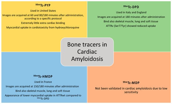
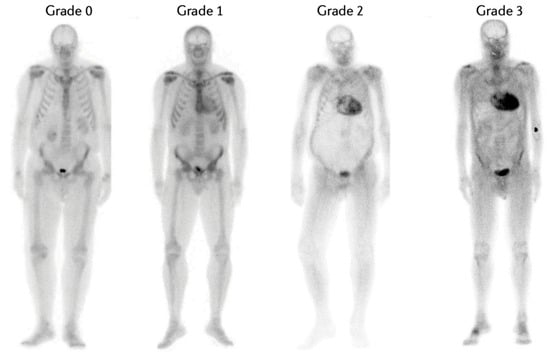
Radionuclide bone scintigraphy is the cornerstone of an imaging-based algorithm for accurate non-invasive diagnosis of transthyretin cardiac amyloidosis (ATTR-CA). In patients with heart failure and suggestive echocardiographic and/or cardiac magnetic resonance imaging findings, the positive predictive value of Perugini grade 2 or 3 myocardial uptake on a radionuclide bone scan approaches 100% for the diagnosis of ATTR-CA as long as there is no biochemical evidence of a clonal dyscrasia. The technetium-labelled tracers that are currently validated for non-invasive diagnosis of ATTR-CA include pyrophosphate (99mTc-PYP); hydroxymethylene diphosphonate (99mTc-HMDP); and 3,3-diphosphono-1,2-propanodicarboxylate (99mTc-DPD). Although nuclear scintigraphy has transformed the contemporary diagnostic approach to ATTR-CA, a number of grey areas remains, including the mechanism for binding tracers to the infiltrated heart, differences in the kinetics and distribution of these radiotracers, differences in protocols of image acquisition worldwide, the clinical significance of extra-cardiac uptake, and the use of this technique for prognostic stratification, monitoring disease progression and assessing the response to disease-modifying treatments.

  • cardiac amyloidosis
  • cardiomyopathies
  • HFpEF
  • bone scintigraphy
  • bone tracers

1. Bone Tracers and Cardiac Amyloidosis

1.1. Pathophysiological Mechanisms of Amyloidogenic Cascade

Amyloidosis is a systemic disease characterized by the deposition of type A or type B fibrillar amyloid proteins (7–13 nm in diameter) with a classic beta-strand hydrogen-bonded structure. Type A fibrils are composed of full-length and C-terminal fragments of transthyretin, while type B fibrils comprise only full-length transthyretin. Amyloid fibrils are insoluble polymers consisting of various protein subunits that are in turn formed from soluble precursors that, through conformational changes, obtain a beta-sheet configuration. Within these deposits, serum amyloid P components and glycosaminoglycans can be found [1]. At the beginning of fibrillogenesis and nucleated polymerization, several molecules, which may be unfolded, partially folded, or completely folded become nuclei and aggregate to generate totally disordered, partially disordered, or structured oligomers. These aggregates can then fragment and trigger second nucleation reactions, thus creating new nuclei. As the aggregation process proceeds, these oligomers may acquire a beta-sheet structure and then continue to grow by aggregating with each other or with other monomers. The process of fibrillogenesis is facilitated by partial folding or unfolding of the precursors, which is accelerated by acidification, proteolysis, primary nucleation, fibril fragmentation, and secondary nucleation.
At this point, the toxicity of amyloid fibrils is due to their deposition in the extracellular space of organs and tissues, undermining their structural integrity [8][2].

1.2. Cardiac Scintigraphy

The diagnosis of CA was previously established by endomyocardial biopsy (EMB), which was associated with procedural risks and the need to be performed in capable hands; the histological preparation that is stained with Congo red and placed under polarized light shows the pathognomonic apple-green birefringence. Bone tracer scintigraphy is a nuclear medicine diagnostic method that uses radiation emitted by a radiopharmaceutical labelled with radioactive isotopes previously injected intravenously and detected using a suitable instrument, the gamma-camera, allowing areas of tracer accumulation to be identified. Irradiation is minimal (average 3.2 mSv), and no side effects are described. Thus, this technique can be performed in anyone, regardless of renal function, with the exception of pregnant or breastfeeding women. Furthermore, contact with children must be avoided 24 h after exposure. Thanks to the contribution provided initially by Perugini and later by the protocol proposed by Gillmore et al. as well as the consolidation of their findings with the publication of the position statement of the European Society of Cardiology and the very recent guidelines on cardiomyopathies, myocardial scintigraphy with bone tracers allows the diagnosis of ATTR-CA without the need for EMB in selected cases [7][3].

1.3. Bone Tracers

Tracers used for bone scintigraphy are diphosphonates or pyrophosphates bound to metastable technetium-99 (99mTc), and their accumulation at the cardiac level has been described since the 1980s. However, they have been studied in a more comprehensive approach for cardiac amyloidosis from the 2000s onwards, especially since Perugini’s work [9,10][4][5].
Bone tracers currently used to identify cardiac amyloid deposits (Figure 1) include 99mTc-pyrophosphate (99mTc-PYP) [11][6], the only one employed in the United States; 99mTc-3,3-diphosphone-1,2-propan-dicarboxylic acid (99mTc-DPD) [12][7], employed in the United Kingdom and Italy (and generally in Europe); and 99mTc-hydroxymethylene-diphosphonate (99mTc-HMDP) [13][8], which is used in France. All of these tracers are essentially analogues from a diagnostic point of view [14][9]. Although other bone tracers exist, they are not validated for non-invasive confirmation of ATTR-CA. For example, 99mTc methylene diphosphonate (99mTc-MDP) has been associated with low sensitivity towards detection of cardiac amyloid infiltration and false negative results. Post-injection, the 99mTc-PYP image is acquired at 60 min and 80 min or 3 h (based on local protocol), and that of 99mTc-HMDP is acquired at 2.5 or 3 h.
Figure 1. Bone tracers currently used to identify cardiac amyloid deposits. 99mTc-PYP = Technetium-99m-pyrophosphate; 99mTc-DPD = Technetium-99m-3,3-diphosphone-1,2-propan-dicarboxylic acid; 99mTc-HMDP = Technetium-99m-hydroxymethylene-diphosphonate; 99mTc-MDP = Technetium-99m-methylene diphosphonate; ATTRv = amyloid transthyretin variant; ATTRwt = amyloid transthyretin wild-type.
However, the mechanism by which these tracers bind amyloid and why uptake is greater in ATTR amyloidosis than in AL remain unknown. It has been suggested that this difference may be due to a greater presence of microcalcifications in ATTR amyloidosis than in AL [15,16][10][11] or to the different type of fibrils associated with the C-terminal fragments of the protein. For example, type A fibrils, which are those associated with the C-terminal fragments TTR, were able to bind 99mTc-DPD, whereas type B did not [17][12]. Interestingly, in this recent and well conducted study [15][10], the authors analyzed three hearts from patients with ATTR amyloidosis and three hearts from patients with AL amyloidosis. On histology, the authors demonstrated the presence of microcalcifications in a dust-like form in all three ATTRwt hearts and in two out of three AL hearts, however, in much smaller amounts; this would explain why cardiac scintigraphy with bone tracers can show a certain degree of uptake even in patients with AL amyloidosis. Surprisingly, the authors pointed out that the presence of microcalcifications is not necessarily associated with cardiac amyloid deposits [15][10].
Compared to other bone tracers, 99mTC-PYP accumulates substantially in myocardial TTR deposits, while it exhibits extremely little extracardiac binding [18][13]. In contrast, with 99mTc-DPD/HMDP, amyloid deposition was also revealed in skeletal muscle, lung and soft tissue [19][14].
There is only a single study designed as a head-to-head comparison of clinical accuracy of different bone tracers in patients with suspected CA and has been conducted at the National Amyloidosis Centre (NAC) in a cohort of patients being scanned with 99mTc-HMDP (locally) and, later, with 99mTc-DPD (at the NAC) [20][15]. The median time interval between the first and second radionuclide scan was less than 5 months. The Perugini grade differed between 99mTc-HMDP and 99mTc-DPD in 33% patients, all of whom had wild-type ATTR-CA, with lower myocardial uptake on 99mTc-HMDP (grade 1) in all such cases compared with 99mTc-DPD (grade 2). Based on these findings, a prospective head-to-head comparison of the three approved radiotracers is required in the near future.

1.4. Cardiac Evaluation Methods

There are several quantitative and qualitative methods utilized to assess the presence and degree of bone tracer accumulation in the heart. The acquisition of both antero-posterior and latero-lateral projection is crucial to discriminate the site of accumulation on planar imaging. The most commonly used is the Perugini score, which qualitatively assesses the degree of cardiac uptake compared to bone uptake on planar images. Tracer uptake is then classified into 4 categories (Figure 2) [4][16]: grade 0, no cardiac uptake; grade 1, mild cardiac uptake that is less than bone uptake; grade 2, moderate cardiac uptake accompanied by attenuated bone uptake; and grade 3, strong cardiac uptake with mild/absent bone uptake [10,21][5][17]. Recently, a new modified Perugini score has been proposed by Dorbala et al., in which radiotracer uptake is compared with bone uptake and in particular with rib uptake. The score is classified as follows: grade 0, no myocardial uptake and normal bone uptake; grade 1, myocardial uptake less than rib uptake; grade 2, myocardial uptake equal to rib uptake; and grade 3, myocardial uptake greater than rib uptake with mild/absent rib uptake. However, clinical application of this modified score in the real world has not been validated to date [22][18].
Figure 2. Degree of cardiac uptake on planar scintigraphy. This figure, which is public, was taken with the author’s consent from the article by Porcari et al. [4][16].
Semi-quantitative methods to assess the degree of myocardial uptake of bone tracers include the heart to contralateral ratio (H/CL), heart to whole body ratio (H/WB), heart to pelvis ratio (H/P), and heart to skull ratio (H/S).

2. Clinical Application of Cardiac Scintigraphy with Bone Tracers

Cardiac scintigraphy with bone tracers has proven to be a crucial technique to allow non-invasive diagnosis of ATTR CA in about 70% of cases [23][19] as recommended by the position statement of the European Society of Cardiology [24][20] and serves as a useful tool in differentiating CA from other diseases that cause increased LV thickness [7][3].
In the presence of a strong clinical, echocardiographic or cardiac MRI suspicion of CA and after ruling out the possible presence of a monoclonal component by serum and urinary immunofixation and serum light chain assay, a cardiac scintigraphy with bone tracers demonstrating a Perugini grade 2 or 3 myocardial uptake confirms the diagnosis of ATTR-CA with an accuracy and specificity of 99% and 100%, respectively [7][3]. Cardiac or extracardiac histological diagnosis is mandatory in all cases with evidence of monoclonal proteins in serum and/or urine. Initially, Gillmore’s protocol [7][3] was included and partially revised in the 2021 position statement of the European Society of Cardiology (ESC) Working Group on Myocardial and Pericardial Diseases on Diagnosis and Treatment of Cardiac Amyloidosis [24][20]. Recently, the ESC guidelines on the management of Cardiomyopathies [25][21] have been published; regarding cardiac amyloidosis, they substantially refer to the position statement mentioned above. Indeed, in these new guidelines on cardiomyopathies, the typical echocardiographic/cardiac magnetic resonance findings associated with a Perugini grade 2–3 obtained by planar scintigraphy and SPECT with bone tracers (99mTc-PYP, 99mTc-DPD and 99mTc-HMDP) are required for the non-invasive diagnosis of cardiac amyloidosis. In addition to these criteria, the exclusion of a monoclonal component from blood and urine is mandatory. The concept remains essentially unchanged except that more weight is rightly given to tomographic scintigraphy, which the authors of these guidelines wrote “should be considered to reduce the number of misclassifications.” This guidance is also based on the article by Asif et al. [26][22] Indeed, single-photon emission computed tomography (SPECT) should always be performed following the acquisition of planar imaging, ideally with a hybrid SPECT/CT technique to increase specificity. SPECT allows three-dimensional images to be obtained to better understand the location of the bone tracer accumulation; planar images are no longer recommended in isolation for the work up of patients with suspected CA. For example, SPECT is useful to differentiate myocardial uptake from the persistence of the bone tracer in the ventricular cavity (“blood pool”) or from uptake of thoracic bone structures overlapping the heart (i.e., rib fracture, metastatic bone lesion) [21,27,28][17][23][24]. In addition, SPECT allows focal myocardial uptake to be identified and helps in cases of a falsely negative H/CL index due to patients with previous infarction and thus reduced vital myocardium.
However, there are further scenarios that must be considered to avoid inappropriate applications of scintigraphy or incorrect interpretations with the risk of important clinical implications, such as fatal misdiagnosis or inappropriate utilization of financial and biological resources [29,30][25][26].

References

  1. Wechalekar, A.D.; Gillmore, J.D.; Hawkins, P.N. Systemic amyloidosis. Lancet 2016, 387, 2641–2654.
  2. Chiti, F.; Dobson, C.M. Protein Misfolding, Amyloid Formation, and Human Disease: A Summary of Progress Over the Last Decade. Annu. Rev. Biochem. 2017, 86, 27–68.
  3. Gillmore, J.D.; Maurer, M.S.; Falk, R.H.; Merlini, G.; Damy, T.; Dispenzieri, A.; Wechalekar, A.D.; Berk, J.L.; Quarta, C.C.; Grogan, M.; et al. Nonbiopsy diagnosis of cardiac transthyretin amyloidosis. Circulation 2016, 133, 2404–2412.
  4. Martinez-Naharro, A.; Baksi, A.J.; Hawkins, P.N.; Fontana, M. Diagnostic imaging of cardiac amyloidosis. Nat. Rev. Cardiol. 2020, 17, 413–426.
  5. Perugini, E.; Guidalotti, P.L.; Salvi, F.; Cooke, R.M.; Pettinato, C.; Riva, L.; Leone, O.; Farsad, M.; Ciliberti, P.; Bacchi-Reggiani, L.; et al. Noninvasive etiologic diagnosis of cardiac amyloidosis using 99mTc-3,3-diphosphono-1,2-propanodicarboxylic acid scintigraphy. J. Am. Coll. Cardiol. 2005, 46, 1076–1084.
  6. Castano, A.; Haq, M.; Narotsky, D.L.; Goldsmith, J.; Weinberg, R.L.; Morgenstern, R.; Pozniakoff, T.; Ruberg, F.L.; Miller, E.J.; Berk, J.L.; et al. Multicenter Study of Planar Technetium 99m Pyrophosphate Cardiac Imaging: Predicting Survival for Patients With ATTR Cardiac Amyloidosis. JAMA Cardiol. 2016, 1, 880–889.
  7. Hutt, D.F.; Quigley, A.M.; Page, J.; Hall, M.L.; Burniston, M.; Gopaul, D.; Lane, T.; Whelan, C.J.; Lachmann, H.J.; Gillmore, J.D.; et al. Utility and limitations of 3,3-diphosphono-1, 2-propanodicarboxylic acid scintigraphy in systemic amyloidosis. Eur. Heart J. Cardiovasc. Imaging 2014, 15, 1289–1298.
  8. Glaudemans, A.W.; van Rheenen, R.W.; van den Berg, M.P.; Noordzij, W.; Koole, M.; Blokzijl, H.; Dierckx, R.A.; Slart, R.H.; Hazenberg, B.P. Bone scintigraphy with 99mtechnetiumhydroxymethylene diphosphonate allows early diagnosis of cardiac involvement in patients with transthyretin-derived systemic amyloidosis. Amyloid 2014, 21, 35–44.
  9. Rapezzi, C.; Gagliardi, C.; Milandri, A. Analogies and disparities among scintigraphic bone tracers in the diagnosis of cardiac and non-cardiac ATTR amyloidosis. J. Nucl. Cardiol. 2019, 26, 1638–1641.
  10. Thelander, U.; Westermark, G.T.; Antoni, G.; Estrada, S.; Zancanaro, A.; Ihse, E.; Westermark, P. Cardiac microcalcifications in transthyretin (ATTR) amyloidosis. Int. J. Cardiol. 2022, 352, 84–91.
  11. Stats, M.A.; Stone, J.R. Varying levels of small microcalcifications and macrophages in ATTR and AL cardiac amyloidosis: Implications for utilizing nuclear medicine studies to subtype amyloidosis. Cardiovasc. Pathol. 2016, 25, 413–417.
  12. Pilebro, B.; Suhr, O.B.; Näslund, U.; Westermark, P.; Lindqvist, P.; Sundström, T. 99mTc-DPD uptake reflects amyloid fibril composition in hereditary transthyretin amyloidosis. Upsala J. Med. Sci. 2016, 121, 17–24.
  13. Sperry, B.W.; Gonzalez, M.H.; Brunken, R.; Cerqueira, M.D.; Hanna, M.; Jaber, W.A. Non-cardiac uptake of technetium-99m pyrophosphate in transthyretin cardiac amyloidosis. J. Nucl. Cardiol. 2019, 26, 1630–1637.
  14. Hutt, D.F.; Gilbertson, J.; Quigley, A.M.; Wechalekar, A.D. 99mTc-DPD scintigraphy as a novel imaging modality for identification of skeletal muscle amyloid deposition in light-chain amyloidosis. Amyloid 2016, 23, 134–135.
  15. Porcari, A.; Hutt, D.F.; Grigore, S.F.; Quigley, A.M.; Rowczenio, D.; Gilbertson, J.; Patel, R.; Razvi, Y.; Ioannou, A.; Rauf, M.U. Comparison of different technetium-99m labelled bone tracers for imaging cardiac amyloidosis. Eur. J. Prev. Cardiol. 2023, 30, E4–E6.
  16. Porcari, A.; Fontana, M.; Gillmore, J.D. Transthyretin cardiac amyloidosis. Cardiovasc. Res. 2022, 118, 3517–3535.
  17. Dorbala, S.; Ando, Y.; Bokhari, S.; Dispenzieri, A.; Falk, R.H.; Ferrari, V.A.; Fontana, M.; Gheysens, O.; Gillmore, J.D.; Glaudemans, A.W.J.M.; et al. ASNC/AHA/ASE/EANM/HFSA/ISA/SCMR/SNMMI expert consensus recommendations for multimodality imaging in cardiac amyloidosis: Part 2 of 2—Diagnostic criteria and appropriate utilization. J. Nucl. Cardiol. 2019, 27, 659–673.
  18. Dorbala, S.; Ando, Y.; Bokhari, S.; Dispenzieri, A.; Falk, R.H.; Ferrari, V.A.; Fontana, M.; Gheysens, O.; Gillmore, J.D.; Glaudemans, A.W.J.M.; et al. ASNC/AHA/ASE/EANM/HFSA/ISA/SCMR/SNMMI expert consensus recommendations for multimodality imaging in cardiac amyloidosis: Part 1 of 2—Evidence base and standardized methods of imaging. J. Nucl. Cardiol. 2019, 26, 2065–2123.
  19. Sinagra, G.; Porcari, A.; Fabris, E.; Merlo, M. Standardizing the role of endomyocardial biopsy in current clinical practice worldwide. Eur. J. Heart Fail. 2021, 23, 1995–1998.
  20. Garcia-Pavia, P.; Rapezzi, C.; Adler, Y.; Arad, M.; Basso, C.; Brucato, A.; Burazor, I.; Caforio, A.L.P.; Damy, T.; Eriksson, U.; et al. Diagnosis and treatment of cardiac amyloidosis: A position statement of the ESC Working Group on Myocardial and Pericardial Diseases. Eur. Heart J. 2021, 42, 1554–1568.
  21. Arbelo, E.; Protonotarios, A.; Gimeno, J.R.; Arbustini, E.; Barriales-Villa, R.; Basso, C.; Bezzina, C.R.; Biagini, E.; Blom, N.A.; de Boer, R.A.; et al. 2023 ESC Guidelines for the management of cardiomyopathies. Eur. Heart J. 2023, 44, 3503–3626.
  22. Asif, T.; Gomez, J.; Singh, V.; Doukky, R.; Nedeltcheva, A.; Malhotra, S. Comparison of planar with tomographic pyrophosphate scintigraphy for transthyretin cardiac amyloidosis: Perils and pitfalls. J. Nucl. Cardiol. 2021, 28, 104–111.
  23. Mattana, F.; Muraglia, L.; Girardi, F.; Cerio, I.; Porcari, A.; Dore, F.; Bonfiglioli, R.; Fanti, S. Clinical application of cardiac scintigraphy with bone tracers: Controversies and pitfalls in cardiac amyloidosis. Vessel Plus 2022, 6, 13.
  24. Porcari, A.; Rossi, M.; Dore, F.; Imazio, M.; Fontana, M.; Merlo, M.; Sinagra, G. Ten questions for the cardiologist about cardiac scintigraphy with bone tracers, amyloidosis and the heart. G. Ital. Di Cardiol. 2022, 23, 424–432.
  25. Hanna, M.; Ruberg, F.L.; Maurer, M.S.; Dispenzieri, A.; Dorbala, S.; Falk, R.H.; Hoffman, J.; Jaber, W.; Soman, P.; Witteles, R.M.; et al. Cardiac Scintigraphy With Technetium-99m-Labeled Bone-Seeking Tracers for Suspected Amyloidosis: JACC Review Topic of the Week. J. Am. Coll. Cardiol. 2020, 75, 2851–2862.
  26. Porcari, A.; Fontana, M.; Gillmore, J.D. Letter by Porcari et al Regarding Article, Association Between Atrial Uptake on Cardiac Scintigraphy With Technetium-99m-Pyrophosphate Labeled Bone-Seeking Tracers and Atrial Fibrillation. Circ. Cardiovasc. Imaging 2022, 15, E014692.
More
Video Production Service