Blood–Brain Barrier Breakdown in Alzheimer’s Disease: Comparison
Please note this is a comparison between Version 1 by AMER AL KHALIFA and Version 2 by Lindsay Dong.

The blood–brain barrier (BBB) is a unique and selective feature of the central nervous system’s vasculature. BBB dysfunction has been observed as an early sign of Alzheimer’s Disease (AD) before the onset of dementia or neurodegeneration. The intricate relationship between the BBB and the pathogenesis of AD, especially in the context of neurovascular coupling and the overlap of pathophysiology in neurodegenerative and cerebrovascular diseases, underscores the urgency to understand the BBB’s role more deeply. Preserving or restoring the BBB function emerges as a potentially promising strategy for mitigating the progression and severity of AD. Molecular and genetic changes, such as the isoform ε4 of apolipoprotein E (ApoEε4), a significant genetic risk factor and a promoter of the BBB dysfunction, have been shown to mediate the BBB disruption. Additionally, receptors and transporters like the low-density lipoprotein receptor-related protein 1 (LRP1), P-glycoprotein (P-gp), and the receptor for advanced glycation end products (RAGEs) have been implicated in AD’s pathogenesis.

  • Alzheimer’s disease
  • blood–brain barrier
  • BBB

1. Introduction

Alzheimer’s disease (AD) is a chronic neurodegenerative disorder that predominantly affects the elderly, leading to cognitive decline, memory loss, and an impaired daily functioning [1]. As the most common cause of dementia, AD accounts for 60–80% of all cases, making it a significant health concern and the sixth leading cause of death for Americans aged 65 and above [2], with an estimated 6.7 million Americans are currently living with the disease [3]. Furthermore, with the aging of the global population, the prevalence of AD is on the rise, positioning AD as a formidable global healthcare challenge. In light of this growing health concern, the U.S. Food and Drug Administration (FDA) has granted an accelerated approval to a novel treatment of AD, Aducanumab, and the full approval to Lecanemab. These drugs are monoclonal antibodies targeting amyloid beta (Aβ) that is implicated in the AD pathology [4][5][4,5]. However, the other used medications, including the acetylcholinesterase inhibitors (galantamine, rivastigmine, and donepezil) and the N-methyl-D-aspartate (NMDA) antagonist, i.e., memantine, only provide symptomatic relief by improving memory and the ability to perform daily functions without curing the disease [6].
AD comprises two primary neuropathological hallmarks: extracellular amyloid-β (Aβ) accumulation and aggregated neurofibrillary tangles (NFTs) that are spread across the brain [7][12]. These pathological events induce neuronal atrophy and synaptic loss, culminating in neurodegeneration. Multiple hypotheses have been formulated to explain the development of AD, including the amyloidogenic cascade, tauopathy, neurovascular dysfunction, oxidative stress, and neuroinflammation [8][13]. Out of these, neurovascular dysfunction has received significant importance; several studies have implied that neurovascular dysfunction plays a vital role in the initiation and progression of AD, which suggests an association between alterations in cerebrovascular function and neurodegeneration [9][14]. In line with this, the two-hit vascular hypothesis of AD suggests cerebrovascular damage (hit 1) as an initial insult that is self-sufficient to initiate neuronal injury and neurodegeneration. Additionally, it promotes the buildup of Alzheimer’s Aβ toxin in the brain (hit 2) [10][15].
It is crucial to highlight the role of genetic factors in neurovascular dysfunction and their consequential influence on the development and progression of AD and cerebral amyloid angiopathy (CAA). CAA is a progressive Aβ build-up in the walls of small leptomeningeal and cortical arteries and cortical capillaries [11][16]. Amyloid plaques from CAA tend to accumulate in cortical and leptomeningeal vessels, whereas those caused by AD gather in the parenchyma [12][17]. The ε4 allele of the apolipoprotein E (ApoE) gene (ApoEε4) plays a crucial role in Aβ deposition as a senile plaque. It is associated with brain vascular damage, leading to AD and CAA pathogeneses [13][14][18,19]. Moreover, AD patients with homozygous ApoEε4 exhibit thinner capillary basement membranes and an increased plasma protein leakage into the cortex [15][20]. The ApoEε4 isoform has been linked to the blood–brain barrier (BBB) breakdown, reduced cerebral blood flow (CBF), neuronal loss, and behavioral deficits independent of Aβ [16][21]. The BBB dysfunction is increasingly recognized as a significant factor contributing to AD [17][22]. It has been suggested that the BBB breakdown may precede cognitive decline and neurodegeneration, highlighting the critical need for further exploration of the BBB’s role in AD and its potential as a therapeutic target [17][22]. The BBB endothelium is a specialized system of brain microvascular endothelial cells that separate circulating blood from the brain’s extracellular fluid, maintaining the brain’s homeostatic environment [18][23]. The BBB endothelium plays a crucial role in the protection and functioning of the brain, allowing the selective passage of nutrients and molecules essential for brain function while simultaneously preventing the entry of potentially neurotoxic substances [17][22].

2. NVU and the BBB

A schematic presentation of the NVU and the BBB are shown in Figure 1. The NVU is a crucial anatomical and functional unit that safeguards the homeostasis and optimal functioning of the central nervous system (CNS) [19][24]. Comprising several cell types, the NVU forms an interactive and dynamic system with its cellular constituents (Figure 1A). Neurons, the principal functional entities orchestrating signal transmission throughout the nervous system, are essential for the NVU [20][25]. Their close interaction with astrocytes, star-shaped glial cells, is pivotal for maintaining extracellular ion balance, facilitating synaptic transmission, and regulating CBF [21][26]. Microglia, the CNS’s resident immune cells, continually patrol the neural milieu and are ready to respond to alterations or threats [22][27]. Microglia participate in inflammatory responses and contribute to the NVU’s overall functionality by interacting with other cellular entities, including neurons and astrocytes [23][28].
Figure 1. Schematic representation of the NVU (A) and the BBB (B). B is a magnification of the Box surrounding the capillary in A to demonstrate the cellular components of the BBB. The Box in B is magnified to show the connection between endothelial cells represented by tight and adherence junctions.
The extracellular matrix (ECM), a non-cellular component of the NVU, provides structural support and influences cellular functions, including cell adhesion, migration, differentiation, and proliferation [24][29]. The NVU further comprises pericytes and endothelial cells, which are critical cellular components and contributors to the BBB’s integrity and function [25][30]. Pericytes, encapsulating the endothelial cells within capillaries, play a key role in vascular stability, angiogenesis, and the BBB’s permeability [26][31]. The BBB is a highly selective semipermeable barrier crucial in maintaining brain homeostasis [17][27][22,32]. BBB acts as an essential protective and regulatory shield for the CNS, restricting the entry of neurotoxic substances from the blood circulation [28][33]. The BBB is more than a passive barrier; it performs a dynamic orchestration of the CBF to meet the metabolic demands of the neurons [29][34]. Its role is not only limited to maintaining CNS homeostasis by regulating crucial nutrients such as glucose and oxygen but also includes the selective removal of metabolic waste products from the brain [30][35].  The endothelial cells of the BBB are interconnected to form a polarized monolayer with unique luminal (apical) and abluminal (basolateral) compartments separating the brain parenchyma from the peripheral system (Figure 1B) [28][33]. These distinct compartments play a critical role in maintaining the physical and functional integrity of the BBB [28][31][33,38]. The endothelial cells contribute significantly to the BBB functionality [28][33]. Primarily, on the apical side, the membrane connecting CNS endothelial cells creates a paracellular barrier, restricting the diffusion of small hydrophilic molecules and ions [28][32][33,39]. Secondly, the passive and active receptors, channels, and transport proteins located on the luminal and abluminal sides govern the transportation of endogenous molecules into and out of the brain. Lastly, the endothelial cells function as a communication medium between the CNS and the peripheral system by controlling the migration of circulating immune cells into the brain’s microenvironment [28][32][33,39].

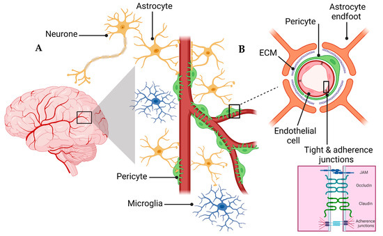
3. BBB Dysregulation with Aging

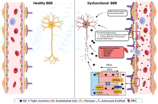
The dysregulation of BBB in aging involves both a loss of selective transport mechanisms and a reduction in structural integrity [33][44]. This is exemplified by a global shift in the pattern of protein transcytosis, transitioning away from a receptor-mediated transport (RMT) to an increased caveolar transcytosis, which allows the entry of potentially neurotoxic proteins such as albumin and fibrinogen that are otherwise restricted in youth [33][34][44,45]. This change in the BBB’s permeability is implicated in the pathophysiology of neurodegenerative diseases, including AD, by facilitating neuroinflammation through unregulated protein entry [35][36][46,47]. Moreover, the age-related decrease in pericyte coverage further compromises the BBB, impairing blood flow and neuronal function [37][48]. The observed upregulation of endothelial alkaline phosphatase, namely ALPL, in AD patients, suggests it to be a potential therapeutic target, as its inhibition could modify the BBB’s permeability and influence the disease progression [38][49]. Overall, these changes underscore a “two-hit” hypothesis for vascular contributions to AD, where both pericyte loss and dysfunctional transcytosis act synergistically with other pathogenic factors like Aβ accumulation. Understanding the evolving nature of the BBB’s communication with the periphery is crucial for deciphering the impact of aging on the brain in health and disease [38][49].

4. BBB Dysfunction in AD

4.1. Effect of Comorbidities on BBB

The BBB disruption is related to multiple comorbidities, including vascular comorbidities such as atherosclerosis, where both diseases are characterized by inflammation and vascular dysfunction [39][58]. Small vessel dysfunction is responsible for the BBB breakdown, CBF and Aβ clearance reduction, and neuronal dysfunction [39][40][58,59]. However, findings from the Baltimore Longitudinal Study of Aging (BLSA) Cohort, which investigated the relationship between Alzheimer’s and atherosclerosis, found no association between the degree of Alzheimer’s pathology and atherosclerosis in the aorta, heart, and intracranial vessels, though only intracranial atherosclerosis correlated with dementia [41][60]. Moreover, comorbidities, including Type 2 Diabetes Mellitus (T2DM), hypertension, obesity, sleep disorders, and hypercholesterolemia, are increasingly recognized as contributing factors in the disruption of the BBB and the progression of AD [42][43][61,62]. In the case of T2DM, which is characterized by insulin resistance and chronic hyperglycemia, it causes endothelial dysfunction and inflammation that could ultimately compromise the BBB’s integrity. These alterations potentially facilitate the entry of neurotoxic molecules like Aβ, thereby contributing to AD pathology [44][45][63,64].  Obesity, especially as a part of the metabolic syndrome, often precipitates systemic inflammation and endothelial dysfunction, contributing to a compromised BBB function [46][66]. Sleep disorders, particularly those impacting the glymphatic system, are emerging as critical players in hindering the efficient clearance of neurotoxic products such as Aβ, ultimately fostering an environment conducive to AD progression [47][67].  The cumulative impact of these comorbidities cannot be overlooked, as they often co-occur and may have synergistic effects on BBB dysfunction and AD pathology. These comorbidities exacerbate the degradation of TJs, thereby compounding the risk of BBB dysfunction and the subsequent development and progression of AD [48][49][50][51][69,70,71,72]. The interplay between the degradation of TJs by proteases and the risk factors for AD suggests a vicious cycle where the compromised barrier function of the BBB can lead to further neuronal damage and progression of AD. At the same time, the disease state promotes a further BBB breakdown [51][72]

4.2. ApoE and BBB Dysfunction in AD

ApoE is mainly expressed in astrocytes and microglia in the brain; its functions are associated with the endocytosis of lipoproteins, the deposition and transport of Aβ, membrane integrity, neurotoxicity, and synaptic plasticity [14][19]. It is produced in the brain, liver, kidneys, skin, adipose tissue, and many other organs [52][74]. Within the CNS, ApoE is produced de novo separately and independently from peripheral ApoE [53][75]. ApoE protein has three different isoforms: ApoEε2, ApoEε3, and ApoEε4. These isoforms have functional and structural differences, inconsistencies, and discrepancies in their interaction with low-density lipoprotein (LDL) receptors [52][74]. Within the cell, ApoE plays a role in cellular processes involving the maintenance of the cytoskeleton, mitochondria, and dendrites, leading it to play a prominent role in the overall health of neurons [52][74]. ApoE amino acids vary at locations 112 and 158 on chromosome 19, and there are six different genotypes, i.e., three homozygous and three heterozygous [54][76]. ApoEε2 is protective against the pathology of AD [55][77]. ApoEε2 expression clears Aβ normally and may suppress inflammation, which can protect against AD [54][76]. To investigate the role of ApoEε2 in AD, using post-mortem human cortices, Fernández-Calle, and colleagues reported that the abundance of ApoEε2 and its ability to bind to LDLRs increased the efficiency of Aβ clearance. In addition, they found that the expression of mRNA molecules related to ECM increased, which could contribute to the protection of the BBB’s integrity. These effects of ApoEε2 point toward its protective abilities against AD [56][79]. ApoEε3, the most common form of the gene, on the other hand, does not present a risk for or protection from AD relative to the ε4 and ε2 alleles, respectively. ApoEε3 protein functions well in binding to LDL, very low-density lipoprotein (VLDL), and low-density lipoprotein-related protein-1 (LRP1) receptors, which play a role in clearing Aβ [56][79]. The most heavily researched isoform is ApoEε4. ApoEε4 is associated with an earlier onset of AD due to a higher plaque density earlier in life [14][19]. ApoEε4 likely contributes to AD pathology by increasing neurotoxicity and decreasing the protection of the CNS [53][75]. However, the mouse apoE gene exists only in one isoform and is located on chromosome 7 rather than 19. For this reason, mice are often artificially introduced to human ApoE to make the study applicable to human AD [56][79]. ApoEε4 correlates with a decreased BBB integrity and cognitive impairment due to disturbed homeostasis and neurotoxin extravasation into the brain [57][80]. ApoEε4 knock-in mice displayed the loss of pericytes and a subsequent BBB degradation as they aged [58][81]. This loss of pericytes was also found in human ApoEε4 carriers [53][75].

4.3. Tight Junctions’ Role in BBB Health and Disease

The barrier function of TJs in the BBB is multifaceted. They restrict the intercellular space, preventing the unregulated diffusion of substances between the blood and the brain. This role is critical in preserving the specialized environment required for an optimal neural function [59][84]. Concurrently, TJs maintain cellular polarity by ensuring an asymmetrical distribution of proteins across the cell, a decisive factor for a directional transport across the endothelium [59][84]. On the paracellular front, TJs exert tight control over the diffusion pathways by leveraging an array of integral proteins, such as claudins, occludin, and JAMs. The specific assembly of these proteins determines the permeability properties of the junctions, allowing selective passage of ions and molecules while safeguarding against harmful substances [60][85]. For the transcellular route, the asymmetrical distribution of transporters and channels underpins a regulated transport mechanism, ensuring that essential nutrients reach the brain while metabolic waste products are efficiently evacuated. The concerted action of these transcellular elements with TJs establishes the BBB as a dynamic regulatory interface, which is adept at adjusting to physiological needs and protecting against pathological insults. In AD, one of the critical pathways by which TJs can be compromised involves the activity of matrix metalloproteinases (MMPs) and other proteases [61][86]. MMPs are a family of zinc-dependent endopeptidases capable of degrading various components of the extracellular matrix (ECM). They are known to modulate the permeability of the BBB by targeting TJ proteins [62][87]. MMPs, particularly MMP2 and MMP9, have been demonstrated to cleave occludin, claudins, and ZO proteins, leading to the disassembly of TJs and an increased BBB permeability [62][87]. The activity of MMPs is tightly regulated under normal physiological conditions but can be upregulated in response to inflammation, ischemia, and oxidative stress, which are common in the AD pathology [61][86].

4.4. Transporters’ Role in BBB Health and Disease

The functionality of the BBB is partly enabled by the presence of diverse influx and efflux transporters expressed in the endothelial cells, notably from the solute carrier (SLC) and ATP-binding cassette (ABC) superfamilies [63][64][65][91,92,93]. The BBB restricts drug access to the brain by permitting only lipophilic molecules of low molecular weight into the brain via the transcellular pathway from the bloodstream, where most researchers consider a cut-off point of 400–600 kDa. However, deviations from this cut-off point have been published [66][94]. Small, lipid-soluble drugs, such as antidepressants, penetrate the BBB via passive diffusion across the BBB [31][38]. Transcytosis transports macromolecules, such as insulin and amino acids [31][38]. Efflux transporters counteract the passive diffusion by forcing foreign substances, toxic metabolites, and other waste products out of the brain [31][38]. In contrast, recombinant proteins, therapeutic antibodies, and nucleic acid drugs are too large to cross the BBB [31][38]. The BBB disruption could be due to, at least in part, the modulation of transporters’ function or expression, accumulating waste products, and the inadequate nutrient delivery to the brain [31][38]. Overall, more than 50 transporters are expressed in the BBB, and their modulation by aging or AD could alter the BBB function, thus contributing to AD pathology [67][95]

5. BBB Breakdown Mechanisms

A schematic presentation of the discussed mechanisms is presented in Figure 2. The disruption of the BBB could impair any of the NVU cellular components. The disruption of endothelial cells might result in a decreased TJs’ expression. It has been shown that occludin, claudin-5, and ZO1 expressions are lower in the Aβ-laden capillaries of patients with capillary CAA [31][38], implying that reduced TJs may increase the vascular permeability in an AD brain. Multiple cellular signaling pathways, such as calcium signaling, could mediate the disruption of TJs. In AD, the RAGE–Aβ42 interaction disrupts TJs via a calcium-calcineurin signaling pathway [68][133]. In addition, in the monolayer culture of bEnd3 cells, adding Aβ42 induces structural alterations in ZO1 [68][133]. Other TJ proteins, such as claudin-5 and occludin, were also structurally altered, and their expression was reduced by Aβ42 [68][133]. This result was confirmed as neutralizing antibodies against RAGEs and calcineurin and MMP inhibitors prevented Aβ42-induced changes in the ZO1 expression [68][133]. Furthermore, the expression of TJs is negatively correlated with Aβ40 levels in cortical areas and positively correlated with synaptic markers in patients with AD, highlighting the role of TJs in the AD pathology development [69][134].
Figure 2.
A representative scheme demonstrates a healthy BBB and a dysfunctional BBB.
Transport protein expression alterations in endothelial cells could be a mechanism or a consequence of BBB disruption [70][54]. In AD, the failure of Aβ clearance through transport across the BBB is caused by decreased levels of LRP1 and P-gp and an increased RAGE expression. For example, in an in vitro experiment, a high cholesterol level decreased the LRP1 expression, increased the RAGE expression, and increased Aβ40 levels in cerebral microvascular endothelial cells [71][135]. Other transport disruptions include a reduced expression of the GLUT1 transporter [70][72][54,136]. This effect was seen with a lower expression of GLUT1 in endothelial cells but not in astrocytes of the BBB in GLUT1-deficient APPsw-mice [73][102]. The LRP1 protein is vital to maintain the BBB’s integrity; it acts as a co-activator of peroxisome proliferator-activated receptor gamma (PPARγ), transports cholesterol associated with ApoE [74][137], plays a role in glucose metabolism [75][138], and interacts with Aβ to transport it across the BBB [74][137]. The LRP1 activation can stimulate PPARγ and increase TJs’ expression. This effect has been investigated in mouse models where a selective brain endothelial LRP1 knockout reduced the expression of TJ proteins and P-gp, increased the MMP activity, and decreased the TEER, leading to endothelial cell disruptions [76][122]. Endothelial cell disruption causes neuroinflammation due to neurotoxic substances infiltrating into the brain, causing inflammation and neurodegeneration. Fibrinogen leakiness in the brain activates the macroglia and initiates inflammation [77][78][139,140]
Aside from endothelial cell disruptions, neuroinflammation and the activation of glial cells can further drive AD pathology. Microglial activation can release pro-inflammatory factors, resulting in neuroinflammation [79][141]. Although inflammation is a protection mechanism, an overly aggressive inflammatory response can cause or contribute to tissue damage. In AD, astrocyte degeneration causes the BBB disruption. Using tamoxifen-astrocyte-depleted mice, large molecules such as fibrinogen and smaller molecules such as cadaverine, an exogenous labeled small molecule, were detected in the brain as soon as the astrocytes were depleted, which was accompanied by reduced levels of TJ proteins, and lower expression of GLUT1 [80][142]. Aquaporin-4 (AQP-4), a water channel facilitating bidirectional water transfer, is expressed by astrocytes at the BBB [81][56]
The nucleotide-binding oligomerization domain-like receptor pyrin domain-containing 3 (NLRP3) inflammasome has a significant role in AD-related neuroinflammation. The interaction of Aβ with astrocytes and macroglia can activate NLRP3 inflammasome, causing the release of chemokines and inflammatory mediators and activating the caspase-1 cascade [82][145]. The activation of NLRP3 inflammasome results in the BBB breakdown by the generated cytokines, whereas reducing the inflammasome activation reduces the inflammatory response and improves the BBB function [83][146]. NF-κB is another inflammatory transcription factor that is increased in AD. The activation of NF-κB by Aβ plaques, NFT, or oxidative stress causes the release of proinflammatory cytokines and ROS and promotes neuroinflammation. Like neuroinflammation, oxidative stress plays a significant role in the BBB disruption [79][141]. Oxidative stress is a condition produced by an imbalance between the generation and accumulation of ROS in cells and tissues and the ability of a biological system to detoxify these reactive products [84][147]. Neuroinflammation and oxidative stress result in neuronal cell death, altered neurotransmitter production and activity, and decreased synaptic functioning, all of which can lead to cerebral injury and dysfunction [85][86][117,148]
In AD, Aβ deposits cause pericyte degeneration [87][88][151,152]. Aβ-pericytes signaling causes vascular constriction and decreases CBF through ROS generation in humans and rodents [89][153]. This degeneration is affected by the ApoE isoform where ApoEε4 accelerates the BBB breakdown; ApoEε4 also reduces the calcineurin–nuclear factor of activated T cells (NFAT) signaling in pericytes and causes the BBB disruption [90][154]. ApoEε4 iPSC-derived-mural cells expressed higher levels of cytoplasmic and nuclear NFATc1 protein and exhibited a higher gene expression of calcineurin catalytic subunits (PPP3CA and PPP3CC). Increased levels of NFATc1 induce the CAA pathology by interacting with ApoEε4 promoter in ApoEε4-expressing pericytes. Inhibiting calcineurin in iPSC-derived mural cells reduced ApoE expression in ApoE4-expressing cells and NFATc1 in ApoE3-expressing cells [90][154]
In AD, the dysfunction of the Wnt/β-catenin signaling pathway has been associated with the BBB breakdown. Under normal conditions, Wnt/β-catenin signaling is essential for the CNS angiogenesis [91][156]. Reduction in this signaling pathway reduces the expression of TJs and modulates the expression of transporters such as P-gp and GLUT1, leading to an increased BBB permeability [91][156]. Suppressing Wnt/β-catenin signaling in the APPswe/PS1dE9 (APP/PS1) microvessels increased glycogen synthase kinase-3 beta (GSK-3β), whereas its activation, using LRP6-optogenetic tool, restored the BBB TJs, and prevented Aβ-induced endothelial cells’ dysfunction [92][157]. The same effect has been shown in human iPSCs where the activation of the Wnt/β-catenin pathway using CHIR 99021 (CHIR), an inhibitor of GSK-3β and an agonist of Wnt/β-catenin, induced GLUT1 and claudin-5 expressions, and downregulated levels of plasma lemma vesicle-associated protein (PLVAP) [93][158]. Wnt/β-catenin signaling also regulates paracellular and transcellular permeabilities [94][159]
The genetic underpinnings of the BBB dysfunction in AD provide a fascinating, yet relatively understudied, angle to the ongoing research in the field. A significant proportion of genes implicated in the AD risk have been linked to the brain’s vascular and perivascular systems, further suggesting the integral role of the BBB’s integrity in the AD pathogenesis. The human brain vasculature expresses 30 out of the top 45 genes linked to the AD risk based on genome-wide association studies (GWAS) [95][160]. Remarkably, several genes identified in GWAS, including PICALM, known for its involvement in endocytosis facilitating the internalization of cell receptors, have variants that can increase the Aβ accumulation in the brain, exacerbating the AD pathology [96][161], as shown by triggering clathrin-mediated endocytosis through its interaction with LRP1 [97][162]
Mutations in the OCLN gene, responsible for encoding occludin [98][167], and the junctional adhesion molecule-C (JAM-C) gene, responsible for encoding the junctional molecule JAM-C, are directly implicated in the compromise of the BBB’s integrity [99][100][168,169]. Furthermore, NOTCH3 gene, vital for vascular integrity through smooth muscle cell differentiation, presents mutations that affect blood vessel walls and the BBB’s integrity, further linking it to AD and vascular dementia [101][170]. Mice with the CADASIL-associated Notch3-R169C mutation exhibit an accumulation of the NOTCH ectodomain within pericytes. This accumulation is concomitant with the pericyte degeneration and a subsequent compromise of the BBB’s integrity [102][171]
TREM2, which encodes the triggering receptor expressed on myeloid cells 2 protein, was highlighted as an AD susceptibility gene in two GWAS [103][104][175,176]. Notably, these studies identified the R47H missense mutation, which presents a heightened AD risk comparable to possessing one ApoEε4 allele [103][175]. Predominantly expressed in brain microglia, TREM2 is pivotal in mediating the neuroinflammatory reactions associated with AD [105][177]. In-depth functional analyses of TREM2 have elucidated its involvement in the modulation of Aβ plaque accumulation in brain parenchyma, the advancement of tau-related pathology, and BBB dysfunction [104][106][107][108][176,178,179,180]. TREM2 may affect the integrity of the BBB by affecting inflammation, the microglial oxidative response, and insulin resistance. Moreover, many studies have found that soluble TREM2 (sTREM2) may disrupt the BBB’s integrity in AD by interacting with pro-inflammatory proteins such as TNF receptor 1 and TNF receptor 2, and their effectors like intercellular adhesion molecule 1 and vascular cell adhesion molecule 1 [109][181]

6. The BBB as a Therapeutic Target

The BBB dysfunction has been implicated in the pathogenesis of AD and many other neurodegenerative diseases [25][110][30,189]. While there are new medications targeting Aβ, such as the recent FDA-approved monoclonal antibody Lecanemab, a curative therapy for AD remains elusive. Furthermore, these medications are associated with severe adverse reactions, including edema and cerebral microhemorrhages [111][190]. Such concerns underscore the potential need for combined therapies that simultaneously address the BBB function and other key aspects of AD. Accordingly, there has been a growing interest in developing therapeutic strategies that target the BBB to enhance drug delivery, improve the clearance of toxic molecules, and restore the barrier function [112][191]. Strategies to modulate the BBB function in AD can be broadly categorized into three main areas: enhancing the Aβ clearance across the BBB, improving the BBB’s integrity and function, and addressing neuroinflammation [25][110][30,189]. Indeed, the intricate regulation of Aβ transporters, both influx and efflux, plays a crucial role in the Aβ accumulation. The dysregulation of these transporters has been recognized as a potential therapeutic target of AD [112][191].
ScholarVision Creations