Flavonols on Cognitive Functions in Diabetic Animals: Comparison
Please note this is a comparison between Version 2 by Catherine Yang and Version 1 by Maja Jazvinšćak Jembrek.

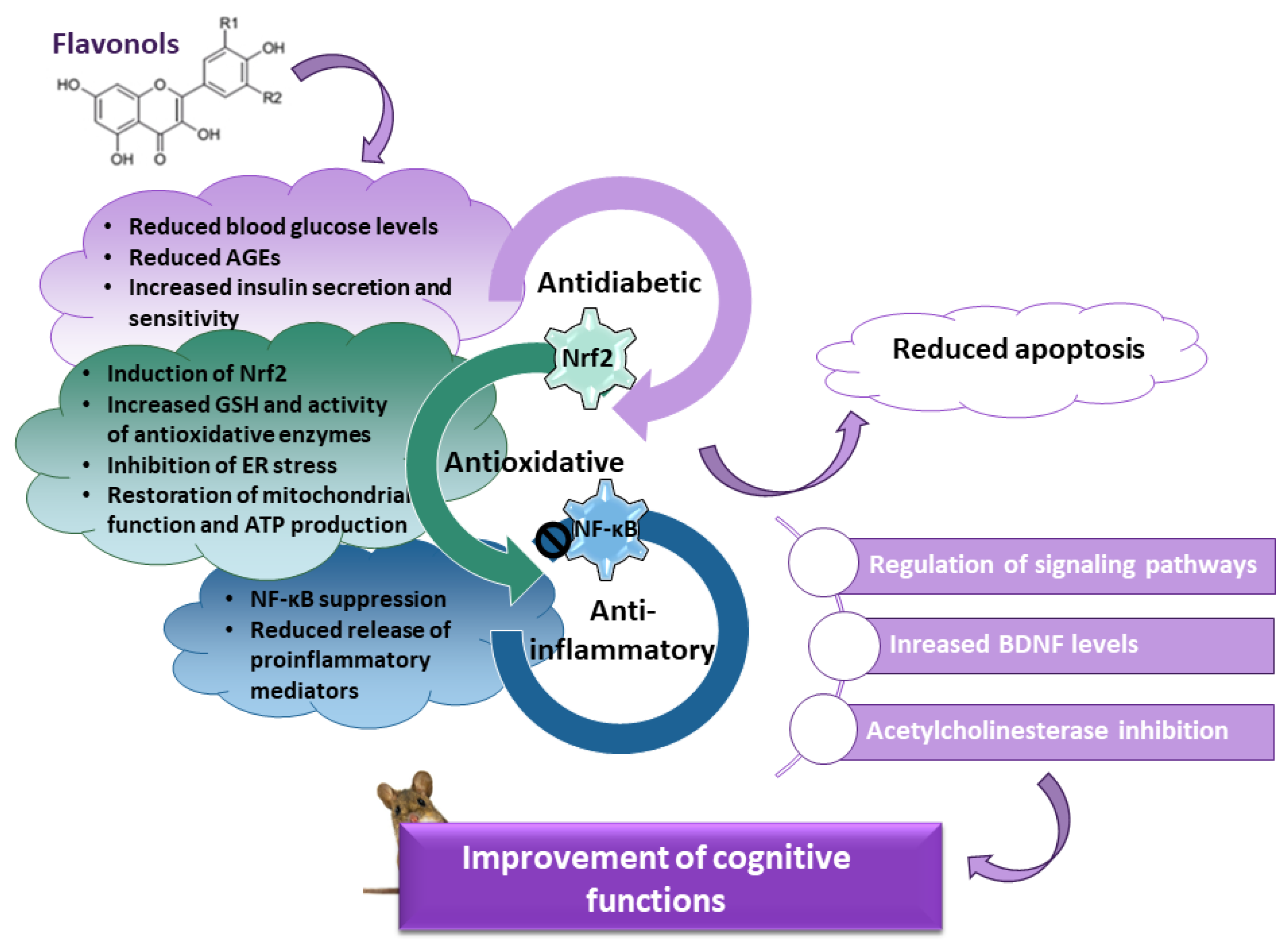
Diabetes mellitus is a complex metabolic disease associated with reduced synaptic plasticity, atrophy of the hippocampus, and cognitive decline. Cognitive impairment results from several pathological mechanisms, including increased levels of advanced glycation end products (AGEs) and their receptors, prolonged oxidative stress and impaired activity of endogenous mechanisms of antioxidant defense, neuroinflammation driven by the nuclear factor kappa-light-chain enhancer of activated B cells (NF-κB), decreased expression of brain-derived neurotrophic factor (BDNF), and disturbance of signaling pathways involved in neuronal survival and cognitive functioning. Flavonols, a highly abundant class of flavonoids in the human diet, are appreciated as a potential pharmacological intervention against cognitive decline in diabetes. 

  • diabetes
  • cognitive functions
  • flavonols
  • oxidative stress

1. Introduction

Foods rich in flavonoids may have beneficial effects on cognitive functioning [14,49][1][2]. Flavonoids comprise a large group of secondary plant metabolites with well-documented antioxidative, anti-inflammatory, and neuroprotective effects, as well as antidiabetic and memory-enhancing properties [14,53,209,210,211][1][3][4][5][6]. Structurally, all flavonoids have benzene and phenyl rings (rings A and B), which are bridged by the heterocyclic pyrene ring C. Based on the diversity of chemical structure (degree of oxidation and saturation, pattern of attached groups, and position at which the B-ring is connected to the C-ring), they are categorized into several subclasses [211,212][6][7]. The most important subtypes of flavonoids are flavonols, flavanols, flavones, flavanones, anthocyanins, and isoflavones [212,213][7][8].
The main dietary sources of flavonoids are fruits and vegetables, tea, and red wine [214,215][9][10]. Accordingly, there is great interest in the potential health-promoting effects of a diet enriched with fruit and vegetables. The Mediterranean diet is characterized by a high consumption of fruits, vegetables, and beverages rich in flavonoids, and this type of diet has been repeatedly associated with a lower risk of chronic diseases, including diabetes [216,217,218][11][12][13]. The composition of the various flavonoids in different foods can be assessed using the available databases on flavonoid content. However, the estimated dietary intake of flavonoids varies widely between studies. The estimation of the total dietary intake of flavonoids depends on the number of foods considered, the geographical and seasonal origin of the foods, the environmental conditions, agricultural practices, the degree of ripeness, the storage and processing of the foods, the analytical methods used for quantification, as well as the population studied, as intake may vary according to age, gender, dietary habits, income level, and ethnicity [214,215,219][9][10][14]. In two US studies, the total daily intake of flavonoids ranged from 189.7 mg/day to 251 mg/day. Apart from flavan-3-ols, which accounted for about 80% of the total flavonoid intake, flavonols were the second most abundant flavonoids in these studies. Their content is estimated at 6.8% and 8% of all flavonoids [214,215][9][10]. However, in a Greek plant-based weekly menu, the daily intake of flavonoids was estimated at 118.6 mg, of which flavonols accounted for 22% [220][15]. Flavonols are mainly found in onions, kale, broccoli, apples, cherries, berries, tea, and red wine. The most abundant flavonols in the diet are quercetin, kaempferol, and myricetin [219,220][14][15]. Structurally, flavonols have an oxo group at position 4, a hydroxyl group at position 3, and a 2,3-double bond on ring C, which is particularly relevant in the context of their redox properties and health-promoting effects [62][16]. Flavonols are one of the most studied flavonoids. Their great potential against various chronic diseases has been demonstrated in many studies [62,211,221][6][16][17]. Moreover, it has been suggested that the health-promoting effects of flavonols are superior to those of other subclasses of flavonoids due to their complex mechanisms of action [221][17].
Based on numerous preclinical studies, restoration of redox homeostasis is appreciated as a promising therapeutic option to manage cognitive impairment in diabetes. Flavonols in general possess remarkable antioxidant abilities. They exert antioxidant effects via several mechanisms, including (i) direct ROS scavenging, (ii) induction of the endogenous mechanisms of antioxidative defense, (iii) chelation of metal ions that otherwise may initiate ROS formation via Fenton chemistry, and (iv) regulation of aberrant redox-sensitive signaling pathways [211,222,223,224][6][18][19][20]. In humans, plasma levels of antioxidants and total antioxidant status are positively correlated with the general intake of fruits and vegetables [225,226,227,228][21][22][23][24]. Evidence is growing that increased consumption of foods rich in antioxidants, or supplementation with antioxidants, may reduce levels of oxidative stress markers [229][25] and even improve cognitive performance in various conditions, both in humans and animals [230,231][26][27].
In addition, possessing powerful antioxidant abilities, flavonols are capable of regulating oxidative and inflammatory signaling cascades involved in neuronal survival. They also target various proteins important for cognitive processes, such as BDNF, thus preserving the structural integrity and functionality of neural circuits, which is ultimately beneficial for overall cognitive performance [206,211,224,231][6][20][27][28]. As will be shown later, the results of many preclinical studies indicate that individual flavonols are effective against cognitive decline in diabetes. On the contrary, there are only a few studies investigating the effects of flavonols and foods rich in flavonols on diabetic patients. In a longitudinal study that lasted for more than 6 years and was performed on more than 10,000 middle-aged participants (45–64 years), dietary intake of food enriched with flavonols slowed down cognitive decline over time [232][29]. By using the combined change score from the delayed word recall test, the Wechsler Adult Intelligence Scale-Revised (WAIS-R) digit symbol subtest, and the word fluency test, it was found that intake of flavonols, particularly myricetin, kaempferol, and quercetin, positively correlates with the preservation of cognitive abilities [232][29]. The effect was the most pronounced for the digit symbol subtest; in the word recall test, it was not significant and was marginal in the word fluency test. The association between a diet rich in flavonols and the slower progression of cognitive disabilities was also demonstrated in an American study. This study was performed on almost 1000 participants aged 60–100 years whose cognitive abilities were evaluated with a battery of 19 cognitive tests. The obtained results revealed better global cognition, working memory, episodic and semantic memory, and perceptual speed in participants with a higher dietary intake of flavonols, particularly quercetin and kaempferol [233][30]. Regarding diabetes, in a study lasting for app. 12 years that included almost 3000 individuals, the intake of flavonols and flavan-3-ol reduced the incidence of diabetes by 26% and 11%, respectively. Associations between the intake of flavonoids from other classes (flavones, flavanones, anthocyanins, and polymeric flavonoids) and diabetes risk were not found [56][31].

2. Mechanisms Underlying the Beneficial Effects of Quercetin in Diabetic Animals

Quercetin (3,3′,4′,5,7-pentahydroxyflavone) is one of the most widely distributed flavonols in the human diet. In large amounts, it can be found in apples, onions, broccoli, berries, kale, green tea, red wine, seeds, and nuts [234][32]. It possesses powerful antioxidant and anti-inflammatory abilities [210,211,235][5][6][33] that underlie its antidiabetic effects. Based on a 100-item food frequency questionnaire, daily quercetin intake in a Chinese population has been estimated to be 20.9 ± 2.32 mg/day. More importantly, dietary quercetin consumption negatively correlated with diabetes prevalence, suggesting the protective role of quercetin in the development of the disease [236][34].
The effects of quercetin on memory dysfunction have been documented in animal models of streptozotocin-induced diabetes. In the MWM test, diabetic rats exhibited higher escape latency during training trials and reduced time spent in the target quadrant in the probe trial compared to control animals, whereas in the elevated plus maze task, diabetic animals showed increased latency. Upon diabetes induction, the changes observed in the MWM and the elevated plus maze tasks were reversed in rats receiving quercetin (5, 10, and 20 mg/kg, twice daily for 30 days) [206][28]. In another experiment in the same study, rats received 20 and 40 mg/kg quercetin (twice daily) during training trials (from days 31 to 35). Acute treatment with the higher dose reduced the escape latency and increased the time spent in the quadrant with a platform [206][28], indicating that even the short-term administration of quercetin may improve cognitive performance.
Numerous preclinical studies have shown that quercetin reduces blood glucose levels in diabetic animals [237,238,239,240][35][36][37][38] and increases sensitivity to insulin [241,242][39][40]. Accordingly, it has been suggested that the beneficial effects of quercetin on the improvement of cognitive functions at least partially rely on its anti-hyperglycemic properties, preventing the hyperglycemia-mediated effects on the induction of oxidative stress and neuroinflammation via the AGEs/RAGE axis [206,243][28][41]. The restoration of glucose metabolism is usually accompanied by the prominent antioxidative effects of quercetin at the systemic level and in specific tissues, particularly in the pancreas and liver. Thus, quercetin administration reduced the extent of lipid peroxidation, improved the activities of antioxidative enzymes, mainly SOD, catalase, and GPx, and restored levels of small antioxidants such as GSH and vitamins C and E [237,244,245,246,247,248][35][42][43][44][45][46]. Likewise, quercetin-mediated effects on cognitive functions were associated with improved activity of antioxidative enzymes and reduced amounts of oxidative stress markers [249,250][47][48].
In addition to the mentioned activities, quercetin acts as an acetylcholinesterase (AChE) inhibitor, which very likely contributes to its memory-enhancing abilities [209,243,249][4][41][47]. Quercetin-mediated attenuation of cholinergic dysfunction has been demonstrated in the hippocampus and cerebral cortex of streptozotocin-induced diabetic rats (quercetin was applied at doses of 25 and 50 mg/kg for 40 days) [249][47]. In addition, quercetin reduced the MDA levels in a dose- and brain region-specific manner and prevented impairment of enzymes involved in the regulation of purinergic transmission, at least in the synaptosomes from the cerebral cortex [249][47]. Likewise, in mice administered with streptozotocin, quercetin (2.5, 5, and 10 mg/kg, p.o. for 21 days) reduced the increase in AChE activity, decreased MDA and nitrite levels, and increased GSH content [243][41]. In addition, attenuating oxidative stress and cholinergic dysfunction, quercetin restored cerebral blood flow and ATP levels and prevented memory impairment. Effects of quercetin on streptozotocin-induced memory impairment were evaluated in the MWM test (quercetin at doses 5 and 10 mg/kg decreased the mean latency time after the third session) and the passive avoidance test (quercetin increased the transfer latency time in retention trials in comparison with acquisition trials) [243][41].
Another study has shown that the mechanism underlying the beneficial effect of quercetin on cognitive performance is mediated by the increased activation of deacetylase sirtuin 1 (SIRT1) and inhibition of ER stress. Namely, among other functions, SIRT1 regulates insulin secretion and blood glucose levels and may have an inhibitory effect on signaling pathways implicated in ER stress and cognitive dysfunction. In female db/db mice, quercetin at a dose of 70 mg/kg (applied for 12 weeks) attenuated glucose tolerance and insulin resistance and improved cognitive performance. It shortened the time needed to find the hidden platform in the MWM test, and following platform removal, it increased the time spent in the target quadrant and time engaged in the exploration of the platform area [250][48]. Likewise, quercetin improved performance in the novel object recognition test. Together with these effects, quercetin upregulated SIRT expression as well as the expression of synapse-related proteins and neurotrophic factors, including BDNF and NGF, and reduced the expression of ER stress markers. Quercetin-mediated improvement of cognitive abilities was also accompanied by reduced oxidative stress, reduced expression of proapoptotic proteins, and attenuated neuronal apoptosis and neurodegeneration in the hippocampal and cortical areas, altogether suggesting the promising potential of quercetin for relieving symptoms of diabetes-induced cognitive decline [250][48]. A similar study that was performed in streptozotocin-induced diabetic rats also demonstrated the ability of quercetin (100 mg/kg b.w., applied orally in aqueous solution for 15 days) to reduce serum glucose levels and increase SIRT1 expression [238][36].
In addition to central effects, quercetin also restored activation of the adenosine 5′-monophosphate (AMP) activated protein kinase (AMPK)/peroxisome proliferator-activated receptor-γ coactivator 1α (PGC-1α) pathway and prevented mitochondrial dysfunction in an animal model of diabetic peripheral neuropathy [246][44]. The hyperglycemia-induced impairment of mitochondrial function has been suggested as one of the major pathological mechanisms of diabetic neuropathy [246][44]. This chronic microvascular complication of diabetes results in neuropathic pain due to the activation of purinergic P2X4 receptors in the dorsal root ganglia [251][49]. In the streptozotocin-induced rats, oral administration of quercetin (for 6 weeks at doses of 30 and 60 mg/kg/daily) improved the paw withdrawal threshold (an indicator of mechanical allodynia), the motor nerve conduction velocity, ultrastructural abnormalities of sciatic nerves, loss and morphological changes of neurons in the dorsal root ganglion, axonopathy, expression of myelin proteins, and demyelination of myelin sheet [246][44]. Together with the alleviation of mitochondrial degeneration and restoration of ATP production, quercetin increased expression of phosphorylated AMPK, PGC-1α, SIRT1, nuclear respiratory factor 1 (NRF1), and mitochondrial transcriptional factor A (TFAM), suggesting that its neuroprotective effects are closely related to the restoration of mitochondrial energy metabolism and activation of the AMPK/PGC-1α pathway. In general, the energy status of cells and mitochondrial biogenesis are controlled through the AMPK/PGC-1α pathway, and upregulation of its activity usually preserves mitochondrial function in hyperglycemic conditions [246][44]. Furthermore, in dorsal root ganglion neurons exposed to high glucose, quercetin activated the Nrf2 pathway and inhibited the NF-κB signaling cascade, suppressing the production of proinflammatory cytokines and the expression of iNOS [252][50]. Of note, the SIRT1/PGC-1α axis also participates in the regulation of redox homeostasis in mitochondria, and its stimulation may increase the ROS-detoxifying abilities, which is in line with the observed antioxidative effect of quercetin [246,252][44][50]. In addition, quercetin may prevent upregulation of the P2X4 receptors and activity of the p38 pathway in diabetic rats [253][51], whereby the latter is known to be upregulated in the diabetic brain and likely contributes to cognitive dysfunction [42,254][52][53].
The anti-inflammatory effects of quercetin also play an important role in rescuing cognitive abilities. It was shown that pure quercetin and quercetin-conjugated superparamagnetic iron oxide nanoparticles (applied at 25 mg/kg for a period of 35 consecutive days) may attenuate expression of microRNA-146a (miR-146a), an inflammation-sensitive microRNA, and expression of NF-κB and NF-κB-related downstream targets, such as TNF-α, in the hippocampus of the streptozotocin-induced diabetic rats. Molecular docking revealed that the inhibitory effect of quercetin on the NF-κB pathway could be mediated by targeting the IKK protein [124][54]. Quercetin and quercetin-conjugated superparamagnetic iron oxide nanoparticles also alleviated diabetes-induced overexpression of redox-sensitive miR-27a, which subsequently increased levels of Nrf2 and its target genes SOD and catalase, together with the prevention of memory dysfunction [255][55]. A study performed with db/db mice has shown that quercetin (35 and 70 mg/kg for 12 weeks) may improve cognitive performance by regulating the Sirt1/nucleotide-binding domain-like receptor protein 3 (NLRP3) pathway. In particular, quercetin increased the expression of Sirt1, which is a negative regulator of the inflammatory response, and consequently reduced the expression of inflammation-related proteins, including NLRP3 and the pro-inflammatory cytokines IL-1β and IL-18, thus inhibiting the NLRP3 inflammasome activation and further neurodegenerative processes. Furthermore, quercetin enhanced the expression of synapse-related proteins and neurotrophic factors BDNF and NGF, demonstrating the co-existence of multiple mechanisms that likely function together in the restoration of cognitive abilities [256][56]. The anti-inflammatory potential of quercetin has also been observed in other tissues of diabetic animals and is suggested to have an important role in the antidiabetic effects of quercetin [248][46].
Except in rodents, the antidiabetic effects of quercetin were demonstrated in the streptozotocin-induced Arbor Acre broilers supplemented with 0.02–0.06% quercetin and were mainly attributed to the antioxidant mechanism of quercetin action. In chickens, quercetin in a dose-dependent manner improved fasting blood glucose and insulin levels, activities of antioxidant enzymes catalase and SOD, MDA and nitric oxide content in serum and liver tissue, and regulated expression of genes related to the PI3K/Akt pathway [257][57].
As mentioned, besides pure quercetin, administration of quercetin-conjugated superparamagnetic iron oxide nanoparticles demonstrated a positive effect on learning and memory dysfunction in streptozotocin-induced diabetic animals. These nanoparticles were used to investigate the possibility of overcoming the high clinical doses of quercetin due to its limited permeability across the blood-brain barrier, low bioavailability, and rapid metabolic changes in the gastrointestinal system. In diabetic rats, nanoparticles demonstrated better efficacy in the improvement of cognitive abilities in comparison to free quercetin (both were applied at a dose of 25 mg/kg for 35 days). In the MWM test, rats that received quercetin-conjugated superparamagnetic iron oxide nanoparticles showed reduced escape latencies in the repeated training trials compared to diabetic animals and the free quercetin group, the shortest swimming path length before finding the hidden platform in subsequent trial days, and spent the highest amount of time in the target quadrant. Likewise, free and conjugated quercetin increased the step-through latency in the passive avoidance task, indicating the beneficial effect of quercetin on the retrieval of the fear memory. Treatment with quercetin also prevented the apoptotic cell death of granular neurons in the hippocampus [239][37]. As the side-effects of nanoparticles were not noticed, and considering that quercetin in the conjugated form was more effective in improving memory performances compared to free quercetin, it is likely that novel therapeutic strategies for oral quercetin delivery in managing cognitive decline in diabetes should be oriented towards the development of conjugated formulations and better characterization of their mode of action, stability in the circulation, brain distribution, and safety profile.
The anti-diabetic effects of quercetin have also been investigated in humans. Ostadmohammadi and co-authors performed a meta-analysis of randomized controlled trials, concluding that supplementation for a period longer than 8 weeks at doses higher than 500 mg/day may reduce fasting plasma glucose [258][58]. In patients with type 2 diabetes, aged 30–60 years, daily oral intake of lower doses of quercetin (250 mg/day, every 2 weeks for 8 weeks) was sufficient to increase the total antioxidant status and antioxidative defense, although this dose was insufficient to regulate glycemic parameters (fasting blood glucose and serum insulin) [259][59]. Studies focused on cognitive abilities following the quercetin supplementation protocol are not numerous. In a randomized, double-blind, placebo-controlled clinical trial including 70 Japanese men and women aged 60–80 years, intake of quercetin-rich onion (the daily quercetin intake was estimated to be 50 mg as aglycone equivalent) for 24 weeks improved the Mini-Mental State Examination scores. Although the mechanism behind the quercetin action was not elucidated, the authors suggested that quercetin reduces cognitive decline by improving emotional functions and motivation [260][60]

3. Effects of Other Flavonols on Diabetes-Induced Cognitive Decline in Animal Models

In contrast to quercetin, whose effects are relatively well-studied, the potential of other flavonols against cognitive impairment in diabetes has yet to be explored. Although there are studies that investigate the antidiabetic effects of other flavonols in different tissues, studies demonstrating their effects on the brain and cognitive abilities in preclinical diabetic settings are not numerous.
Rutin (quercetin-3-O-rutinoside) is flavonol with the attached disaccharide rutinose on the quercetin backbone. In various neuropathological conditions, rutin displayed antioxidative, anti-inflammatory, and neuroprotective effects [261,262,263][61][62][63].
Protective and regulatory effects of rutin based on antioxidative and anti-inflammatory mechanisms have been demonstrated in non-brain tissues of diabetic (streptozotocin-induced and db/db) animals [264,265,266,267,268,269,270][64][65][66][67][68][69][70]. Thus, rutin reduced blood glucose level [264[64][65][66],265,266], increased insulin secretion and glycogen storage [264[64][66],266], improved lipid profile and biochemical parameters [267[67][69],269], reduced generation of AGEs [270][70], regulated the PI3K/Akt/GSK-3β pathway [270][70], prevented degenerative changes [264[64][67][68],267,268], reduced the MDA levels [268][68], increased the GSH content and non-enzymatic and enzymatic antioxidative defense [264[64][65][66][68],265,266,268], and attenuated production of proinflammatory cytokines TNF-α and IL-6 [269][69].
In the brain of the streptozotocin-induced rats, rutin (applied for 45 days at a dose of 100 mg/kg) improved the antioxidant status by increasing the non-enzymatic (GSH) and enzymatic antioxidant protection (SOD, catalase, GPx, glutathione reductase) and preventing morphological changes [266][66]. Effects of rutin on cognitive performance have been investigated in the intracerebroventricular-streptozotocin (ICV-STZ)-infused rats, a model of sporadic AD that overlaps in many symptoms with diabetes-induced pathological changes, such as learning and memory deficits, impaired glucose metabolism, oxidative stress, and neuroinflammation. Pretreatment for 3 weeks with rutin (25 mg/kg) attenuated oxidative stress, as evidenced by the reduced lipid peroxidation and nitrite accumulation and the increased GSH content and activity of the GPx, glutathione reductase, and catalase in the hippocampus. In the same study, rutin demonstrated remarkable anti-inflammatory activity by suppressing microglial activation and expression of iNOS and NF-κB, which likely contributed to the preservation of hippocampal morphology and improved performance in the MWM task [271][71]. Regarding diabetes, the effects of rutin have been studied in diabetic retinas. Administration of rutin (100 mg/kg) for 5 weeks increased levels of BDNF and NGF, increased GSH levels, attenuated lipid peroxidation, and demonstrated an antiapoptotic effect by decreasing levels of caspase-3 and upregulating expression of Bcl-2 [272][72]. This may suggest that similar protective mechanisms may be effective against diabetes-induced cognitive complications in the brain.
Troxerutin is a semi-synthetic flavonol derived from rutin that has powerful antioxidant properties. Much evidence suggests that it may improve cognitive decline in animals treated with streptozotocin. In the hippocampus of diabetic rats, troxerutin (150 mg/kg/day for 6 weeks, starting 4 weeks after streptozotocin administration) reduced lipid peroxidation and increased SOD activity, attenuated gene expression of the NADPH oxidase subunits (NADPH oxidase is an important source of ROS), stimulated the nuclear translocation of Nrf2, and increased the cytosolic fraction of the antioxidant enzymes heme oxygenase-1 (HO-1) and NAD(P)H:quinone oxidoreductase (NQO1). Although troxerutin did not regulate blood glucose levels at this dose, the memory-enhancing effect of troxerutin was confirmed in the MWM test and mainly attributed to the antioxidant mechanisms of its action. Taken together, these results suggest that the effects of troxerutin on improving spatial memory were mediated through the regulation of the Nrf2/ARE pathway and suppression of oxidative stress [273][73]. In rats treated for 6 weeks, but starting at 12 weeks after streptozotocin administration, troxerutin also improved cognitive performance in the MWM test, regulated oxidative stress-related parameters (SOD activity, GSH, and MDA levels), and stimulated expression of the catalytic and modifier subunits of the glutamate cysteine ligase that catalyze the rate-limiting step of GSH synthesis [274][74]. Similar results were shown when troxerutin was used in the early stages of diabetes, which resembles prophylactic use. Rats were injected with 60 mg/kg of troxerutin for 12 weeks, starting 72 h after streptozotocin injection. This type of treatment improved learning and memory abilities, increased Nrf2 expression in the hippocampus, stimulated SOD activity, and decreased lipid peroxidation. In addition, it preserved normal hippocampal morphology, altogether indicating that troxerutin is effective in delaying cognitive decline when used as a preventive intervention [275][75]. The contribution of the anti-inflammatory mechanisms to the memory-enhancing effects of troxerutin has also been revealed. In the streptozotocin-induced diabetic rats, troxerutin (administered at 150 mg/kg for 1 month, from 7th to 10th weeks after streptozotocin injection) inhibited expression of NF-κB and its adaptor proteins TRAF-6 and IRAK-1, most likely by targeting regulatory miR-146a [276][76]. In high-fat diet-fed rats, troxerutin attenuated the neuroinflammatory response (as evidenced by the decreased production of proinflammatory cytokines) and increased BDNF levels, which may be beneficial in diabetic conditions as well [277][77]. Furthermore, in AD preclinical models, troxerutin inhibited AChE activity and stimulated the PI3K/Akt pathway, demonstrating its ability to target the key molecular mechanisms underlying the cognitive dysfunction in diabetes [278][78].
Like quercetin, myricetin (3,3′,4′,5,5′,7-hexahydroxylflavone) is a flavonoid from the flavonol class that is present in various fruits and vegetables, nuts, berries, and beverages [279][79]. Animal and clinical studies revealed its hypoglycemic effect and ability to increase the sensitivity to insulin and enhance glycogen synthesis and glucose utilization, together with the prominent antioxidative and anti-inflammatory activities that are most likely essential for its protective effects against diabetic complications in various tissues [280,281,282,283,284,285][80][81][82][83][84][85]. Myricetin possesses five hydroxyl groups that underlie its potent antioxidative activity. On the other hand, the glucoregulatory activity of myricetin is probably based on its ability to act as an agonist of the glucagon-like peptide 1 (GLP-1) receptor. Briefly, GLP-1 stimulates insulin secretion and regulates blood glucose levels but shows extremely low stability. Hence, other natural or synthetic GLP-1 receptor agonists are suggested as a more promising therapeutic approach in diabetes. In that regard, it is considered that molecular modeling and chemical modifications of myricetin molecules could greatly advance a search for more potent GLP-1 receptor agonists for further clinical evaluation [286][86]. In the streptozotocin-induced diabetic rats lacking insulin, it was suggested that the acute administration of myricetin increases the release of β-endorphin and lowers plasma glucose levels by activating the opioid μ receptors in peripheral tissues [287][87]. In diabetic peripheral neuropathy, myricetin (0.5–2.0 mg/kg/day, injected intraperitoneally for 2 weeks from the 21st day after streptozotocin administration) was able to reduce abnormal sensations and improve nerve morphology and conduction velocity, as well as the blood flow. More importantly, myricetin reduced the generation of AGEs and ROS, increased the activity of the antioxidant enzymes, activated the Nrf2 pathway, and improved the antioxidant defense, suggesting its potential therapeutic value for other diabetic complications as well [288][88]. In addition, myricetin inhibited AChE activity [209][4] and enhanced memory functions by protecting hippocampal neurons in a rat model of AD, which may suggest its potential against diabetes-induced cognitive decline [289][89]. Similarly, in the senescence-accelerated mouse-prone 8 (SAMP8) mice displaying accelerated aging, intake of myricetin improved performance in the novel object recognition test and the Y-maze test and upregulated expression of BDNF and NGF [290][90]. However, regarding the safety issues of myricetin supplementation, detailed studies should be carried out. Specifically, elevated levels of systemic and hippocampal copper are found in patients with diabetes, together with the positive correlation between serum levels of copper and deregulation of glycemic control [291,292][91][92]. On the other hand, in certain environmental conditions, such as the presence of high levels of copper, myricetin demonstrated prooxidative and cytotoxic effects [293][93], which theoretically may negatively affect cognitive abilities.
Dihydromyricetin is a flavonoid with great structural similarity to myricetin, although it is not classified as a flavonol (2,3-double bond of myricetin is hydrogenated in dihydromyricetin). In a diabetic mouse model (mice were fed a high-sugar and high-fat diet for 8 weeks and then administered a low dose of streptozotocin), dihydromyricetin improved spatial learning and working memory, likely by suppressing oxidative stress (i.e., it decreased MDA accumulation and increased activity of SOD, catalase, and GPx) and increasing BDNF levels, which together exerted neuroprotective effects in the hippocampus [294][94].
Myricitrin, a 3-O-rhamnoside of myricetin, belongs to flavonols and can be found in large amounts in the root bark of Myrica cerifera and Myrica esculenta [295][95]. In the streptozotocin-nicotinamide model of type 2 diabetes, the encapsulated myricitrin restored blood glucose and insulin levels and improved the total antioxidant capacity and levels of MDA, antioxidant enzymes (SOD, catalase), and apoptotic markers in the pancreas [296][96]. Similarly, in the high-fat diet and streptozotocin-induced diabetic mice, myricitrin decreased fasting blood glucose and attenuated gene expression of the proinflammatory cytokines in the liver [297][97], which suggests the antidiabetic potential of myricetin, but its effects on the brain and cognitive abilities still need to be investigated.
Kaempferol is yet another flavonol abundantly present in the human diet. It can be found in various fruits, vegetables, and beverages, such as broccoli, beans, apples, and strawberries [298,299][98][99]. In various neurological conditions, including the experimental model of sporadic AD, kaempferol regulated the NF-κB, p38, and Akt signaling cascades, attenuated ROS production, stimulated the activity of endogenous antioxidants, and increased BDNF levels. Thus, the neuroprotective effects were largely mediated by its remarkable antioxidative and anti-inflammatory activities and ability to modulate intracellular signaling pathways [300,301][100][101]. The beneficial effects of kaempferol that were based on antioxidative, anti-inflammatory, and signaling-related mechanisms have also been observed in diabetes and diabetes-induced complications [302,303,304,305,306,307][102][103][104][105][106][107]. In addition, restoring glucose and insulin levels [302[102][103][104][107],303,304,307], kaempferol suppressed activation of the AGEs/RAGE axis [304][104], attenuated lipid peroxidation [303[103][104][105][107],304,305,307], improved effectiveness of the enzymatic and non-enzymatic systems of antioxidative defense [303[103][104][105][107],304,305,307], reduced ROS levels [305[105][107],307], and stimulated Nrf2 activity [305,307][105][107] in the plasma and various organs of the streptozotocin-induced diabetic animals. Furthermore, kaempferol suppressed transactivation of the NF-κB [304,307][104][107] and attenuated release of proinflammatory cytokines such as TNF-α and IL-6 [304,305,307][104][105][107], inhibited apoptosis [304[104][105][106][107],305,306,307], regulated expression of the autophagy-related proteins and activity of the AMPK/mTOR pathway [306][106], restored activity of the p38 and JNK pathways [304,307][104][107] and upregulated deacetylase activity and levels of SIRT1 [305][105]. Regarding complications affecting neuronal cells, the effects of kaempferol were studied in streptozotocin-induced diabetic neuropathy. Kaempferol partially reduced the nociceptive pain and increased motor nerve conduction velocity by attenuating AGE formation, oxidative and nitrosative stress, and the inflammatory response [308,309][108][109]
Fisetin is a flavonol that has shown antinociceptive effects against diabetic neuropathic pain. It reduced thermal hyperalgesia and mechanical allodynia and demonstrated prominent antioxidative activity (based on targeting the Nrf2 pathway) and the ability to attenuate the inflammatory response (based on regulation of NF-κB activity) [310,311][110][111]. In diabetic rats, fisetin also regulated glucose levels, reduced expression of the NF-kB p65 unit (in the pancreas), and levels of IL-1β and NO in the blood [312][112]. The promising potential of fisetin in alleviating cognitive dysfunction was shown in old SAMP8 mice (a model for sporadic AD). In these mice, fisetin improved cognitive impairment by targeting specific proteins involved in synaptic function, cellular response to stress, brain inflammation, and modulation of the stress-activated protein kinase (SAPK)/JNK pathway [313][113]. All these mechanisms of action may also be relevant for improving cognitive impairment in diabetes, but further studies are needed to determine if fisetin may enhance cognitive functions in diabetic conditions.
Flavonol morin has shown neuroprotective effects in diabetic neuropathy, as evidenced by the improvement of motor and sensory nerve conduction velocities and nerve blood flow. Based on the results obtained in vitro, the authors proposed the regulatory role of morin along the Nrf2 and NF-κB signaling pathways [314][114]. Another study also suggested the therapeutic potential of morin in diabetic neuropathy based on the reduced cytokine levels and lipid peroxidation, increased levels of NGF and GSH, and improved activity of antioxidative enzymes in sciatic nerves [315][115]. Regarding cognitive functions, morin applied at doses of 50 and 100 mg/kg for 60 days reduced the escape latency time in the MWM task in rats treated with streptozotocin. This behavioral improvement was accompanied by reduced oxidative damage of proteins and membrane lipids in the brain, inhibition of apoptosis, increased BDNF levels, and regulation of the TrkB/Akt pathway [316][116].
The main mechanisms contributing to the observed beneficial effects of flavonols on the improvement of cognitive abilities in preclinical models are summarized in Figure 21 and Table 1.
Figure 21.
The main mechanisms contributing to the memory-enhancing effects of flavonols.
Table 1.
Effects of flavonols associated with the improvement of cognitive functions in diabetic animals.
Flavonol Experimental Model Route of Administration, Dose, and Treatment Duration The Mechanisms Contributing to the Improvement of Cognitive Functions in the Neuronal Tissue Reference(s)
Quercetin STZ-induced diabetic rats p.o., 5, 25 and

50 mg/kg/day,

for 40 days
↓ MDA levels

↓ ADA activity

↓ AChE activity

↑ NTPDase activity
[239][37]
  female diabetic (db/db) mice p.o., 70 mg/kg/day,

for 12 weeks
↑ expression of synapse-related proteins (PSD93, PSD95)

↑ neurotrophic factors (BDNF, NGF)

↑ SIRT1 protein expression

↓ expression of ER stress markers

(PERK, IRE-1α, ATF6, eIF2α, BIP, and PDI)

↓ oxidative stress levels
[240][38]
  STZ-induced diabetic rats p.o., 30 and

60 mg/kg/day,

for 6 weeks
restoration of the mitochondrial

energy metabolism

↑ ATP production

↑ pAMPK, PGC-1α, SIRT1, NRF1, and

TFAM expression

↑ AMPK/PGC-1α pathway
[236][34]
  High-fat diet- and STZ-induced diabetic rats i.p., 50 mg/kg/day,

for 14 days
↓ P2X4 receptor expression

↓ P2X
4 and GFAP coexpression

↓ p38MAPK pathway

↓ p-p38MAPK
[243][41]
  STZ-induced diabetic rats p.o., 25 mg/kg/day

quercetin or QCSPIONs,

for 35 days
normalized total antioxidant capacity

↓ miR-27a expression

↑ Nrf2 and expression of responsive

antioxidant genes
[229,245][25][43]
  Diabetic (db/db)

mice
p.o., 70 mg/kg/day,

for 12 weeks
↑ expression of synapse-related proteins (PSD93, PSD95)

↑ neurotrophic factors (BDNF, NGF)

↑ SIRT1 expression

↓ expression of NLRP3 inflammation-

related proteins

↓ NLRP3, adaptor protein ASC

↓ cleaved caspase-1,

↓ expression of pro-inflammatory cytokines IL-1β and IL-18

↓ NLRP3 inflammasome activation

↓ expression of proapoptotic proteins
[246][44]
Rutin STZ-induced diabetic rats p.o., 100 mg/kg/day,

for 5 weeks
↑ BDNF and NGF levels

↑ GSH levels

↓ lipid peroxidation

antiapoptotic effect

↓ caspase-3 levels

↑ Bcl-2
[262][62]
Troxerutin STZ-induced diabetic rats p.o., 150 mg/kg/day,

for 6 weeks
↓ lipid peroxidation

↓ oxidative stress

↑ SOD activity

↓ expression of NADPH oxidase subunits

↑ nuclear translocation of Nrf2

↑ cytosolic fraction of HO-1 and NQO1
[263][63]
  STZ-induced diabetic rats i.p., 60 mg/kg/day,

for 6 weeks
↑ SOD activity

↑ GSH level

↑ GCLM and GCLC subunits expression

↓ MDA level
[264][64]
  STZ-induced diabetic rats i.p., 60 mg/kg/day,

for 12 weeks
↑ Nrf2 expression

↑ SOD activity

↓ lipid peroxidation
[265][65]
Myricetin STZ-induced diabetic rats i.p., 0.5, 1 or

2 mg/kg/day,

for 2 weeks
↓ generation of AGEs and ROS

↑ Na+, K+-ATPase activity

↑ activity of antioxidative enzymes

↑ H
2S, HO-1 and Nrf2 levels

↑ Nrf2 pathway
[278][78]
  STZ-induced diabetic rats i.p., 5 or

10 mg/kg/day,

for 21 day
↑ number of hippocampal CA3 pyramidal neurons [279][79]
Dihydromyricetin High-sugar, high-fat, and STZ-induced diabetic mice p.o., 125 or

250 mg/kg/day,

for 16 weeks
↓ oxidative stress

↓ MDA accumulation

↑ SOD, catalase and GPx

↑ BDNF
[284][84]
Morin STZ-induced diabetic rats 50 mg/kg/day,

for 60 days
↓ oxidative damage of proteins and membrane lipids

↓ apoptosis

↑ BDNF levels

↑ TrkB/Akt pathway
[306][106]
↑—increase; ↓—decrease; AChE—acetylcholinesterase; ADA—adenosine deaminase; AGEs—advanced glycation end-products; ATF6—activating transcription factor 6; BDNF—brain-derived neurotrophic factor; eIF2α—eukaryotic initiation factor 2α; ER—endoplasmic reticulum; HO-1—heme oxygenase-1; i.p.—intraperitoneal; IRE-1α— inositol-requiring transmembrane kinase/endoribonuclease 1α; GFAP—glial fibrillary acidic protein; GCLC—glutamate-cysteine ligase catalytic; GCLM—glutamate-cysteine ligase modifier; GPx—glutathione peroxidase; GSH—glutathione; MDA—malondialdehyde; NADPH—nicotinamide adenine dinucleotide phosphate; NGF—nerve growth factor; pAMPK—phosphorylated AMP-activated protein kinase; PDI—protein disulfide isomerase; PERK—protein kinase R-like endoplasmic reticulum kinase; PGC-1α—peroxisome proliferator-activated receptor-gamma coactivator-1alpha; NLRP3—NOD-, LRR- and pyrin domain-containing 3; NRF1—nuclear respiratory factor 1; Nrf2—nuclear factor erythroid 2–related factor 2; NQO1—NAD(P)H:quinone oxidoreductase; p.o.—per os; PSD93—postsynapticdensity 93; PSD95—postsynapticdensity 95; p38MAPK—p38 mitogen-activated protein kinase; p-p38MAPK—phosphorylated p38 mitogen-activated protein kinase; QCSPIONs—quercetin conjugated with superparamagnetic iron oxide nanoparticles; ROS—reactive oxygen species; SOD—superoxide dismutase; STZ—streptozotocin; TFAM—mitochondrial transcriptional factor A.

References

  1. Sharma, G.; Parihar, A.; Talaiya, T.; Dubey, K.; Porwal, B.; Parihar, M.S. Cognitive impairments in type 2 diabetes, risk factors and preventive strategies. J. Basic Clin. Physiol. Pharmacol. 2020, 31, 20190105.
  2. Godos, J.; Caraci, F.; Castellano, S.; Currenti, W.; Galvano, F.; Ferri, R.; Grosso, G. Association Between Dietary Flavonoids Intake and Cognitive Function in an Italian Cohort. Biomolecules 2020, 10, 1300.
  3. Bakoyiannis, I.; Daskalopoulou, A.; Pergialiotis, V.; Perrea, D. Phytochemicals and cognitive health: Are flavonoids doing the trick? Biomed. Pharmacother. 2019, 109, 1488–1497.
  4. Szwajgier, D. Anticholinesterase activities of selected polyphenols—A short report. Pol. J. Food Nutr. Sci. 2014, 64, 59–64.
  5. Zubčić, K.; Radovanović, V.; Vlainić, J.; Hof, P.R.; Oršolić, N.; Šimić, G.; Jazvinšćak Jembrek, M. PI3K/Akt and ERK1/2 Signalling Are Involved in Quercetin-Mediated Neuroprotection against Copper-Induced Injury. Oxidative Med. Cell. Longev. 2020, 2020, 9834742.
  6. Jazvinšćak Jembrek, M.; Oršolić, N.; Mandić, L.; Sadžak, A.; Šegota, S. Anti-Oxidative, Anti-Inflammatory and Anti-Apoptotic Effects of Flavonols: Targeting Nrf2, NF-κB and p53 Pathways in Neurodegeneration. Antioxidants 2021, 10, 1628.
  7. Ku, Y.-S.; Ng, M.-S.; Cheng, S.-S.; Lo, A.W.-Y.; Xiao, Z.; Shin, T.-S.; Chung, G.; Lam, H.-M. Understanding the Composition, Biosynthesis, Accumulation and Transport of Flavonoids in Crops for the Promotion of Crops as Healthy Sources of Flavonoids for Human Consumption. Nutrients 2020, 12, 1717.
  8. Wang, L.; Lee, I.M.; Zhang, S.M.; Blumberg, J.B.; Buring, J.E.; Sesso, H.D. Dietary intake of selected flavonols, flavones, and flavonoid-rich foods and risk of cancer in middle-aged and older women. Am. J. Clin. Nutr. 2009, 89, 905–912.
  9. Chun, O.K.; Chung, S.J.; Song, W.O. Estimated dietary flavonoid intake and major food sources of U.S. adults. J. Nutr. 2007, 137, 1244–1252.
  10. Sebastian, R.S.; Wilkinson Enns, C.; Goldman, J.D.; Martin, C.L.; Steinfeldt, L.C.; Murayi, T.; Moshfegh, A.J.A. New Database Facilitates Characterization of Flavonoid Intake, Sources, and Positive Associations with Diet Quality among US Adults. J. Nutr. 2015, 145, 1239–1248.
  11. Yammine, A.; Namsi, A.; Vervandier-Fasseur, D.; Mackrill, J.J.; Lizard, G.; Latruffe, N. Polyphenols of the Mediterranean Diet and Their Metabolites in the Prevention of Colorectal Cancer. Molecules 2021, 26, 3483.
  12. Gantenbein, K.V.; Kanaka-Gantenbein, C. Mediterranean Diet as an Antioxidant: The Impact on Metabolic Health and Overall Wellbeing. Nutrients 2021, 13, 1951.
  13. Al-Aubaidy, H.A.; Dayan, A.; Deseo, M.A.; Itsiopoulos, C.; Jamil, D.; Hadi, N.R.; Thomas, C.J. Twelve-Week Mediterranean Diet Intervention Increases Citrus Bioflavonoid Levels and Reduces Inflammation in People with Type 2 Diabetes Mellitus. Nutrients 2021, 13, 1133.
  14. Hollman, P.C.; Katan, M.B. Dietary flavonoids: Intake, health effects and bioavailability. Food. Chem. Toxicol. 1999, 37, 937–942.
  15. Vasilopoulou, E.; Georga, K.; Joergensen, M.; Naska, A.; Trichopoulou, A. The antioxidant properties of Greek foods and the flavonoid content of the Mediterranean menu. Curr. Med. Chem. Immunol. Endocr. Metab. Agents 2005, 5, 33–45.
  16. Panche, A.N.; Diwan, A.D.; Chandra, S.R. Flavonoids: An overview. J. Nutr. Sci. 2016, 5, e47.
  17. Barreca, D.; Trombetta, D.; Smeriglio, A.; Mandalari, G.; Romeo, O.; Felice, M.R.; Gattuso, G.; Nabavi, S. Food Flavonols: Nutraceuticals with Complex Health Benefits and Functionalities. Trends Food Sci. Technol. 2021, 117, 194–204.
  18. Kicinska, A.; Jarmuszkiewicz, W. Flavonoids and Mitochondria: Activation of Cytoprotective Pathways? Molecules 2020, 25, 3060.
  19. Kelsey, N.A.; Wilkins, H.M.; Linseman, D.A. Nutraceutical antioxidants as novel neuroprotective agents. Molecules 2010, 15, 7792–7814.
  20. Kumar, S.; Pandey, A.K. Chemistry and biological activities of flavonoids: An overview. Sci. World J. 2013, 2013, 162750.
  21. Svilaas, A.; Sakhi, A.K.; Andersen, L.F.; Svilaas, T.; Ström, E.C.; Jacobs, D.R., Jr.; Ose, L.; Blomhoff, R. Intakes of antioxidants in coffee, wine, and vegetables are correlated with plasma carotenoids in humans. J. Nutr. 2004, 134, 562–567.
  22. Hamer, M.; Chida, Y. Intake of fruit, vegetables, and antioxidants and risk of type 2 diabetes: Systematic review and meta-analysis. J. Hypertens. 2007, 25, 2361–2369.
  23. Dauchet, L.; Péneau, S.; Bertrais, S.; Vergnaud, A.C.; Estaquio, C.; Kesse-Guyot, E.; Czernichow, S.; Favier, A.; Faure, H.; Galan, P.; et al. Relationships between different types of fruit and vegetable consumption and serum concentrations of antioxidant vitamins. Br. J. Nutr. 2008, 100, 633–641.
  24. Aune, D.; Keum, N.; Giovannucci, E.; Fadnes, L.T.; Boffetta, P.; Greenwood, D.C.; Tonstad, S.; Vatten, L.J.; Riboli, E.; Norat, T. Dietary intake and blood concentrations of antioxidants and the risk of cardiovascular disease, total cancer, and all-cause mortality: A systematic review and dose-response meta-analysis of prospective studies. Am. J. Clin. Nutr. 2018, 108, 1069–1091.
  25. Bacchetti, T.; Turco, I.; Urbano, A.; Morresi, C.; Ferretti, G. Relationship of fruit and vegetable intake to dietary antioxidant capacity and markers of oxidative stress: A sex-related study. Nutrition 2019, 61, 164–172.
  26. Perrig, W.J.; Perrig, P.; Stähelin, H.B. The relation between antioxidants and memory performance in the old and very old. J. Am. Geriatr. Soc. 1997, 45, 718–724.
  27. Tuzcu, M.; Baydas, G. Effect of melatonin and vitamin E on diabetes-induced learning and memory impairment in rats. Eur. J. Pharmacol. 2006, 537, 106–110.
  28. Bhutada, P.; Mundhada, Y.; Bansod, K.; Bhutada, C.; Tawari, S.; Dixit, P.; Mundhada, D. Ameliorative effect of quercetin on memory dysfunction in streptozotocin-induced diabetic rats. Neurobiol. Learn. Mem. 2010, 94, 293–302.
  29. Root, M.; Ravine, E.; Harper, A. Flavonol Intake and Cognitive Decline in Middle-Aged Adults. J. Med. Food 2015, 18, 1327–1332.
  30. Holland, T.M.; Agarwal, P.; Wang, Y.; Dhana, K.; Leurgans, S.E.; Shea, K.; Booth, S.L.; Rajan, K.B.; Schneider, J.A.; Barnes, L.L. Association of Dietary Intake of Flavonols with Changes in Global Cognition and Several Cognitive Abilities. Neurology 2023, 100, e694–e702.
  31. Jacques, P.F.; Cassidy, A.; Rogers, G.; Peterson, J.J.; Meigs, J.B.; Dwyer, J.T. Higher dietary flavonol intake is associated with lower incidence of type 2 diabetes. J. Nutr. 2013, 143, 1474–1480.
  32. Kawabata, K.; Mukai, R.; Ishisaka, A. Quercetin and related polyphenols: New insights and implications for their bioactivity and bioavailability. Food Funct. 2015, 6, 1399–1417.
  33. Jazvinšćak Jembrek, M.; Oršolić, N.; Karlović, D.; Peitl, V. Flavonols in Action: Targeting Oxidative Stress and Neuroinflammation in Major Depressive Disorder. Int. J. Mol. Sci. 2023, 24, 6888.
  34. Yao, Z.; Gu, Y.; Zhang, Q.; Liu, L.; Meng, G.; Wu, H.; Xia, Y.; Bao, X.; Shi, H.; Sun, S.; et al. Estimated daily quercetin intake and association with the prevalence of type 2 diabetes mellitus in Chinese adults. Eur. J. Nutr. 2019, 58, 819–830.
  35. Mahesh, T.; Menon, V.P. Quercetin alleviates oxidative stress in streptozotocin-induced diabetic rats. Phytother. Res. 2004, 18, 123–127.
  36. Iskender, H.; Dokumacioglu, E.; Sen, T.M.; Ince, I.; Kanbay, Y.; Saral, S. The effect of hesperidin and quercetin on oxidative stress, NF-κB and SIRT1 levels in a STZ-induced experimental diabetes model. Biomed. Pharmacother. 2017, 90, 500–508.
  37. Ebrahimpour, S.; Esmaeili, A.; Beheshti, S. Effect of quercetin-conjugated superparamagnetic iron oxide nanoparticles on diabetes-induced learning and memory impairment in rats. Int. J. Nanomed. 2018, 13, 6311–6324.
  38. Dini, S.; Zakeri, M.; Ebrahimpour, S.; Dehghanian, F.; Esmaeili, A. Quercetin-conjugated superparamagnetic iron oxide nanoparticles modulate glucose metabolism-related genes and miR-29 family in the hippocampus of diabetic rats. Sci. Rep. 2021, 11, 8618.
  39. Kannappan, S.; Anuradha, C.V. Insulin sensitizing actions of fenugreek seed polyphenols, quercetin & metformin in a rat model. Indian J. Med. Res. 2009, 129, 401–408.
  40. Ansari, P.; Choudhury, S.T.; Seidel, V.; Rahman, A.B.; Aziz, M.A.; Richi, A.E.; Rahman, A.; Jafrin, U.H.; Hannan, J.M.A.; Abdel-Wahab, Y.H.A. Therapeutic Potential of Quercetin in the Management of Type-2 Diabetes Mellitus. Life 2022, 12, 1146.
  41. Tota, S.; Awasthi, H.; Kamat, P.K.; Nath, C.; Hanif, K. Protective effect of quercetin against intracerebral streptozotocin induced reduction in cerebral blood flow and impairment of memory in mice. Behav. Brain Res. 2010, 209, 73–79.
  42. Chiş, I.C.; Mureşan, A.; Oros, A.; Nagy, A.L.; Clichici, S. Protective effects of Quercetin and chronic moderate exercise (training) against oxidative stress in the liver tissue of streptozotocin-induced diabetic rats. Physiol. Int. 2016, 103, 49–64.
  43. Roslan, J.; Giribabu, N.; Karim, K.; Salleh, N. Quercetin ameliorates oxidative stress, inflammation and apoptosis in the heart of streptozotocin-nicotinamide-induced adult male diabetic rats. Biomed. Pharmacother. 2017, 86, 570–582.
  44. Zhang, Q.; Song, W.; Zhao, B.; Xie, J.; Sun, Q.; Shi, X.; Yan, B.; Tian, G.; Liang, X. Quercetin Attenuates Diabetic Peripheral Neuropathy by Correcting Mitochondrial Abnormality via Activation of AMPK/PGC-1α Pathway in vivo and in vitro. Front. Neurosci. 2021, 15, 636172.
  45. El-Shaer, N.O.; Hegazy, A.M.; Muhammad, M.H. Protective effect of quercetin on pulmonary dysfunction in streptozotocin-induced diabetic rats via inhibition of NLRP3 signaling pathway. Environ. Sci. Pollut. Res. Int. 2023, 30, 42390–42398.
  46. Rahmani, A.H.; Alsahli, M.A.; Khan, A.A.; Almatroodi, S.A. Quercetin, a Plant Flavonol Attenuates Diabetic Complications, Renal Tissue Damage, Renal Oxidative Stress and Inflammation in Streptozotocin-Induced Diabetic Rats. Metabolites 2023, 13, 130.
  47. Maciel, R.M.; Carvalho, F.B.; Olabiyi, A.A.; Schmatz, R.; Gutierres, J.M.; Stefanello, N.; Zanini, D.; Rosa, M.M.; Andrade, C.M.; Rubin, M.A.; et al. Neuroprotective effects of quercetin on memory and anxiogenic-like behavior in diabetic rats: Role of ectonucleotidases and acetylcholinesterase activities. Biomed. Pharmacother. 2016, 84, 559–568.
  48. Hu, T.; Shi, J.J.; Fang, J.; Wang, Q.; Chen, Y.B.; Zhang, S.J. Quercetin ameliorates diabetic encephalopathy through SIRT1/ER stress pathway in db/db mice. Aging 2020, 12, 7015–7029.
  49. Teixeira, J.M.; Dos Santos, G.G.; Neves, A.F.; Athie, M.C.P.; Bonet, I.J.M.; Nishijima, C.M.; Farias, F.H.; Figueiredo, J.G.; Hernandez-Olmos, V.; Alshaibani, S.; et al. Diabetes-induced Neuropathic Mechanical Hyperalgesia Depends on P2X4 Receptor Activation in Dorsal Root Ganglia. Neuroscience 2019, 398, 158–170.
  50. Shi, Y.; Liang, X.C.; Zhang, H.; Wu, Q.L.; Qu, L.; Sun, Q. Quercetin protects rat dorsal root ganglion neurons against high glucose-induced injury in vitro through Nrf-2/HO-1 activation and NF-κB inhibition. Acta Pharmacol. Sin. 2013, 34, 1140–1148.
  51. Yang, R.; Li, L.; Yuan, H.; Liu, H.; Gong, Y.; Zou, L.; Li, S.; Wang, Z.; Shi, L.; Jia, T.; et al. Quercetin relieved diabetic neuropathic pain by inhibiting upregulated P2X4 receptor in dorsal root ganglia. J. Cell. Physiol. 2019, 234, 2756–2764.
  52. Zhou, X.Y.; Ying, C.J.; Hu, B.; Zhang, Y.S.; Gan, T.; Zhu, Y.D.; Wang, N.; Li, A.A.; Song, Y.J. Receptor for advanced glycation end products aggravates cognitive deficits in type 2 diabetes through binding of C-terminal AAs 2-5 to mitogen-activated protein kinase kinase 3 (MKK3) and facilitation of MEKK3-MKK3-p38 module assembly. Aging Cell 2022, 21, e13543.
  53. Farhadi, A.; Totonchi, M.; Nabavi, S.M.; Baharvand, H.; Pakdaman, H.; Hajizadeh-Saffar, E.; Mousavi, S.A.; Hadi, F.; Al-Sinawi, H.; Li, Q.; et al. P38 initiates degeneration of midbrain GABAergic and glutamatergic neurons in diabetes models. Eur. J. Neurosci. 2022, 56, 3755–3778.
  54. Ebrahimpour, S.; Esmaeili, A.; Dehghanian, F.; Beheshti, S. Effects of quercetin-conjugated with superparamagnetic iron oxide nanoparticles on learning and memory improvement through targeting microRNAs/NF-κB pathway. Sci. Rep. 2020, 10, 15070.
  55. Ebrahimpour, S.; Shahidi, S.B.; Abbasi, M.; Tavakoli, Z.; Esmaeili, A. Quercetin-conjugated superparamagnetic iron oxide nanoparticles (QCSPIONs) increases Nrf2 expression via miR-27a mediation to prevent memory dysfunction in diabetic rats. Sci. Rep. 2020, 10, 15957.
  56. Hu, T.; Lu, X.-Y.; Shi, J.-J.; Liu, X.-Q.; Chen, Q.-B.; Wang, Q.; Chen, Y.-B.; Zhang, S.-J. Quercetin protects against diabetic encephalopathy via SIRT1/NLRP3 pathway in db/db mice. J. Cell. Mol. Med. 2020, 24, 3449–3459.
  57. Ying, L.; Chaudhry, M.T.; Xiao, F.; Mao, Y.; Wang, M.; Wang, B.; Wang, S.; Li, Y. The Effects and Mechanism of Quercetin Dietary Supplementation in Streptozotocin-Induced Hyperglycemic Arbor Acre Broilers. Oxidative Med. Cell. Longev. 2020, 2020, 9585047.
  58. Ostadmohammadi, V.; Milajerdi, A.; Ayati, E.; Kolahdooz, F.; Asemi, Z. Effects of quercetin supplementation on glycemic control among patients with metabolic syndrome and related disorders: A systematic review and meta-analysis of randomized controlled trials. Phytother. Res. 2019, 33, 1330–1340.
  59. Mazloom, Z.; Abdollahzadeh, S.M.; Dabbaghmanesh, M.-H.; Rezaianzadeh, A. The effect of quercetin supplementation on oxidative stress, glycemic control, lipid profile and insulin resistance in type 2 diabetes: A randomized clinical trial. J. Health Sci. Surveill. Syst. 2014, 2, 8–14.
  60. Nishihira, J.; Nishimura, M.; Kurimoto, M.; Kagami-Katsuyama, H.; Hattori, H.; Nakagawa, T.; Muro, T.; Kobori, M. The effect of 24-week continuous intake of quercetin-rich onion on age-related cognitive decline in healthy elderly people: A randomized, double-blind, placebo-controlled, parallel-group comparative clinical trial. J. Clin. Biochem. Nutr. 2021, 69, 203–215.
  61. Khan, M.M.; Ahmad, A.; Ishrat, T.; Khuwaja, G.; Srivastawa, P.; Khan, M.B.; Raza, S.S.; Javed, H.; Vaibhav, K.; Khan, A.; et al. Rutin protects the neural damage induced by transient focal ischemia in rats. Brain Res. 2009, 1292, 123–135.
  62. Koda, T.; Kuroda, Y.; Imai, H. Rutin supplementation in the diet has protective effects against toxicant-induced hippocampal injury by suppression of microglial activation and pro-inflammatory cytokines: Protective effect of rutin against toxicant-induced hippocampal injury. Cell. Mol. Neurobiol. 2009, 29, 523–531.
  63. Çelik, H.; Kandemir, F.M.; Caglayan, C.; Özdemir, S.; Çomaklı, S.; Kucukler, S.; Yardım, A. Neuroprotective effect of rutin against colistin-induced oxidative stress, inflammation and apoptosis in rat brain associated with the CREB/BDNF expressions. Mol. Biol. Rep. 2020, 47, 2023–2034.
  64. Stanley Mainzen Prince, P.; Kamalakkannan, N. Rutin improves glucose homeostasis in streptozotocin diabetic tissues by altering glycolytic and gluconeogenic enzymes. J. Biochem. Mol. Toxicol. 2006, 20, 96–102.
  65. Kamalakkannan, N.; Prince, P.S. Antihyperglycaemic and antioxidant effect of rutin, a polyphenolic flavonoid, in streptozotocin-induced diabetic wistar rats. Basic Clin. Pharmacol. Toxicol. 2006, 98, 97–103.
  66. Kamalakkannan, N.; Stanely Mainzen Prince, P. Rutin improves the antioxidant status in streptozotocin-induced diabetic rat tissues. Mol. Cell. Biochem. 2006, 293, 211–219.
  67. Fernandes, A.A.; Novelli, E.L.; Okoshi, K.; Okoshi, M.P.; Di Muzio, B.P.; Guimarães, J.F.; Fernandes Junior, A. Influence of rutin treatment on biochemical alterations in experimental diabetes. Biomed. Pharmacother. 2010, 64, 214–219.
  68. Butchi Akondi, R.; Kumar, P.; Annapurna, A.; Pujari, M. Protective Effect of Rutin and Naringin on Sperm Quality in Streptozotocin (STZ) Induced Type 1 Diabetic Rats. Iran. J. Pharm. Res. 2011, 10, 585–596.
  69. Niture, N.T.; Ansari, A.A.; Naik, S.R. Anti-hyperglycemic activity of rutin in streptozotocin-induced diabetic rats: An effect mediated through cytokines, antioxidants and lipid biomarkers. Indian J. Exp. Biol. 2014, 52, 720–727.
  70. Liang, W.; Zhang, D.; Kang, J.; Meng, X.; Yang, J.; Yang, L.; Xue, N.; Gao, Q.; Han, S.; Gou, X. Protective effects of rutin on liver injury in type 2 diabetic db/db mice. Biomed. Pharmacother. 2018, 107, 721–728.
  71. Javed, H.; Khan, M.M.; Ahmad, A.; Vaibhav, K.; Ahmad, M.E.; Khan, A.; Ashafaq, M.; Islam, F.; Siddiqui, M.S.; Safhi, M.M.; et al. Rutin prevents cognitive impairments by ameliorating oxidative stress and neuroinflammation in rat model of sporadic dementia of Alzheimer type. Neuroscience 2012, 210, 340–352.
  72. Ola, M.S.; Ahmed, M.M.; Ahmad, R.; Abuohashish, H.M.; Al-Rejaie, S.S.; Alhomida, A.S. Neuroprotective Effects of Rutin in Streptozotocin-Induced Diabetic Rat Retina. J. Mol. Neurosci. 2015, 56, 440–448.
  73. Gao, M.; Kang, Y.; Zhang, L.; Li, H.; Qu, C.; Luan, X.; Liu, L.; Zhang, S. Troxerutin attenuates cognitive decline in the hippocampus of male diabetic rats by inhibiting NADPH oxidase and activating the Nrf2/ARE signaling pathway. Int. J. Mol. Med. 2020, 46, 1239–1248.
  74. Zhang, S.; Li, H.; Zhang, L.; Li, J.; Wang, R.; Wang, M. Effects of troxerutin on cognitive deficits and glutamate cysteine ligase subunits in the hippocampus of streptozotocin-induced type 1 diabetes mellitus rats. Brain Res. 2017, 1657, 355–360.
  75. Zhang, S.; Yuan, L.; Zhang, L.; Li, C.; Li, J. Prophylactic Use of Troxerutin Can Delay the Development of Diabetic Cognitive Dysfunction and Improve the Expression of Nrf2 in the Hippocampus on STZ Diabetic Rats. Behav. Neurol. 2018, 2018, 8678539.
  76. Yavari, R.; Badalzadeh, R.; Alipour, M.R.; Tabatabaei, S.M. Modulation of hippocampal gene expression of microRNA-146a/microRNA-155-nuclear factor-kappa B inflammatory signaling by troxerutin in healthy and diabetic rats. Indian J. Pharmacol. 2016, 48, 675–680.
  77. Hoseindoost, M.; Alipour, M.R.; Farajdokht, F.; Diba, R.; Bayandor, P.; Mehri, K.; Nayebi Rad, S.; Babri, S. Effects of troxerutin on inflammatory cytokines and BDNF levels in male offspring of high-fat diet fed rats. Avicenna J. Phytomed. 2019, 9, 597–605.
  78. Zamanian, M.; Bazmandegan, G.; Sureda, A.; Sobarzo-Sanchez, E.; Yousefi-Manesh, H.; Shirooie, S. The Protective Roles and Molecular Mechanisms of Troxerutin (Vitamin P4) for the Treatment of Chronic Diseases: A Mechanistic Review. Curr. Neuropharmacol. 2021, 19, 97–110.
  79. Taheri, Y.; Suleria, H.; Martins, N.; Sytar, O.; Beyatli, A.; Yeskaliyeva, B.; Seitimova, G.; Salehi, B.; Semwal, P.; Painuli, S.; et al. Myricetin bioactive effects: Moving from preclinical evidence to potential clinical applications. BMC Complement. Med. Ther. 2020, 20, 241.
  80. Liu, I.M.; Liou, S.S.; Lan, T.W.; Hsu, F.L.; Cheng, J.T. Myricetin as the active principle of Abelmoschus moschatus to lower plasma glucose in streptozotocin-induced diabetic rats. Planta Med. 2005, 71, 617–621.
  81. Ozcan, F.; Ozmen, A.; Akkaya, B.; Aliciguzel, Y.; Aslan, M. Beneficial effect of myricetin on renal functions in streptozotocin-induced diabetes. Clin. Exp. Med. 2012, 12, 265–272.
  82. Li, Y.; Ding, Y. Minireview: Therapeutic potential of myricetin in diabetes mellitus. Food Sci. Hum. Wellness 2012, 1, 19–25.
  83. Kandasamy, N.; Ashokkumar, N. Protective effect of bioflavonoid myricetin enhances carbohydrate metabolic enzymes and insulin signaling molecules in streptozotocin–cadmium induced diabetic nephrotoxic rats. Toxicol. Appl. Pharmacol. 2014, 279, 173–185.
  84. Niisato, N.; Marunaka, Y. Therapeutic potential of multifunctional myricetin for treatment of type 2 diabetes mellitus. Front. Nutr. 2023, 10, 1175660.
  85. Al-Abbasi, F.A.; Kazmi, I. Therapeutic role of kaempferol and myricetin in streptozotocin-induced diabetes synergistically via modulation in pancreatic amylase, glycogen storage and insulin secretion. Mol. Cell. Biochem. 2023, 478, 1927–1937.
  86. Li, Y.; Zheng, X.; Yi, X.; Liu, C.; Kong, D.; Zhang, J.; Gong, M. Myricetin: A potent approach for the treatment of type 2 diabetes as a natural class B GPCR agonist. FASEB J. 2017, 31, 2603–2611.
  87. Liu, I.M.; Liou, S.S.; Cheng, J.T. Mediation of beta-endorphin by myricetin to lower plasma glucose in streptozotocin-induced diabetic rats. J. Ethnopharmacol. 2006, 104, 199–206.
  88. Ma, J.; Liu, J.; Chen, Y.; Yu, H.; Xiang, L. Myricetin Improves Impaired Nerve Functions in Experimental Diabetic Rats. Front. Endocrinol. 2022, 13, 915603.
  89. Ramezani, M.; Darbandi, N.; Khodagholi, F.; Hashemi, A. Myricetin protects hippocampal CA3 pyramidal neurons and improves learning and memory impairments in rats with Alzheimer’s disease. Neural Regen. Res. 2016, 11, 1976–1980.
  90. Shimada, Y.; Sato, Y.; Kumazoe, M.; Kitamura, R.; Fujimura, Y.; Tachibana, H. Myricetin improves cognitive function in SAMP8 mice and upregulates brain-derived neurotrophic factor and nerve growth factor. Biochem. Biophys. Res. Commun. 2022, 616, 33–40.
  91. Gembillo, G.; Labbozzetta, V.; Giuffrida, A.E.; Peritore, L.; Calabrese, V.; Spinella, C.; Stancanelli, M.R.; Spallino, E.; Visconti, L.; Santoro, D. Potential Role of Copper in Diabetes and Diabetic Kidney Disease. Metabolites 2022, 13, 17.
  92. Philbert, S.A.; Schönberger, S.J.; Xu, J.; Church, S.J.; Unwin, R.D.; Cooper, G.J.S. Elevated hippocampal copper in cases of type 2 diabetes. EBioMedicine 2022, 86, 104317.
  93. Sadžak, A.; Vlašić, I.; Kiralj, Z.; Batarelo, M.; Oršolić, N.; Jazvinšćak Jembrek, M.; Kušen, I.; Šegota, S. Neurotoxic effect of flavonol myricetin in the presence of excess copper. Molecules 2021, 26, 845.
  94. Ling, H.; Zhu, Z.; Yang, J.; He, J.; Yang, S.; Wu, D.; Feng, S.; Liao, D. Dihydromyricetin improves type 2 diabetes-induced cognitive impairment via suppressing oxidative stress and enhancing brain-derived neurotrophic factor-mediated neuroprotection in mice. Acta Biochim. Biophys. Sin. 2018, 50, 298–306.
  95. Zhang, X.; Zhang, K.; Wang, Y.; Ma, R. Effects of Myricitrin and Relevant Molecular Mechanisms. Curr. Stem Cell Res. Ther. 2020, 15, 11–17.
  96. Ahangarpour, A.; Oroojan, A.A.; Khorsandi, L.; Kouchak, M.; Badavi, M. Solid Lipid Nanoparticles of Myricitrin Have Antioxidant and Antidiabetic Effects on Streptozotocin-Nicotinamide-Induced Diabetic Model and Myotube Cell of Male Mouse. Oxidative Med. Cell. Longev. 2018, 2018, 7496936.
  97. Kim, D.Y.; Kim, S.R.; Jung, U.J. Myricitrin Ameliorates Hyperglycemia, Glucose Intolerance, Hepatic Steatosis, and Inflammation in High-Fat Diet/Streptozotocin-Induced Diabetic Mice. Int. J. Mol. Sci. 2020, 21, 1870.
  98. Chen, A.Y.; Chen, Y.C. A review of the dietary flavonoid, kaempferol on human health and cancer chemoprevention. Food Chem. 2013, 138, 2099–2107.
  99. Dabeek, W.M.; Marra, M.V. Dietary Quercetin and Kaempferol: Bioavailability and Potential Cardiovascular-Related Bioactivity in Humans. Nutrients 2019, 11, 2288.
  100. Babaei, P.; Eyvani, K.; Kouhestani, S. Sex-Independent Cognition Improvement in Response to Kaempferol in the Model of Sporadic Alzheimer’s Disease. Neurochem. Res. 2021, 46, 1480–1486.
  101. Silva Dos Santos, J.; Gonçalves Cirino, J.P.; de Oliveira Carvalho, P.; Ortega, M.M. The Pharmacological Action of Kaempferol in Central Nervous System Diseases: A Review. Front. Pharmacol. 2021, 11, 565700.
  102. Alkhalidy, H.; Moore, W.; Wang, Y.; Luo, J.; McMillan, R.P.; Zhen, W.; Zhou, K.; Liu, D. The Flavonoid Kaempferol Ameliorates Streptozotocin-Induced Diabetes by Suppressing Hepatic Glucose Production. Molecules 2018, 23, 2338.
  103. Al-Numair, K.S.; Chandramohan, G.; Veeramani, C.; Alsaif, M.A. Ameliorative effect of kaempferol, a flavonoid, on oxidative stress in streptozotocin-induced diabetic rats. Redox Rep. 2015, 20, 198–209.
  104. Suchal, K.; Malik, S.; Khan, S.I.; Malhotra, R.K.; Goyal, S.N.; Bhatia, J.; Ojha, S.; Arya, D.S. Molecular Pathways Involved in the Amelioration of Myocardial Injury in Diabetic Rats by Kaempferol. Int. J. Mol. Sci. 2017, 18, 1001.
  105. Alshehri, A.S.; El-Kott, A.F.; Eleawa, S.M.; El-Gerbed, M.S.A.; Khalifa, H.S.; El-Kenawy, A.E.; Albadrani, G.M.; Abdel-Daim, M.M. Kaempferol protects against streptozotocin-induced diabetic cardiomyopathy in rats by a hypoglycemic effect and upregulating SIRT1. J. Physiol. Pharmacol. 2021, 72, 339–355.
  106. Sheng, H.; Zhang, D.; Zhang, J.; Zhang, Y.; Lu, Z.; Mao, W.; Liu, X.; Zhang, L. Kaempferol attenuated diabetic nephropathy by reducing apoptosis and promoting autophagy through AMPK/mTOR pathways. Front. Med. 2022, 9, 986825.
  107. Alshehri, A.S. Kaempferol attenuates diabetic nephropathy in streptozotocin-induced diabetic rats by a hypoglycaemic effect and concomitant activation of the Nrf-2/Ho-1/antioxidants axis. Arch. Physiol. Biochem. 2023, 129, 984–997.
  108. Abo-Salem, O.M. Kaempferol Attenuates the Development of Diabetic Neuropathic Pain in Mice: Possible Anti-Inflammatory and Anti-Oxidant Mechanisms. Open Access Maced. J. Med. Sci. 2014, 2, 424–430.
  109. Kishore, L.; Kaur, N.; Singh, R. Effect of Kaempferol isolated from seeds of Eruca sativa on changes of pain sensitivity in Streptozotocin-induced diabetic neuropathy. Inflammopharmacology 2018, 26, 993–1003.
  110. Zhao, X.; Li, X.L.; Liu, X.; Wang, C.; Zhou, D.S.; Ma, Q.; Zhou, W.H.; Hu, Z.Y. Antinociceptive effects of fisetin against diabetic neuropathic pain in mice: Engagement of antioxidant mechanisms and spinal GABAA receptors. Pharmacol. Res. 2015, 102, 286–297.
  111. Sandireddy, R.; Yerra, V.G.; Komirishetti, P.; Areti, A.; Kumar, A. Fisetin Imparts Neuroprotection in Experimental Diabetic Neuropathy by Modulating Nrf2 and NF-κB Pathways. Cell. Mol. Neurobiol. 2016, 36, 883–892.
  112. Prasath, G.S.; Sundaram, C.S.; Subramanian, S.P. Fisetin averts oxidative stress in pancreatic tissues of streptozotocin-induced diabetic rats. Endocrine 2013, 44, 359–368.
  113. Currais, A.; Farrokhi, C.; Dargusch, R.; Armando, A.; Quehenberger, O.; Schubert, D.; Maher, P. Fisetin Reduces the Impact of Aging on Behavior and Physiology in the Rapidly Aging SAMP8 Mouse. J. Gerontol. A Biol. Sci. Med. Sci. 2018, 73, 299–307.
  114. Bachewal, P.; Gundu, C.; Yerra, V.G.; Kalvala, A.K.; Areti, A.; Kumar, A. Morin exerts neuroprotection via attenuation of ROS induced oxidative damage and neuroinflammation in experimental diabetic neuropathy. BioFactors 2018, 44, 109–122.
  115. AlSharari, S.D.; Al-Rejaie, S.S.; Abuohashish, H.M.; Aleisa, A.M.; Parmar, M.Y.; Mohammed, M. A Ameliorative potential of morin in streptozotocin-induced neuropathic pain in rats. Trop. J. Pharm. Res. 2014, 13, 1429–1436.
  116. Shyma, R.L.; Mini, S. Neuroprotective effect of Morin via TrkB/Akt pathway against diabetes mediated oxidative stress and apoptosis in neuronal cells. Toxicol. Mech. Methods 2022, 32, 695–704.
More
Video Production Service