The reasons initiating insulin resistance are not identified. Various metabolic derailments have been characterized. These are the outcome and not the initiation of insulin resistance. In animal models of type 2 diabetes and hypertension, a decreased hormonal stimulation of the synthesis of the cyclic AMP antagonist prostaglandylinositol cyclic phosphate (cyclic PIP) was determined. The resultant imbalance of the action of cyclic AMP and cyclic PIP shifts metabolic regulation to the dominance of catabolism and a decrease in imperative anabolism. This dominance develops gradually since the more cyclic AMP dominates, the more the synthesis of cyclic PIP will be inhibited. Vanishing actions of cyclic PIP are its 10-fold activation of glucose uptake in adipocytes, its inhibition of insulin release from pancreatic β-cells, its inhibition of PKA and its 7-fold activation of protein ser/thr phosphatase.
- cyclic AMP
- cyclic PIP
- insulin resistance
- prostaglandylinositol cyclic phosphate
- protein phosphorylation
- protein kinase A
1. Introduction
2. Cyclic AMP
After its discovery by Earl Sutherland, cyclic AMP was for many years viewed as the unique intracellular regulator that activates the cyclic AMP-dependent protein kinase (PKA). By protein ser/thr phosphorylation, catabolic enzymes are predominantly activated and anabolic enzymes are predominantly inactivated. Further ways of action of cyclic AMP are (a) its activation of guanine exchange factors, (b) its binding to cyclic AMP binding proteins and (c) its regulation of cyclic nucleotide-gated ion channels [10]. Cyclic AMP synthesis is stimulated for instance by glucagon and adrenaline via its β-receptors. Adenylate cyclase is activated by the Gαs subunit of the stimulatory, heterotrimeric Gs protein [11]. The reversal of this activation is controlled by the GTPase activity of the Gαs protein which converts the bound GTP to GDP inactivating adenylate cyclase. Cyclic PIP inhibits adenylate cyclase dose-dependently up to 100% [8]. In contrast, the Gi protein inhibits adenylate cyclase by approximately 30% at maximal hormone concentration [12]. This effect is declared to be an adrenergic α2-receptor action (see below).3. The Natural Cyclic AMP Antagonist Cyclic PIrostaglandylinositol Phosphate
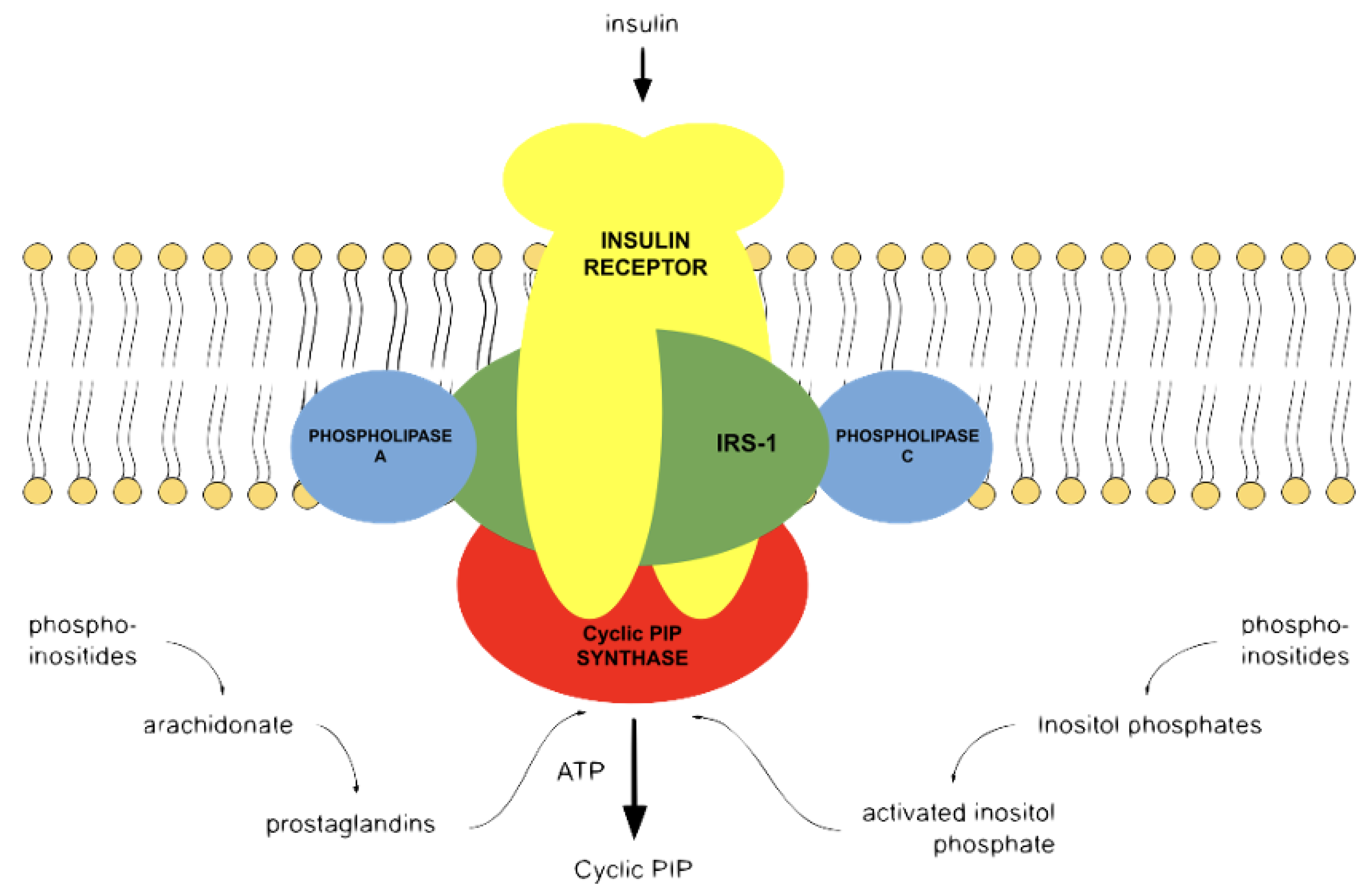
The search for an antagonist to cyclic AMP started in the laboratory of the late Earl Sutherland. Cyclic PIP was primarily isolated from rat livers, which were extra-corporally perfused with buffer and stimulated with noradrenaline or insulin, homogenized within 1 to 3 min, and then put under denaturing conditions to minimize the enzymatic degradation of cyclic PIP. From 2 kg of rat livers, approximately 60 g of water-soluble, low-molecular-weight compounds were obtained, which also contained cyclic PIP. A rough calculation indicated that 500,000 to 1,000,000 molecules of this extract contain 1 molecule of cyclic PIP. But more challenging was the finding that cyclic PIP is one of the most labile molecules of this extract. It decays by 80% within 30 min held at an ionic strength comparable to a 0.5 molar sodium chloride solution, approximately a 3-fold concentrated physiological saline solution. The labilities of cyclic PIP are connected to the tension of the 5-ring phosphodiester, which is the most labile bond of cyclic PIP, as well as the allyl-ether bond combining two secondary alcohols and the beta-keto-hydroxy structure of the C5-ring of prostaglandin E (PGE). Chemically it is O-(prostaglandyl-E)-(15-4′)-(myo-inositol 1′:2′-cyclic phosphate) [8]. It is biosynthesized from PGE and activated inositol phosphate by cyclic PIP synthase [8], which is active in a tyrosine-phosphorylated form [13]. The synthesis of cyclic PIP is stimulated also by adrenergic α1- and α2-receptors [14]. This result contradicts the present view of adrenergic α1- and α2-receptor action, as discussed in [14,15][14][15]. The adrenergic receptors are transmembrane, G protein-coupled receptors; thus, cyclic PIP synthase should also be activated by a G protein [13]. It is not known whether there are two different variants of cyclic PIP synthase, but it is known that the biosynthesis of the substrates for cyclic PIP synthesis involves phospholipase A2 and phospholipase C, both of which are activated by these two modes of activation as summarized in [15]. The synthesis of cyclic AMP from ATP is a one-step reaction, and ATP is generally always present in a high enough amount in all cells to warrant maximal synthesis of cyclic AMP. In contrast, the biosynthesis of cyclic PIP needs at least five reaction steps (Figure 1), and both substrates are obtained from membrane-bound lipids [15].
References
- Nelson, M.E.; Madsen, S.; Cooke, K.C.; Fritzen, A.M.; Thorius, I.H.; Masson, S.W.C.; Carroll, L.; Weiss, F.C.; Seldin, M.M.; Potter, M.; et al. Systems-level analysis of insulin action in mouse strains provides insight into tissue- and pathway-specific interaction that drive insulin resistance. Cell Metab. 2022, 34, 227–239.
- Mejhert, N.; Ryden, M. Understanding the complexity of insulin resistance. Nat. Rev. Endocrinol. 2022, 18, 269–270.
- Petersen, M.C.; Shulman, G.I. Mechanisms of insulin action and insulin resistance. Physiol. Rev. 2018, 98, 2133–2223.
- Galicia-Garcia, U.; Benito-Vicente, A.; Jebari, S.; Larrea-Sebal, A.; Siddiqi, H.; Uribe, K.B.; Ostolaza, H.; Martin, C. Pathophysiology of type 2 diabetes mellitus. Int. J. Mol. Sci. 2020, 21, 6275.
- White, M.F.; Kahn, C.R. Insulin action at a molecular level—100 years of progress. Mol. Metab. 2021, 52, 101304.
- Nagao, H.; Cai, W.; Brandao, B.B.; Wever Albrechtsen, N.J.; Steger, M.; Gattu, A.K.; Pan, H.; Dreyfuss, J.M.; Wunderlich, T.; Mann, M.; et al. Leucine-973 is a crucial residue differentiating insulin and IGF-1 receptor signaling. J. Clin. Invest. 2023, 133, E161472.
- Robison, G.A.; Butcher, R.W.; Sutherland, E.W. Cyclic AMP; Academic Press: New York, NY, USA, 1971.
- Wasner, H.K. Prostaglandylinositol cyclic phosphate, the natural antagonist of cyclic AMP. IUBMB Life 2020, 72, 2282–2289.
- Wasner, H.K. Metformin’s mechanism of action is stimulation of the biosynthesis of the natural cyclic AMP antagonist prostaglandylinositol cyclic phosphate (cyclic PIP). Int. J. Mol. Sci. 2022, 23, 2200.
- Yarwood, S.J. Special issue on “new advances in cyclic AMP signaling”—An editorial overview. Cells 2020, 9, 2274.
- von Keulen, S.C.; Rothlisberger, U. Exploring the inhibition mechanism of adenylyl cyclase type 5 by n-terminal myristoylated Gai1. PLoS. Comput. Biol. 2017, 13, e1005673.
- Sabol, S.L.; Nirenberg, M. Regulation of adenylate cyclase of neuroblastoma x glioma hybrid cells by a-adrenergic receptors. J. Biol. Chem. 1979, 254, 1913–1920.
- Wasner, H.K.; Gebel, M.; Hucken, S.; Schaefer, M.; Kincses, M. Two different mechanisms for activation of cyclic PIP synthase: By a G protein or by protein tyrosine phosphorylation. Biol. Chem. 2000, 382, 145–153.
- Wasner, H.K.; Salge, U.; Gebel, M. The endogenous cyclic AMP antagonist, cyclic PIP: Its ubiquity, hormone-stimulated synthesis and identification as prostaglandylinositol cyclic phosphate. Acta Diabetol. 1993, 30, 220–232.
- Wasner, H.K.; Lessmann, M.; Conrad, M.; Amini, H.; Psarakis, E.; Mir-Mohammad-Sadegh, A. Biosynthesis of the endogenous cyclic adenosine monophosphate (AMP) antagonist, prostaglandylinositol cyclic phosphate (cyclic PIP), from prostaglandin E and activated inositol polyphosphate in rat liver plasma membranes. Acta Diabetol. 1996, 33, 126–138.
- Wasner, H.K.; Salge, U. Prostaglandylinositol cyclic phosphate, a second messenger for insulin. Excerpta Medica Int. Congr. Ser. 1987, 726, 226–231.
- Brautigan, D.L. Protein ser/thr phosphatases—The ugly ducklings of cell signaling. FEBS J. 2013, 280, 324–345.
