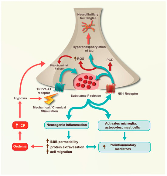
Traumatic brain injury (TBI) is an acquired insult to the brain caused by external mechanical impact and/or acceleration forces that result in transient or permanent neurological dysfunction. Substance P is a member of the tachykinin protein family whose neuronal release after TBI plays a critical role in TBI pathophysiology, including the development of post-traumatic oedema, increased intracranial pressure, neuroinflammation, neuronal cell death, and neurodegeneration. Because substance P release after TBI is dependent on the intensity and frequency of injury-related mechanical stimulation, the degree and anatomical distribution of substance P receptor activation after TBI will vary with injury severity and frequency, resulting in different outcomes for different injuries.
- NK1 receptor
- substance P
- neurotrauma
- oedema
- intracranial pressure
- brain injury
- CTE
1. Introduction
2. Mild TBI
Mild TBI, encompassing concussion, is any traumatic injury to the brain that results in immediate and transient impairment of brain function, including disturbances of vision, hearing, and/or equilibrium and potentially alterations of consciousness, amongst others. While the physical symptoms such as blurred vision, dizziness, and nausea, as well as any cognitive deficits and behavioural changes, usually resolve in a relatively short time frame, these latter symptoms may nonetheless persist in some individuals [9][11]. However, because few symptoms are visible to the casual observer in the immediate timeframe after impact, concussion was historically considered as somewhat of an innocuous event. Recent evidence, however, has shown that its occurrence can produce persistent functional disability in some individuals [9][11], and even later neurodegeneration [10][12]. Particular attention has been directed to a specific form of neurodegeneration known as chronic traumatic encephalopathy (CTE), which has been widely reported in professional athletes and military personnel who have been exposed to repeated head impacts [10][12]. The first report of substance P in concussion [11][13] simply demonstrated that the neuropeptide was released after a concussive impact and could be observed by immunohistochemistry in brain perivascular neurones and in astrocytes. Given the known role for substance P in oedema formation [12][14], the authors proposed that release of the neuropeptide from perivascular neurones sensitises cerebral blood vessels for increased permeability in the event of a second concussive impact, thus facilitating potentially profound oedema formation (second impact syndrome). Subsequent studies in concussion have focussed on the potential role of substance P in repeated impacts and the development of CTE. CTE is characterised by the accumulation of phosphorylated tau protein tangles, initially appearing in perivascular neurones in areas of the brain which are subject to high mechanical stress with head impact (such as the depths of the sulci) and then spreading to other regions of the brain with time [13][15]. The severity of this tau pathology correlates with cognitive impairments that are observed as the disease progresses [14][16]. Notably, increased vascular permeability has been reported after exposure to repeated head impacts [15][17], implying a role for neuroinflammation in the pathophysiology. In a comprehensive study using a variety of experimental TBI models, Corrigan and colleagues [16][18] demonstrated that brain substance P expression was increased following a single concussive injury but did not result in tau hyperphosphorylation. However, following three concussive injuries inflicted within a 10-day period, brain substance P expression was markedly increased and was associated with profound perivascular tau hyperphosphorylation, the hallmark of CTE. Indeed, the level of tau hyperphosphorylation after three closely timed concussive events was equivalent to that observed following a moderate TBI. Administration of a TRPVI receptor (mechanoreceptor) antagonist prior to the induction of head injury prevented tau phosphorylation, presumably by inhibiting the release of substance P. However, when the TRPV1 antagonist was administered after the head injury, there was no such inhibition, suggesting that substance P release had already been initiated by the mechanical insult. In contrast, administration of the substance P NK1 receptor antagonist, EUC001 [17][19], after the brain injury attenuated tau hyperphosphorylation by moderating the activity of several key kinases associated with tau phosphorylation, including Akt, ERK1/2, and JNK [18][19][20][20,21,22]. Notably, confocal microscopy images of the cortical neurones demonstrated co-localisation of the neuronal phosphorylated tau and NK1 receptors (Figure 1), supporting the pharmacologic data demonstrating a link between NK1 receptors and tau hyperphosphorylation. Notably, administration of the NK1 antagonist at 30 min after mild, blast-induced injury in mice (a known cause of CTE) attenuated the post-traumatic tau phosphorylation for up to 28 days after the injury [16][18].
3. Moderate TBI
Moderate TBI was where a role for neurogenic inflammation was first identified [24][25][26,27]. In these studies, transient depletion of neuropeptides from neuronal C fibres using capsaicin pre-treatment led to reduced BBB permeability, reduced oedema formation, and improved functional outcome after TBI compared to untreated controls. While substance P was strongly implicated in these events, a series of subsequent studies characterised the precise role of substance P and NK1 receptors both in experimental models of TBI and in clinical TBI [26][27][28][29][28,29,30,31]. These studies demonstrated that substance P is released by perivascular neurones, particularly those that have been stressed by the mechanical events associated with TBI [29][31]. Substance P preferentially binds to NK1 receptors in the vicinity, including those located on the vascular endothelium, leading to a localised increase in BBB permeability and an associated movement of blood proteins such as albumin from the vasculature into the brain parenchyma. This creates an osmotic gradient which drives movement of water from the vasculature into the brain tissue causing vasogenic oedema [30][32]. Not only does albumin translocation across the barrier create an osmotic gradient, translocation of albumin into the brain is also known to activate a tissue inflammatory response [31][33], which further exacerbates the BBB permeability, enhances recruitment of inflammatory cells, and further increases oedema formation [32][34]. Together with the primary mechanical insult, this neuroinflammatory response ultimately leads to neuronal cell damage and functional deficits, both motor and cognitive. There is considerable evidence suggesting that chronic neuroinflammation involving a variety of glial cells may also lead to later neurodegeneration, including the tau phosphorylation typically observed in CTE [16][32][18,34]. With clear evidence that substance P is increased after moderate TBI and binds to NK1 receptors to mediate some of its effects, several experimental studies have characterised the potential role of NK1 antagonists as neuroprotective agents [33][35]. A variety of substance P NK1 receptor antagonists have been used in these studies to demonstrate the central role of the NK1 receptor in TBI and its efficacy as a therapeutic intervention. Initial studies focussed on the highly selective NK1 receptor antagonist N-acetyl L-tryptophan 3,5-bis (trifluoromethyl) benzyl ester (L-732,138) and its non-permeable form, N-acetyl L-tryptophan (NAT). Both were shown to inhibit BBB permeability after TBI in an identical dose-dependent manner and were equally effective at improving post-traumatic oedema and post-traumatic functional outcomes when administered after trauma. Notably, the D isomer of NAT was an ineffective relative to the L isomer, suggesting that the neuroprotective effects were receptor mediated [27][29]. However, the effectiveness of the non-permeable NAT was limited to the period immediately after TBI when the BBB was transiently open. Later experimental TBI studies have adopted other, more cell-permeable NK1 antagonists including the Merck compound L-733,060 [34][36] and the Roche compound EUC-001 [17][19]. The move away from NAT in experimental studies was useful not only to address its inability to cross the intact BBB but also because of controversies surrounding its classification as an NK1 antagonist. While some studies have definitively shown that NAT binds to and inhibits the NK1 receptor as well as presynaptic substance P release [35][37], others have been unable to confirm NK1 receptor binding [36][38]. In circumventing these issues, studies using alternative BBB permeable NK1 antagonists may better facilitate clinical translation, particularly EUC-001, which is a highly selective antagonist of the human NK1 receptor, is a water-soluble yet membrane-permeable compound, and has few potential drug–drug interactions [17][19]. Irrespective of the NK1 antagonist that has been utilised in the experimental studies, these antagonists have universally shown profound neuroprotective effects when administered following moderate TBI (see [37][39] for review). Moreover, these neuroprotective effects were apparent irrespective of the animal species used or the model of injury (focal versus diffuse) that was employed. When administered as a parenteral bolus (either IV or IP) at 30 min after injury, NK1 antagonists significantly improved both motor and cognitive deficits over the first 2 weeks after trauma [28][38][30,40]. This neuroprotection was independent of sex, with females also responding positively to the NK1 antagonist [26][28]. In mice, knockout of SP synthesis was equivalent to administering an NK1 antagonist in terms of neuroprotective efficacy [38][40], supporting previous studies showing that preinjury SP depletion with capsaicin was profoundly neuroprotective [24][26]. NK1 antagonists reduced cell death in both the hippocampus and in the cortex, as well as axonal injury in the corpus collosum [26][27][28,29], and were shown to reduce lesion volumes in focal models of injury [38][40]. While initial studies all administered a bolus of NK1 antagonist in the first hour after injury, it has been shown that cell-permeable forms of the NK1 antagonist are efficacious even when administered as late as 12 h after injury [27][29]. Notably, efficacy with a non-membrane-permeable antagonist was limited to the first 5 h, corresponding to when the BBB after TBI is known to be open to large protein molecules [39][41]. Thereafter, the BBB gradually closes to large protein molecules, thus necessitating the use of membrane-permeable versions of the antagonist. Various mechanisms have been identified by which NK1 antagonists might improve cell survival after TBI and improve functional outcomes [38][40]. In addition to reducing aspects of neurogenic inflammation (such as increased BBB permeability and vasogenic oedema), NK1 antagonists have been shown to reduce oxidative stress, preserve mitochondrial membrane potential, inhibit mitochondrial cytochrome c release, inhibit apoptosis, inhibit classical neuroinflammation, preserve ATP levels, inhibit non-apoptotic programmed cell death [40][42], and increase intracellular free magnesium concentration [41][43]. Thus, NK1 antagonists act on multiple known secondary-injury pathways to confer neuroprotective effects after moderate TBI (Figure 2).
