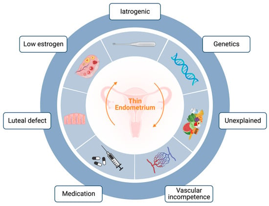
Conventional therapies, including estradiol or combined hormonal therapy
[14], growth hormone
[15], Granulocyte colony-stimulating factor (G-CSF)
[16][17][16,17], sildenafil citrate
[18], and several vasoactive substances
[19] such as aspirin, pentoxifylline, tocopherol, and L-Arginine have shown limited and inconsistent efficacy regarding an increase in EMT increase and restoration of endometrial function
[20][21][20,21]. Consequently, there is a growing demand for novel adjuvant therapies. In recent years, cell-based therapy and other innovative treatment options have emerged as promising strategies in various medical fields
[22][23][24][22,23,24]. Considering the encouraging results in numerous experimental studies, these novel approaches may hold great potential as treatment options for thin endometria.
2. Experimental Therapeutic Strategies
2.1. Platelet-Rich Plasma
Platelet-rich plasma (PRP) is an autologous plasma-based concentration of platelets, offering a host of therapeutic advantages
[25]. Its autologous nature significantly reduces the risk of immune rejection, pathogen transmission, and cancer development, making it an attractive treatment option
[26][27][28][26,27,28]. Rich in growth factors (GFs) and cytokines, PRP has demonstrated pro-regenerative properties, particularly in healing injured tissues. Recently, there has been growing interest in its potential for treating endometrial disorders, such as thin endometria
[29].
In 2018, Molina et al.
[30] administrated PRP through intrauterine infusions to 19 patients with refractory thin endometria undergoing IVF. After the second PRP injection, the endometrial thickness in each patient exceeded 9 mm. Notably, this resulted in an impressive 73.7% positive pregnancy test rate and 26.2% live births. Later in the same year, the first randomized controlled trial
[31] reported significantly higher rates of implantation and clinical pregnancy rates in the PRP group (27.94% vs. 11.67%; 44.12% vs. 20%;
p < 0.05, respectively), along with improved endometrial thickness (
p = 0.001). Subsequently, an increasing number of prospective clinical studies
[32][33][34][35][36][37][38][32,33,34,35,36,37,38] further support these findings, showing that intrauterine infusion of PRP effectively thickened the endometrial lining and improved clinical pregnancy outcomes to different extents. Although growing evidence indicates that PRP treatment is beneficial for treating thin endometria, the positive effects only manifest for certain parameters.
2.2. Stem Cell Therapy
During the last decades, stem cell therapy has rapidly evolved and been applied in the treatment of various diseases. Stem cells possess unique characteristics, such as high self-renewal capacity and the ability to differentiate into multiple cell types. Stem cell therapy has emerged as a promising frontier in reproductive medicine giving its potential to restore endometrial function for patients with a thin endometrium
[39][40][42,43].
2.2.1. BMDSCs
Bone-marrow-derived stem cells (BMDSCs) are multipotent stem cells that can differentiate into different functional cells. Due to their easy acquisition in adult bone marrow, these cells have emerged as an important candidate for stem cell therapy in various diseases
[41][42][43][44,45,46]. In 2011, Nagori et al.
[44][47] reported a successful pregnancy in a patient with refractory intrauterine adhesions through the autologous transplantation of BMDSCs. Later in 2013, Zhao et al.
[45][48] transplanted autologous BMDSCs into the uterine cavity under ultrasound guidance in a woman with severe intrauterine adhesions, resulting in a spontaneous pregnancy after three months. Moreover, Singh et al. reported that autologous transplantation of BMDSCs significantly increased endometrial thickness at 3, 6, and 9 months compared to pre-treatment thickness in six patients with refractory intrauterine adhesions. Menstruation was also restored in five out of six patients
[46][49].
2.2.2. ADSCs
Adipose-derived stem cells (ADSCs) are another abundant and easily accessible stem cell source that are available in large quantities and have the benefit of allowing for isolation and production using a minimally invasive lipectomy procedure
[47][56]. These cells have been tested in repairing injured endometrium in a few clinical trials. In 2019, one study
[48][57] recruited 25 women with thin endometria (EMT < 5 mm) who had embryo implantation failure at least three times. After subendometrial injection of ADSCs, the EMT increased in 80% (20/25) of patients, leading to 13 pregnancies and 9 healthy live births. Another pilot study in 2020
[49][58] examined the effectiveness of restoring functional endometrium in patients with severe Asherman’s syndrome (intrauterine adhesions) using autologous adipose-derived stromal vascular fraction (AD-SVF) containing adipose stem cells (ASCs). Five out of six infertile women with severe intrauterine adhesions achieved increased endometrial thickness along with an increased volume of menstrual bleeding. One of the five women who underwent ART treatment and transferred an embryo became pregnant but spontaneously miscarried at nine weeks.
2.2.3. UCMSCs
In contrast to other stem cell sources, umbilical cord mesenchymal stem cells (UCMSCs) stand out as a highly promising cell source for cell therapy due to their abundance, non-controversial nature, painless collection procedure, and rapid self-renewal properties
[50][60]. Particularly noteworthy is the fact that UCMSCs exhibit negligible or undetectable HLA class I expression, indicating the potential for allograft transplantation without the need for immunosuppression
[51][61]. This unique characteristic enhances the appeal of UCMSCs as a viable option for therapeutic applications. In 2018, a phase I clinical trial implanted UCMSCs in biodegradable collagen scaffolds into the uterine cavity of 26 patients with recurrent intrauterine adhesions, resulting in an increase in EMT in all cases from 4.46 ± 0.85 to 5.74 ± 1.2 mm (
p < 0.01), which is linked to pregnancy in 10 women (38%), 8 of whose pregnancies resulted in live births
[52][62].
2.2.4. Other Stem Cell Sources
Several other sources of stem cells have also been introduced and emerged as potential sources for the treatment of thin endometrium. One such source is menstrual-blood-derived stromal cells (MenSCs), which comprise a mixed population of mesenchymal stem cells and stromal fibroblasts. In 2016, Tan et al.
[53][64] transplanted autologous MenSCs from the menstrual blood of women with intrauterine adhesions back into the uterine cavity. A significant increase in endometrial thickness to 7 mm in five out of seven cases was observed along with successful pregnancies in two out of four women. However, this source of stem cells has certain limitations such as not being applicable for those patients with hypomenorrhea. To explore the endometrial repair mechanism of transplanted MenSCs, one study
[54][65] demonstrated that MenSCs could increase the microvascular density (MVD) of an injured endometrium in a mouse model by activating the ART and ERK pathways; inducing the upregulation of eNOS, VEGFA, VEGFR1, VEGFR2, and Tie2; and promoting cell proliferation, migration, and angiogenesis in vitro. Additionally, uterine-derived cells have been successfully transplanted in a rat model to repair damaged uterine endometrium, leading to promising outcomes
[55][66]. Through transplantation of endometrium-like cells derived from human embryonic stem cell lines (hESCs), researchers demonstrated that these cells could significantly restore the structure and functionality of severely damaged uterine horns in a rat model
[56][67]. Though most of these studies are at a pre-clinical stage, these promising results have encouraged researchers to develop cell-based biomedical approaches for repairing damaged endometria.
2.2.5. Stem-Cell-Derived Extracellular Vesicles
Stem cells possess the ability to secrete a wide range of regenerative cytokines, and these cellular secretions have been proposed to contribute to the positive therapeutic effects observed in different diseases
[57][58][59][68,69,70]. Extracellular vesicles (EVs) are an important component of these secretions that have cytoprotective, antiapoptotic, and angiogenic effects on injured tissues and can promote progenitor and stem cell homing
[60][71]. Building on this idea, researchers are aiming to develop stem-cell-derived exosome-based therapy that focuses on the EVs that the cells secrete, including exosomes. While the clinical research on EVs is still in an early stage, the results from animal studies are encouraging and highlight the potential of EV-based therapies for thin endometria. In 2020, exosomes derived from adipose mesenchymal stem cells were discovered to be able to preserve normal uterine structure, stimulate endometrial regeneration, support collagen remodeling, and elevate the expression of endometrial receptivity markers including integrin β3, LIF, and VEGF
[61][72].
2.3. Tissue Bioengineering
Bioengineering approaches have demonstrated promising outcomes in the field of regenerative medicine over recent years. Various biomedical materials and techniques, such as collagen scaffolds, decellularized scaffolds, hydrogels, and microfluidics, have been employed to facilitate tissue repair and regeneration
[62][63][76,77]. The rapid advancement of biomaterials has also enabled new possibilities for therapeutic strategies in thin endometria. Hydrogels, nanostructured delivery systems, bioactive degradable scaffolds, and other innovations have emerged as promising tools to enhance the survival and function of stem cells
[64][65][78,79]. In 2021, one study
[66][80] investigated the effects of transplanting UCMSCs, seeded on a human acellular amniotic matrix (AAM), to an endometrial injured site in a rat model. The results showed that endometrial thickness significantly increased, and the expression of vimentin, cytokeratin, and integrin β3 were higher in treated rats compared to untreated rats. The UCMSC–AAM combination might potentiate the endometrial repair effect of UCMSCs. The extracellular matrix is indeed an important component in tissue remodeling. Hydrogel, a highly hydrated collagen-based material, has shown great benefits in facilitating constructive and functional tissue remodeling
[67][81].

