Your browser does not fully support modern features. Please upgrade for a smoother experience.
Please note this is a comparison between Version 1 by Dacheng Wei and Version 2 by Rita Xu.
Coronavirus disease 2019 (COVID-19) is a disease caused by the infectious agent of severe acute respiratory syndrome coronavirus type 2 (SARS-CoV-2). Graphene field-effect transistor (GFET) biosensors have become the most promising diagnostic technology for detecting SARS-CoV-2 due to their advantages of high sensitivity, fast-detection speed, label-free operation, and low detection limit.
- GFET biosensor
- COVID-19
- biological sensors
1. Introduction
In 2019, a novel coronavirus as a kind of severe acute respiratory syndrome coronavirus (SARS-CoV-2) swept the world, causing a respiratory disease which is named coronavirus disease 2019 (COVID-19) [1][2][1,2]. COVID-19 is reportedly transmitted by airborne and contact-transmission respiratory droplets [3][4][3,4]. The methods for diagnosing COVID-19 can be roughly divided into three categories [5]: chest CT scanning [6], serological testing [7][8][7,8], and nucleic acid testing [9]. Chest CT scanning is limited to recognizing the virus type and is only available in hospitals. Serological testing is not suitable for early diagnosis of infection because the number of antibodies in our bodies gradually increases for at least a week after being infected with COVID-19 [10][11][12][10,11,12]. The nucleic acid detection method requires skilled professionals, and it takes 4–6 h to obtain the results [13]. Real-time reverse transcription-polymerase chain reaction (RT-PCR) remains the gold standard for COVID-19 diagnosis. However, RT-PCR requires a gene amplification process, tedious preparation steps, expensive equipment, specialized laboratories, and technicians, which reduces the efficiency of the test [14]. Therefore, developing a real-time, fast, accurate, easy-to-operate, and low-cost diagnostic technology is one of the key challenges in the fight against COVID-19.
Biosensors with simple operation and rapid detection have been widely considered a superior alternative detection technology [15]. In recent years, an endless stream of biosensors has been studied [16]. There are many types of biosensors, such as field-effect transistor (FET) biosensors [17], optical biosensors [18], plasmon resonance biosensors [19], and electrochemical biosensors [20]. These biosensors have become a powerful new means of detecting various biomolecules for diagnostics. In particular, GFET biosensors have the advantages of high sensitivity, fast detection speeds, no labels, and low detection limits and have become the most promising technology for COVID-19 detection [21][22][21,22]. GFET biosensors detect SARS-CoV-2 through specific targets on the surface of the graphene channel. SARS-CoV-2 is a segmented, enveloped coronavirus family with a single-stranded RNA structure [23]. The genome inside the particle encodes four structural proteins. Viral RNA is coated with nucleocapsid (N) protein. In addition to nucleocapsid (N) proteins, there are spike proteins (S), membrane proteins (M), and envelope proteins (E), which are embedded in the lipid bilayer [24]. Therefore, SARS-CoV-2 can be detected by using nucleic acid molecules or structural proteins as targets for the specific reaction (Figure 1). Currently, GFET biosensors for detecting COVID-19 mainly use nucleic acid hybridization or antigen–antibody-specific reaction for detection.
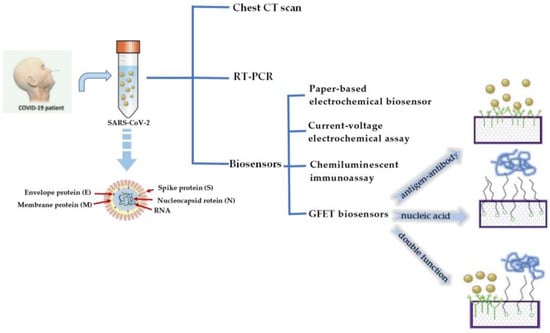
Figure 1. Several detection methods for COVID-19 and the schematic diagram of a GFET biosensor for detecting SARS-CoV-2.
2. Application of GFET Biosensors in the Diagnosis of COVID-19
GFET biosensors have great potential for diagnosing and controlling disease transmission in COVID-19 and other biomolecular detections. Compared with traditional detection technology, GFET biosensors have certain advantages in the bedside maintenance of biomolecules because of their excellent performance.
2.1. GFET Biosensors Detect SARS-CoV-2 Based on Specific Antigen–Antibody Binding
The coronavirus S protein is a large, multifunctional transmembrane fusion glycoprotein of the class I virus. The S protein is attached to the surface of the viral particle and determines the shape of the virus’ crown-like appearance. The coronavirus N protein promotes the assembly of viral particle and plays a role in the formation of the viral genome. After being infected with SARS-CoV-2, B lymphocytes or B cells produce five types of antibodies, IgA, IgG, IgM, IgD, and IgE, known as immunoglobulins [10]. Therefore, many researchers have detected SARS-CoV-2 by targeting antibodies. An ultra-sensitive GFET biosensor was prepared by Wei’s group [25][51]. The SARS-CoV-2 spike S1 protein was modified on the surface of the sensor to detect the spike S1 antibody (Figure 2). The SARS-CoV-2 spike S1 protein was immobilized on the surface of the graphene channel to realize biological functionalization. The strong specific binding between antibodies and proteins affected the concentration of the graphene channel medium and obtained a measurable electrical response.
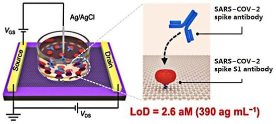
Figure 2. Schematic diagram of the GFET biosensor for detecting SARS-CoV-2 spike antibodies.
In recent years, laser-induced graphene technology has become a new way of preparing GFET biosensors. Cui’s group [26][52] first used lasers to manufacture a graphene channel, using a 450 nm UV laser of 840 MW, and the electrode region was manufactured using a UV laser of 900 MW. The graphene channel region obtained after laser radiation showed a spongy porous shape, which expanded its binding area with biomolecules. This research group has developed a one-step, simple, sensitive, and suitable method for the large-scale preparation of a laser-induced GFET biosensor. The SARS-CoV-2 spike antibody immobilized in graphene channels achieved rapid detection of the SARS-CoV-2 spike protein in 15 min at a detection limit of 1 pg/mL in phosphate-buffered saline (PBS) and 1 ng/mL in human serum, with high specificity for the target virus (Figure 3).
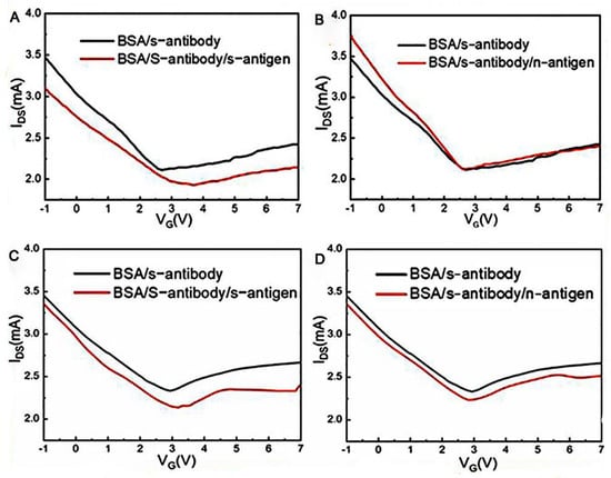
Figure 3. The virus detection performance of the laser-induced GFET. (A) Transfer characteristics of the laser-induced GFET biosensor responding to the complementary 1 pg/mL spike protein in PBS solution and (B) responding to the noncomplementary 1 pg/mL nucleocapsid protein in PBS solution. (C) Transfer characteristics of the laser-induced GFET biosensor responding to 1 pg/mL of complementary spike protein in human serum and (D) responding to 1 pg/mL noncomplementary nucleocapsid protein in human serum.
Shahdeo’s group [21] prepared GFET sensors on SiO2/Si substrates using transparent tape. The internally generated SARS-CoV-2 spike S1 antibody was immobilized on the carboxylic acid-activated graphene surface. The change in resistance generated by antibody–antigen interaction was monitored in response to the assay results (Figure 4). The results indicated that the device has high sensitivity and specificity and detects SARS-CoV-2 spike S1 proteins under conditions with a detection limit as low as 10 fM.
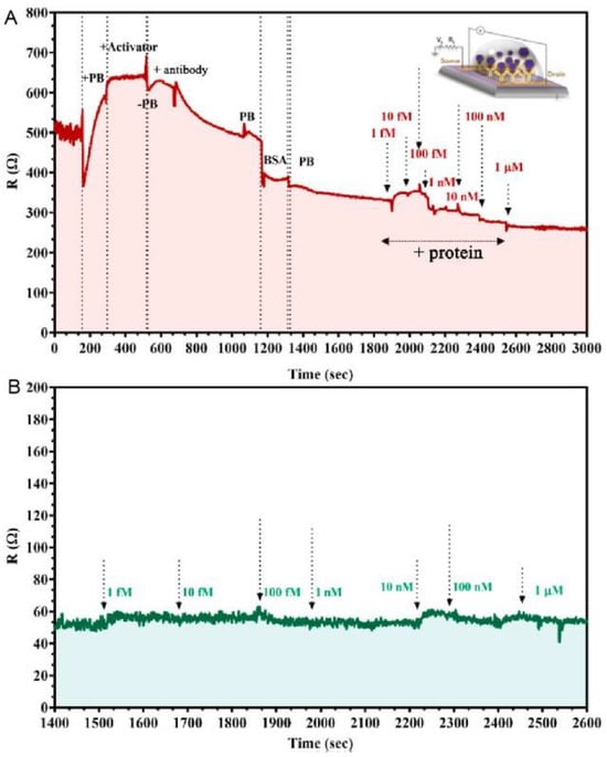
Figure 4. The kinetic response of the GFET device functionalized with the SARS-CoV-2 spike antibody at various concentrations of (A) SARS-CoV-2 spike protein added, ranging from 1 fM to 1 μM in 50 mM phosphate buffer (PB) (pH 7.2) and (B) MERS-CoV protein of various concentrations added (1 fM to 1 μM) in PB.
2.2. GFET Biosensors Based on Nucleic Acid Hybridization Detection of SARS-CoV-2
Nucleic acid detection is the most sensitive detection method for early viral infection and plays a key role in diagnosing and treating disease [27][53]. The RT-PCR method is the gold standard for detecting SARS-CoV-2, but the diagnostic process is complicated and time consuming. Therefore, many scholars tend to develop methods based on nucleic acid as a probe for detecting SARS-CoV-2. How to improve the sensitivity of the probe is an important problem. A lot of work has been carried out, mainly focused on the design of the probe and the development of sensing materials and new sensing mechanisms. In bioassays, different configurations are designed to improve the binding affinity with the target.
In recent years, the development of structural DNA nanotechnology has provided an accurate and controllable method for synthesizing various DNA nanostructures with specific functions. Different DNA nanostructures have different properties for biosensors. Compared with the single-probe nucleic acid hybridization detection method, the two recognition sites of dual probes can improve the sensitivity of virus detection. Wei’s group [28][54] developed a direct acid nucleic assay using a GFET with Y-shaped DNA dual probes (Figure 5). These Y-shaped DNA dual probe GFET biosensor could simultaneously identify ORF1ab and N gene regions, improving the sensitivity for identifying SARS-CoV-2. The synergistic effect of the two recognition sites of Y-shaped DNA dual probes improved the combination of DNA dual probes and targets. Therefore, the Y-type dual-probe GFET biosensor with an average of 40 s for response speed had excellent performance in terms of diagnosis time and detection limit.
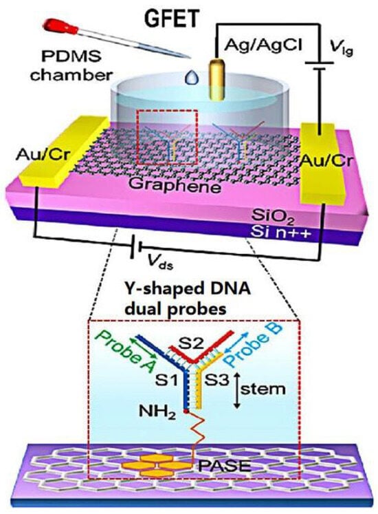
Figure 5.
Schematic diagram of a Y-dual probe GFET biosensor. The dotted box is the structural schematic diagram of Y-shaped DNA dual probes.
Graphene surface-modified probe recognition sites greatly impact the performance of GFET biosensors. Therefore, improving the structural design of the probe is an important factor. Creatures with a multi-tentacle structure have strong olfactory sensitivity and capture and hunting ability. Inspired by these organisms, the sensitivity of multi-probe sensors can often be improved. Wei’s team designed the DNA nanostructure as a probe-tunable TDF dimer. The synergistic action of three probes improves the binding affinity and the sensitivity of the GFET biosensor. Wei’s group modified the GFET biosensor with a triple-probe tetrahedral DNA framework (TDF) to study its detection performance for SARS-CoV-2 RNA [29][55]. The triple-probe TDF dimer was modified on the surface of the graphene channel to form reaction targets (Figure 6). The sensor had highly specific recognition of RNA in the SARS-CoV-2 ORF1ab gene, RdRp gene, and E gene regions. As shown in Figure 7, under the same conditions, the response of the triple-probe TDF dimer was faster than that of dual-probe and single-probe TDF dimer sensors. The sensor identified all 14 positive cases in 30 nasopharyngeal swabs within an average diagnosis time of 74 s, showing promising prospects for real-time and centralized detection screening.
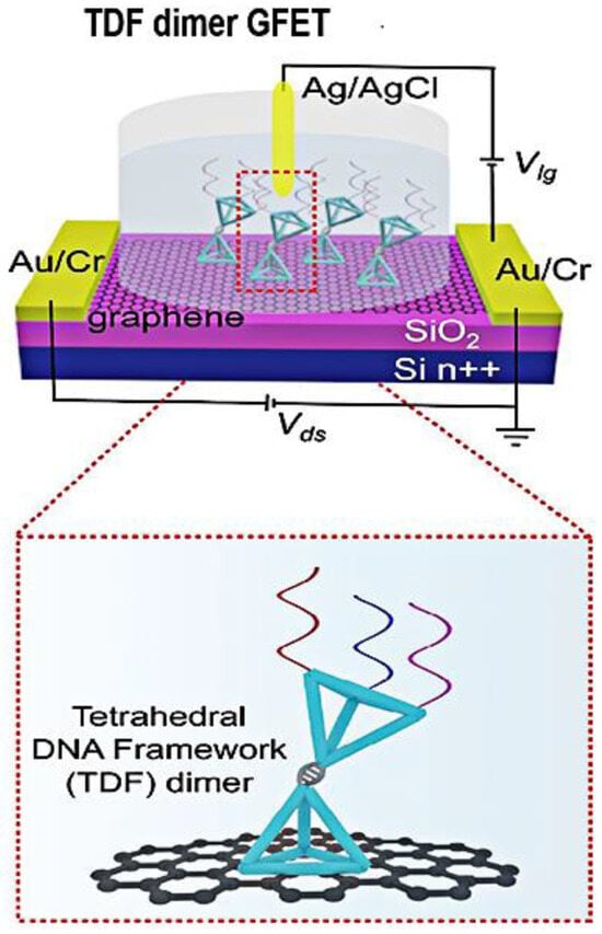
Figure 6. Schematic diagram of the triple-probe TDF dimer GFET sensor for SARS-CoV-2 RNA testing. The dotted box is the structural schematic diagram of TDF dimer.
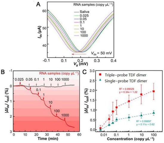
Figure 7. SARS-CoV-2 RNA testing. (A) Transfer curve measurement of adding different concentrations of target RNA (Ids−Vg response curve). (B) Real-time |ΔIds/Ids0| response upon different concentrations of target RNA (red line, modified with triple-probe TDF dimer; gray line, without immobilized probes). (C) |ΔIds/Ids0| responses of single- and triple-probe TDF dimer GFET sensors to different concentrations of target RNA.
2.3. Double Function of GFET Biosensors in Response to the Detection of SARS-CoV-2
Outstanding achievements have been made in detecting SARS-CoV-2 by single-response nucleic acid hybridization and antigen–antibody-specific reactions. The dual response of GFET biosensors to detecting SARS-CoV-2 has also received attention. Ke ’s group [30][56] reported a highly sensitive, specific, and convenient bi-functional GFET biosensor for detecting SARS-CoV-2 with detection limits as low as ~0.1 and ~1 fg·mL−1. The research group immobilized the SS-DNA probe or SARS-CoV-2 antigen protein on the surface of the graphene channel through π-π interaction. Detection results could be obtained in 5–10 min using SS-DNA probe-specific hybridization with a viral RNA polymerase target or SARS-CoV-2 antigen–antibody-specific recognition to convert biochemical effects into electrical signals. In order to verify the sensitivity and accuracy of the sensor for COVID-19 diagnosis, 18 volunteers were recruited for nucleic acid detection and 9 were recruited volunteers for immune detection.
Hwang’s group [31][57] was able to achieve high sensitivity by optimizing the crumpling ratio of the graphene sensing film. The results show that the crumpled GFET biosensor designed by Hwang’s group obtained good sensitivity and high reproducibility at a crumpling rate of about 55% [32][58]. The SARS-CoV-2 spike protein antibody and nucleocapsid protein antibody were immobilized on the surface of the graphene channel by π-π stacking, which could diagnose these two SARS-CoV-2 proteins at a lower detection limit. A field-effect transistor based on graphene oxide/graphene van der Waals heterostructures (GO/Gr heterostructure FET) was developed by Gao’s group [33][37]. Graphene oxide had abundant functional groups on its surface, and graphene oxide was superimposed onto graphene by π-π stacking, which enhances SARS-CoV-2 spike and nucleoprotein adsorption, improving the detection sensitivity of the sensor. The GO/Gr heterostructure FET sensor detects the SARS-CoV-2 protein in the range of 10 to 100 pg/mL with a limit detection as low as ~8 fg/mL. Meanwhile, as shown in Figure 8, the experimental data show ~3 × sensitivity enhancement compared with the GFET biosensor, which indicates its great potential for practical references in diagnosing SARS-CoV-2.
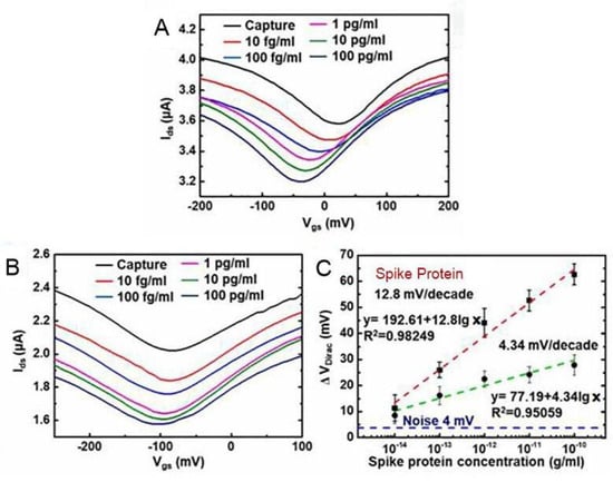
Figure 8. The SARS-CoV-2 spike protein concentrations dependent transfer curves of (A) GO/Gr FET biosensor and (B) Gr FET biosensor. (C) The SARS-CoV-2 spike protein concentrations dependent ΔVDirac shifts for both GO/Gr FET (red line) and Gr FET (green line) biosensors.