Your browser does not fully support modern features. Please upgrade for a smoother experience.
Please note this is a comparison between Version 1 by Zhenyang Li and Version 3 by Camila Xu.
Heterogeneous nuclear ribonucleoproteins (hnRNPs) are a superfamily of RNA-binding proteins consisting of more than 20 members. These proteins play a crucial role in various biological processes by regulating RNA splicing, transcription, and translation through their binding to RNA.
- hnRNPs
- alternative splicing
- muscle development
- muscle disorders
1. Introduction一、简介
Various post-transcriptional modifications are required for the maturation of mRNA in eukaryotic cells. These modifications include the addition of 7-methylguanosine (m7G) at the 5′ end, the formation of a polyadenylic acid tail at the 3′ end, and RNA splicing. Alternative splicing allows for the production of different mature mRNAs from a single pre-mRNA molecule. Heterogeneous nuclear ribonucleoproteins (hnRNPs) are a superfamily of RNA-binding proteins that play a key role in regulating the alternative splicing of pre-mRNA [1].
2. Overview of hnRNPs
2.1. Composition of hnRNPs
Heterogeneous nuclear ribonucleoproteins (hnRNPs) are a class of RNA-binding proteins (RBPs). Through immunopurification and two-dimensional gel electrophoresis, 42 kinds of hnRNPs have been identified in Hela cells. These hnRNPs consist of more than 20 major hnRNPs that exhibit relatively higher abundance, along with other hnRNPs [2][3][2,3]. The main hnRNPs have a molecular weight ranging from 34 kDa to 120 kDa and are named hnRNP A1 to hnRNP U [4][5][6][7][4,5,6,7] based on their molecular weight and structural and functional characteristics. Due to the strong association between hnRNP A1, A2/B1, B2, C1, and C2 and hnRNA, they are referred to as “core proteins” [8].2.2. hnRNPs: RNA-Binding Domains and Structural Insights
The analysis of cDNA sequences has revealed that hnRNPs possess a modular structure comprising at least one RNA-binding motif and auxiliary domains [9][10][9,10] (Figure 1). As recent investigations into the structure of hnRNPs have delved into greater detail, it has been confirmed that hnRNPs consist of three distinct RNA-binding motifs: the RNA recognition motif (RRM) [11][12][11,12], the RNA-binding domain consisting of Arg-Gly-Gly repeats (RGG domain) [13], and the K-homology domain (KH domain) [14].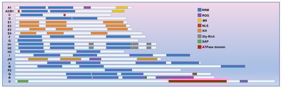
Figure 1. hnRNPs consist of three types of RNA binding motifs: RRM, RGG, and KH domains. The RRM motif is the most prevalent among these motifs in hnRNPs. Apart from RNA-binding motifs, certain hnRNPs may also possess auxiliary domains, namely the M9, NLS, SAP, and ATPase domains. These auxiliary domains play crucial roles in various processes such as nucleocytoplasmic shuttling, DNA binding, and the oligomerization of hnRNPs with caRNAs.
2.3. Functions of hnRNPs
Due to the diverse types and complex structural domains of hnRNPs, they can perform a variety of functions in cells. These functions include processing pre-mRNA into mature mRNA and serving as trans-acting factors that regulate gene expression. hnRNPs also participate in the processing of pre-mRNA alongside other RNPs, playing a crucial role in regulating mRNA transport, localization, translation, and stability (Figure 2). As a result, hnRNPs play a key role in various biological processes within cells. Contemporary research on hnRNPs primarily focuses on their involvement in muscle and neurodegenerative diseases (such as amyotrophic lateral sclerosis) as well as their role in cancer progression [40][41][42][40,41,42].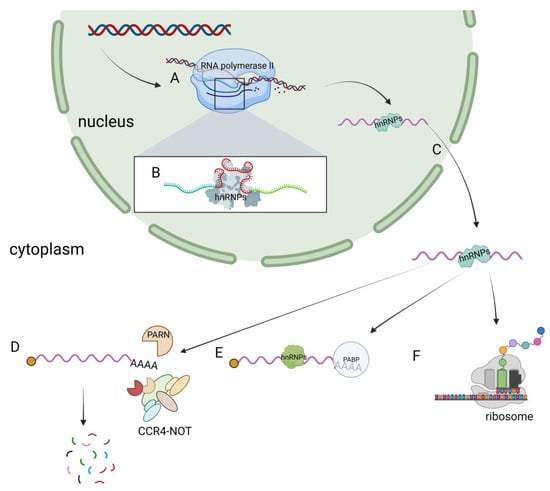
Figure 2. Functions of hnRNPs in cells. (A) hnRNPs have transcriptional regulatory functions. (B) hnRNPs regulate the alternative splicing of pre-mRNA. (C) The nucleocytoplasmic shuttling function of hnRNPs. (D) hnRNPs are involved in the degradation of mRNA. (E) hnRNPs participate in the regulation of mRNA stability. (F) hnRNPs regulate the translation process.
2.3.1. Regulation of Alternative Splicing
The pre-mRNA splicing process in cells relies on the interaction between splicing factors and splice sites. This mainly includes the following steps: (1) The U1 snRNP complex binds to the 5′ splice site [43][44][43,44]. (2) U2AF65 binds to the polypyrimidine site (Py-tract) [45]. (3) U2AF35 binds to the AG bases at the 3′ splice site. (4) The U2AF heterodimer binds to the 3′ splice site [46] (Figure 3). Most hnRNPs typically function as inhibitory splicing regulators, and their mechanism is as follows.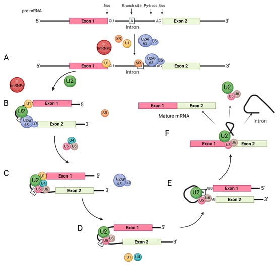
Figure 3. General splicing process. The regulation of RNA-binding proteins such as hnRNPs and SRs involves several steps. (A) The splicing factor U1 snRNP complex binds to the 5′ splice site, U2AF65 binds to the polypyrimidine site (Py-tract), and U2AF35 binds to the AG bases at the 3′ splice site. The U2AF heterodimer combines with the 3′ splicing site to recognize the intron splicing signal. (B) U2 snRNP binds to the branch site with the assistance of U2AF. (C) snRNP U4, U6, U5 join the complex, while U2AF dissociates from the complex. (D) U1 snRNP and U4 snRNP subsequently leave the complex through a series of conformational transitions. (E) The first transesterification reaction connects the 5′ss to the branch site and cleaves the RNA strand, forming a lariat structure. (F) In the second transesterification reaction, the exons are ligated to each other to form the mRNA, and the introns are released in a lariat structure.
-
Splice site identification and shelter: hnRNP proteins have the ability to bind to pre-mRNA splice sites, which can impact the formation of spliceosomes. This binding can mask or hinder the recognition of splice sites, leading to differential alternative splicing. Additionally, the binding of hnRNPs can competitively affect the binding of other splicing factors. For instance, hnRNP I’s RRM1 and RRM2 specifically bind to the polypyrimidine sequence of the fourth internal loop of the U1 snRNA stem loop, thereby inactivating the splicing complex A formed by U1 snRNP [47]. Studies have also shown that hnRNP I can target ITSN1 [48], β-tropomyosin [49], Fas [50], and alpha-actinin [51], competitively inhibiting the binding of U2AF65 and preventing the formation of the U2AF heterodimer complex by binding to the polypyrimidine sequence of pre-mRNA. hnRNP A1 interacts with the 3′ splice site of MAPT exon 10 and facilitates the exclusion of exon 10 [52]. Similarly, hnRNP L can bind to pre-mRNA polypyrimidine sequences, competitively inhibiting the binding of U2AF65 to RNA [53]. Furthermore, hnRNP H1/H2 counteracts the activation of the 3′ splice site [54] (Figure 4B).
-
Splicing inhibition: hnRNPs can impact RNA structure by interacting with pre-mRNA, resulting in the exclusion of specific exons. This inhibitory effect primarily operates by altering the structure of the splice site. For instance, the RRM3 and RRM4 domains of hnRNP I bind to the polypyrimidine sequence (e.g., CUCUCU) near the pre-mRNA exon, forming an RNA ring structure that hinders the binding of other splicing factors and the formation of splicing complexes [51][55][51,55] (Figure 4C).
-
Competitive splicing inhibition: In certain cases, hnRNP proteins and other RNA-binding proteins may competitively bind to the same pre-mRNA, which can impact the inclusion or exclusion of specific exons. The majority of competitive splicing occurs between hnRNPs and SRs. For instance, hnRNP H1 can compete with SRSF3 for binding to PRMT5 pre-mRNA, thereby inhibiting the exclusion of PRMT5 exon 3 by SRSF3 [56]. Additionally, hnRNPA1 can competitively bind to the G-rich sequence downstream of β-tropomyosin exon 6B, along with ASF/SF2, leading to the inhibition of exon 6B exclusion. Simultaneously, hnRNP A1 and ASF/SF2 competitively bind to the 5′ splice site of C175G pre-mRNA (C175G is a synthetic 533 nt pre-mRNA sequence that is frequently employed as a standard model for investigating 5′ splice sites). hnRNP A1 competitively inhibits the binding of U1 snRNP to the 5′ splice site of C175G pre-mRNA, while ASF/SF2 enhances the binding of U1 snRNP to the 5′ splice site of C175G pre-mRNA [57] (Figure 4D).

Figure 4. Schematic diagram of hnRNPs regulating variable splicing. The process of exon excision (left) and intron excision (right) shown in panel (A) relates to when hnRNPs are not involved in variable splicing. Panel (B) demonstrates how hnRNPs prevent splicing factors such as U2AF heterodimer and U1 snRNPs from binding to pre-mRNA. This prevention is achieved through the recognition of splicing sites and masking effects. In panel (C), hnRNPs are shown to promote exon (left) and intron (right) retention by binding to pre-mRNA, forming a special pre-mRNA loop structure. This structure prevents splicing factors from recognizing splice sites on pre-mRNA. Finally, in panel (D), hnRNPs bind to SRs and hinder the promotion of splicing by SRs.
2.3.2. Regulation of mRNA Stability
hnRNPs play a role in regulating mRNA stability through various mechanisms, including the poly(A) tail, AU-rich elements (AREs), and the 3′UTR. For instance, hnRNP H1 and hnRNP F can enhance the stability of APP mRNA by binding to it through cytoplasmic shuttling mediated by the gly Rich motif, thereby increasing its half-life [63]. Additionally, hnRNP F has been found to regulate the TTP/BRF-mediated degradation of ARE-mRNAs [64]. The RRM2 domain of hnRNP A2/B1 acts as an m6A reader [65], recognizing the N6-methyladenosine (m6A) site on TCF7L2 mRNA to stabilize the poly(A) tail and maintain its mRNA stability. Moreover, hnRNP A2/B1 and hnRNP A1 play a role in the regulation of mRNA deadenylation via the CCR4-NOT deadenylation complex. They achieve this by binding to the UAASUUAU sequence present in the mRNA 3′UTR, thereby influencing the degradation of the transcript [66] (Figure 5B). Interestingly, hnRNP A2/B1 also binds to its own 3′UTR to regulate the ratio of nonsense-mediated RNA decay (NMD) (both for sensitive and insensitive types). When the levels of hnRNP A2/B1 protein increase, it combines with its own pre-mRNA 3′UTR, leading to alternative splicing and the production of a higher proportion of NMD-sensitive mRNAs. These NMD-sensitive mRNAs are subsequently degraded during translation [67]. This auto-regulatory mechanism demonstrates how splicing factors control their own expression levels, providing one possible explanation [68]. As an ARE-binding protein, hnRNP D has the ability to recognize and bind to various ARE-mRNA sequences by targeting uridine residues [69]. Subsequently, it recruits transporters such as eIF4G, PABP, Hsp70, Hsc70, and Hsp27 to form the signal transduction regulatory complex (ASTRC). ASTRC plays a crucial role in initiating the 3′ to 5′ deadenylation-dependent mRNA degradation pathway, thereby promoting mRNA degradation. Among the isoforms of hnRNP D, the specific mechanism of action involves AUF1 interacting with the AU-rich element (ARE) in mRNA, which serves as a recognition site for AUF1. While the mRNA is being translated, AUF1 interacts with the translation initiation factor eIF4G [70], causing AUF1 to be released from the ARE by the ribosome. This enables the ribosome to access the mRNA and initiate translation. Simultaneously, AUF1 forms a complex with the polyadenine nucleic acid-binding protein (PABP), which exposes the polyadenine nucleic acid tail and allows the mRNA to be degraded by nuclease. The p40 isoform is involved in the decay of ARE-mRNA, which regulates lymphokine mRNA stability [71] (Figure 5A). Additionally, studies have demonstrated [72] that the KH3 domain of hnRNP K can bind to the poly(C) site of LAPTM5 3′UTR, thereby enhancing the stability of its transcripts. Different hnRNPs exhibit variations in their regulation of mRNA stability. For instance, hnRNP D, K, I, and Q [73] can all bind to the 3′UTR of mPer3, but they differ in their impact on mPer3 stability. hnRNP K maintains the stability of mPer3, while hnRNP D and Q accelerate its degradation. hnRNP I, on the other hand, does not affect the stability of mPer3. Additionally, hnRNP A1 [74], C [75], U [76], and L [77] can bind to the 3′UTR of certain mRNAs and influence their stability, although the mechanism underlying this remains unclear.
Figure 5. hnRNPs-mediated mRNA degradation mechanism. (A) In the translation process, eIF4G binds to AUF1, promoting the dissociation of AUF1 from the ARE sequence. The released AUF1 then binds to PABP, leading to the exposure of the polyadenine nucleic acid tail for mRNA degradation. Both AUF1 and PABP are subsequently degraded through the proteasome. (B) hnRNP A2/B1 and hnRNP A1 interact with the UAASUUAU sequence located in the 3′UTR of mRNA. This interaction plays a role in controlling mRNA deadenylation, which is carried out by the CCR4-NOT deadenylation complex. Ultimately, this process influences the degradation of the transcript.