Novel Cellular and Immunotherapy: Comparison
Please note this is a comparison between Version 1 by Alessandro De Camilli and Version 3 by Dean Liu.

Targeted cellular and immunotherapies have welcomed a new chapter in multi-modal cancer therapy. These agents harness our innate immune system and destroy malignant cells in a precise way as compared with “legacy” chemotherapeutic agents that largely rely on abolishing cell division. New therapies can augment the T-cell recognition of tumor antigens and effectively prevent tumor cells from their historically successful ability to evade immune recognition. These novel agents cause acute and chronic toxicities to a variety of organ systems (enteritis, pneumonitis, hypophysitis, and hepatitis), and this may masquerade as other chronic illnesses or paraneoplastic effects. As the perioperative footprint of cancer patients increases, it is essential that perioperative providers—anesthesiologists, surgeons, nurse anesthetists, and inpatient hospital medicine providers—be up to date on the physiologic mechanisms that underlie these new therapies as well as their acute and subacute toxicity profiles. Immunotherapy toxicity can significantly impact perioperative morbidity as well as influence perioperative management, such as prophylaxis for adrenal insufficiency, preoperative pulmonary assessment, and screening for thyroid dysfunction, among others.

  • immunotherapy
  • cellular therapy
  • perioperative risk
  • hypophysitis

1. Introduction

The last decade has brought about a revolution in cancer therapies that harness the immune system to target cancer-specific antigens. These agents were once reserved for use in palliative chemotherapy for patients with unresectable or refractory metastatic disease. They have now firmly established their intention-to-cure role and play a significant role in neo-adjuvant therapy [1][2][3][4][5][6][7][8][9][10][11][12][13][1,2,3,4,5,6,7,8,9,10,11,12,13]. Immunotherapies have greatly improved quality of life for patients with advanced cancer. More than ever, it is necessary for the perioperative provider to be up to date on the physiology underlying these therapies as well as their unique toxicity profiles.
Early-generation chemotherapeutics relied on nonspecific destruction of cell division, with toxicities mostly related to en masse destruction of rapidly dividing cells. Newer immunotherapies have a more benign acute safety profile but nonetheless can exert chronic toxicities that may be more insidious and require a higher index of suspicion for detection. The predominant cause of toxicities results from unwanted upregulated immune system activity or molecular mimicry, leading to destruction of “bystander” tissue.
To provide an example, ipilumumab and nivolumab are two immune-checkpoint inhibitors (ICIs) that have recently been shown to improve survival when included as part of neoadjuvant therapy for squamous cell esophageal cancer [14]. However, the two drugs in combination can lead to a non-negligible incidence of ICI pneumonitis and ICI hypophysitis. It is thus becoming more important for thoracic surgeons and anesthesiologists who care for these patients to be aware of the potential risks of adrenal insufficiency and reduced pulmonary capacity. A thorough therapeutic history and close collaboration with oncologists are necessary to provide safe care.

2. Immune-Checkpoint Inhibitors

Immune-checkpoint inhibitors have earned their keep as tremendously successful adjuncts to all manner of chemotherapeutic regimens. TheOur innate immune system relies on T-cell activation and proliferation to destroy foreign cells. T-cell activation requires both antigen–receptor coupling as well as co-stimulation by other immune effectors cells. This two-fold process underpins the versatility of theour immune system but is also the process by which cancer cells can evade detection. T-cell activation can be inhibited by two pathways: the cytotoxic T-lymphocyte-associated antigen 4 (CTLA-4) pathway and the programmed cell death protein (PD-LA1) pathway. This interplay of T-cell activation and inhibition is referred to as an “immune checkpoint.” Cancer cells have evolved to thwart T-cell activation by taking advantage of the inhibition role of this immune checkpoint since CTLA-4 and PD-LA1 antigens are commonly found on the surfaces of tumor cells. Activation of these ligands by tumor cells allows them to evade detection. Immune-checkpoint inhibitors (ICIs), as a therapeutic target, prevent tumor cell evasion by acting as monoclonal antibodies against the CTLA-4 and PD-LA1 ligands. In preventing their ability to inactivate T cells, ICIs ensure that tumor cells are vulnerable to destruction (see Figure 1).
Figure 1. Above: normal immune checkpoint activity means that cancer (as tumor cells directly or as tumor antigen presented via an APC) can inhibit T-cell activation via the CTLA-4 and PD-1 ligands. Below: immune checkpoint inhibitors prevent this inhibition, leading to T-cell activation.
This has proven to be a very effective therapeutic target, with successes in the treatment of melanoma, breast cancer, and lymphoma, to name a few. Checkpoint inhibition technology has continued to advance as new agents are developed that target additional T-cell pathways and as existing ones are used on new tumor subtypes. The first immune-checkpoint inhibitors approved for human use were Ipilimumab and Nivolumab (2011 and 2014, respectively). Ipilimumab acts as a monoclonal antibody against CTLA-4 and Nivolumab against PD-1. Ipilimumab was initially shown to dramatically improve survival in patients with metastatic melanoma [15]. The subsequent arrival of Nivolumab showed promise for the two used as combination therapy both for metastatic melanoma and several other cancers—renal cell carcinoma, unresectable non-small-cell lung cancer (NSCL), prostate cancer, and esophageal cancer [15][16][15,16]. Immune-checkpoint inhibitors have shown promise in treating tumors that have been historically resistant to other chemo or immunotherapeutic agents, but there are highly specific indications for the use of each. Up to 45% of patients with cancer are candidates for ICI therapy [17], so the perioperative footprint of these patients continues to rise. Their efficacy is determined by the rate of target PD-L1 or CTLA-4 receptor expression on the tumor cells, the mutational burden of tumors, and the relative inefficacy of classic chemotherapy agents for the tumor in question. ICI therapy in the perioperative period is an area of ongoing study. Thus far, a few studies at major cancer centers have shown no association with perioperative morbidity of any type [18], and in fact, most show greatly improved results when added as neo-adjuvant therapy [9][10][11][9,10,11].

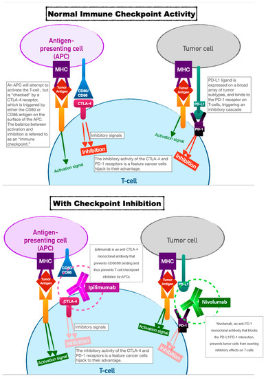
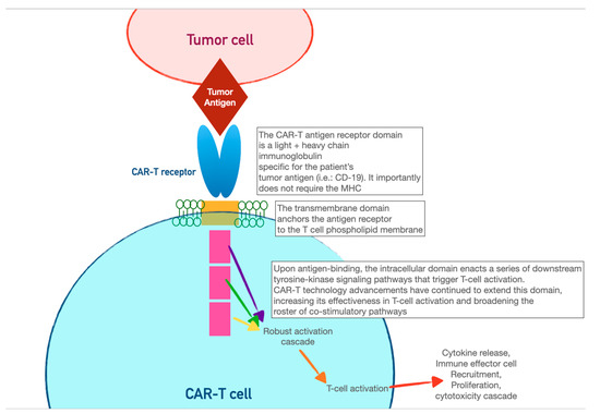
3. Chimeric Antigen-Receptor T Cells (CAR-T)

CAR-T cells harness the adaptive immunity offered by theour T-lymphocytes to target tumor-specific antigens. As the first foray into cellular therapeutics, they represent one of the most consequential discoveries in cancer therapy in the last two decades owing to their relative ease of administration and robust efficacy against highly fatal hematologic malignancies. As discussed in the previous section, the immune system relies on T-cell activation via (1) antigen presentation via an MHC complex and (2) co-stimulation via other immune effector cells. Cancer cells have historically evaded this process of T-cell activation by thwarting antigen recognition. Enter CAR-T cells: the genetically modified host T cells that force this process to occur without the ability of tumor-cell evasion. First, T cells are extracted from the patient (or an allogeneic donor) and modified via a retroviral vector to express a new antigen receptor and co-stimulatory domains (see Figure 2). The term “chimeric” refers to the fact that this engineered receptor both activates and co-stimulates the T cell [19][20][49,50]. The patient is then leuko-reduced to increase the relative proportion of CAR-T cells, and CAR-T cells are infused into the patient. The CAR-T cells will recognize antigens and activate and recruit immune effector cells to destroy tumor cells.
Figure 2. Above: normal T-cell activation requires antigen recognition and co-stimulation. Below: a re-engineered CAR-T cell enables simultaneous antigen recognition and co-stimulation, leading to T-cell-mediated destruction of tumor cells.
The first CAR-T cells used clinically targeted the CD19 receptor, making them effective for the treatment of ALL or diffuse large B-cell lymphoma [21][51]. Trials have consistently shown a 50–90% remission rate for relapsed or refractory disease, which continues to improve with refinement of technique [22][52]. CAR-T for solid tumors is a growing area of research, where re-modeled CAR-T cells can be introduced intra-tumorally or intra-pleurally [23][53]. Once infused, CAR-T cell activity can be controlled via embedded biochemical switches. An “on” switch or, conversely, a “suicide gene” can induce CAR-T cell activation or apoptosis. These external controls allow CAR-T technology to be more pliable in the face of toxicities or other adverse events.

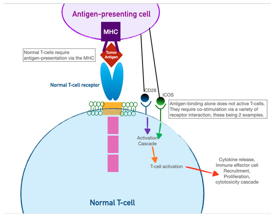
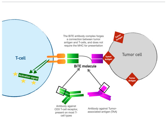
4. Bispecific T-Cell Engager (BiTE) Therapy

BiTE therapy is one of the newest innovations in targeted immunomodulatory chemotherapy. BiTE molecules are an antibody with two domains (hence “bispecific”): one that recognizes tumor-specific antigens and another that recognizes the universal CD-3 receptor on T cells (see Figure 3). The binding sites are essentially two monoclonal antibodies bound together. In essence, the BiTE “forces” a recognition and activation of the T cell, so the tumor cell does not have a chance to present its own antigen and possibly release inhibitory signal receptors. Specific tumor antigen targets of BiTE molecules include CD19 (broadly expressed on B-cell malignancies), B-cell maturation antigen (expressed highly on malignant cells involved in multiple myeloma), and CD33 (expressed in acute myeloid leukemia, myelodysplastic syndrome, and chronic myeloid leukemia). Under development are BiTE targets that include solid tumor antigens such as prostate-specific member antigen and delta-like protein 3, which is highly prevalent in small-cell lung cancer.
Figure 3. The Bispecific T-cell engager (BiTE) molecule has shown promise as a targeted immunotherapy but, unlike CAR-T, does not require harvesting of the patient’s native lymphocytes.
The most widely used BiTE agent at this time is Blinatumomab, approved for use in acute lymphoblastic leukemia (ALL). Trial response rates in children and adults with ALL have shown a resounding 90% complete remission rate, with a 6-month event-free survival of 67% and a 78% overall survival rate [24][54]. It is now approved for use around the globe. Solitumab is a BiTE molecule with a binding domain for the epithelial cell adhesion molecule (EpCAM), which is being investigated for therapy against gastrointestinal, lung, and other solid tumors [25][55]. These molecules are administered as continuous IV infusion over several days owing to their 2–4-h half-life. Not unsurprisingly, owing to their similar mechanisms, they have an adverse event profile that parallels that of CAR-T cells. In the original study for Blinatumomab, the most common adverse events were neutropenia, infection, elevated LFTs, neurotoxicity, and CRS [26][56]. CRS, neurotoxicity, and acute anaphylaxis are the most significant for the acute care and perioperative provider. There is perhaps a lower propensity than CAR-T for high-grade neurotoxicity [27][57]. Treatment for these adverse events follows the same protocol: corticosteroids, IL-6 inhibition, and supportive care.
Video Production Service