Percutaneous Cryoablation in Bone and Soft Tissue Tumors: Comparison
Please note this is a comparison between Version 1 by Nicolas Papalexis and Version 2 by Lindsay Dong.

In the rapidly evolving field of interventional oncology, minimally invasive methods, including CT-guided cryoablation, play an increasingly important role in tumor treatment, notably in bone and soft tissue cancers. Cryoablation works using compressed gas-filled probes to freeze tumor cells to temperatures below −20 °C, exploiting the Joule–Thompson effect. This cooling causes cell destruction by forming intracellular ice crystals and disrupting blood flow through endothelial cell damage, leading to local ischemia and devascularization. Coupling this with CT technology enables precise tumor targeting, preserving healthy surrounding tissues and decreasing postoperative complications.

  • interventional radiology
  • metastatic neoplasms
  • orthopedic surgery
  • palliative medicine
  • cryoablation

1. Introduction

Interventional oncology has emerged as an increasingly crucial component of the multidisciplinary team, providing innovative and minimally invasive treatment approaches for various types of musculoskeletal tumors [1][2][1,2]. One such innovative technique, CT-guided cryoablation, is increasingly gaining recognition for its effectiveness, precision, and improved patient outcomes [3][4][3,4].
Cryoablation is a minimally invasive procedure that uses one or more probes filled with compressed gas (usually argon) to cool tumor tissue to very low temperatures [5]. This cooling is achieved by exploiting the Joule–Thompson effect, where rapid decompression of the gas surrounding the probe tip leads to temperatures below −20 °C [6][7][6,7]. The cooling damages the cells by forming intracellular ice crystals that destroy them, as well as impairing the blood supply by damaging the endothelial cells, which results in local ischemia and devascularization (Figure 1). Its integration with CT (Computed Tomography) technology allows for more accurate targeting of tumor cells, reducing collateral damage to surrounding healthy tissues, and minimizing postoperative complications [3][8][9][3,8,9].
Figure 1. Sequence of events showing the cryoablation technique. The cryoprobes are inserted inside the lesion (AD) until it is all covered by ice (E). The cooling damages cells causing ischemia and devascularization, which result in lesion destruction and volume reduction (FH).

2. Malignant Bone and Soft Tissue Tumors

Percutaneous cryoablation is becoming an increasingly accepted option within the multidisciplinary sarcoma board for the treatment of primary bone and soft tissue tumors, applicable for selected cases. Despite surgical intervention being the mainstay for treating primary, non-metastatic bone, and soft tissue tumors, the local control of recurring bone and soft tissue sarcoma (STS) continues to be a challenging task. It mainly hinges on the disease prognosis as per the guidelines of the European Society for Medical Oncology (ESMO) [10][11][10,11]. Surgical resection is the common protocol for localized conditions, while chemotherapy or radiation therapy may be employed for more extensive diseases or recurrences [10][11][12][10,11,12]. Lately, minimally invasive techniques such as radiofrequency ablation, microwave ablation, or cryoablation have been proposed as potential surgical alternatives for some selected recurrent bone and soft tissue tumors [13][14][15][16][17][13,14,15,16,17].

Some initial studies evaluated the therapeutic effect of Cryoablation for the treatment of a variety of primary bone and soft tissue malignancies with promising results; however, the scientific evidence is still limited. Moreover, there is a recognized need for the standardization of selection criteria for percutaneous cryoablation. Lippa et al. [12] aimed to identify these criteria, finding high agreement for all proposed criteria between two readers. Eligibility for cryoablation was significantly associated with tumors located deeply, with great axes ≤ 5 cm, high local tumor aggressiveness, and a diagnosis of differentiated myxoid liposarcoma or myxofibrosarcoma.

2.1. Recurrent Retroperitoneal Soft Tissue Tumors

Some retrospective studies have reported on the effectiveness and safety of percutaneous cryoablation in the treatment of recurring retroperitoneal soft tissue sarcomas (RPSs). RPSs, which make up approximately 0.15% of all cancers, originate in the retroperitoneum but not from its main organs. Their proximity to critical structures makes them challenging to manage. Surgery is the primary therapeutic approach for localized cases, leading to a survival rate of about 60% over five years [18][19][21,23]. Nonetheless, complications with the removal of RPSs can impact survival and lead to recurrence. For recurrent cases, re-operation is recommended, but it is more difficult, and the additional benefits of chemotherapy and radiotherapy are debatable. In some selected cases, cryoablation has proven safe and effective as a palliative treatment for RPSs and could be included in the armamentarium of the sarcoma board.

2.2. Sacrococcygeal Tumors and Chordoma

Kurup et al. [20] documented the use of cryoablation in treating recurrent sacrococcygeal tumors, with the findings suggesting that this method was secure and relatively effective for local management or pain reduction (Figure 2). Similarly, Susa et al. [21][18] assessed the clinical outcomes of CT-guided cryoablation for recurring or metastatic malignant bone and soft tissue tumors, involving nine patients over a median observation period of 24.1 months. Although they reported promising outcomes, the study’s effectiveness was significantly limited by the small patient group.
Figure 2. A 55-year-old man, cryoablation in a recurrent chordoma showed in an axial T2 MR image ((A)—arrowheads) and axial postcontrast CT image ((B)—arrowheads), for local tumor control. The cryoprobe was placed into the lesion under CT (C) and US guidance, with subcutaneous hydro dissection performed to protect the skin. The ice ball encompassed the entire lesion (D). Also, note the aspect of the ice ball on ultrasound ((E)—arrowheads).

3. Bone Metastases

Bone frequently becomes a site of metastases, ranking as the third most common area for metastatic carcinoma after the lungs and liver [22][24]. Given the typically low survival rates of patients, therapeutic options are usually limited, and surgical resection is rare [22][23][24,25]. However, skeletal complications such as severe and chronic pain, spinal cord compression, and pathological fractures can greatly impair a patient’s quality of life [24][25][26][27][28][29][30][26,27,28,29,30,31,32]. The escalating interest in thermal ablation techniques for handling bone metastases has positioned cryoablation as a top-tier approach. This is due to its capacity to treat extensive and irregularly shaped pathological tissues in real time, while causing less pain compared to heat-based ablative methods like radiofrequency and microwaves [31][32][33][33,34,35]. The current body of literature mostly explores the palliative effect and local tumor control of cryoablation for bone metastases, which makes this technique a useful tool in the multidisciplinary management of cancer patients.

3.1. Pain Palliation and Disease Control

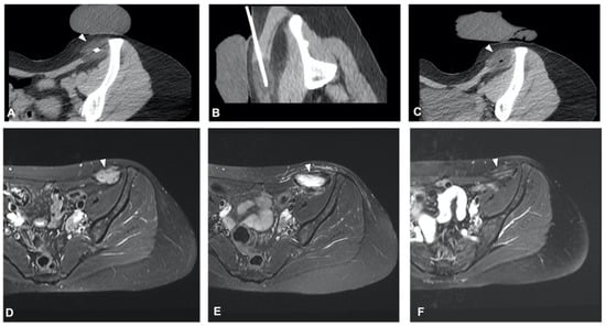
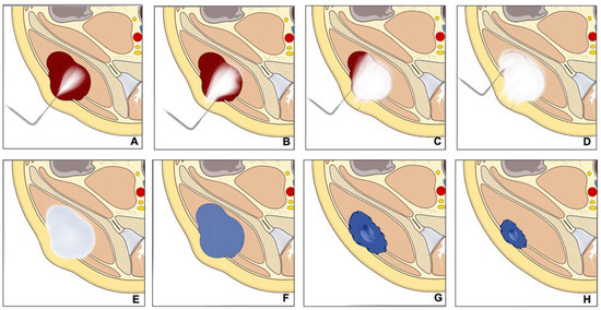
The clinical effect in pain reduction and safety of cryoablation for metastatic bone disease has been investigated by several studies in recent years [34][35][36][37][38][40,43,44,46,48] (Figure 3). A recent multicenter prospective study by Jennings et al. [39][38] assessed the clinical efficacy of cryoablation as a pain palliating method for patients with metastatic bone disease. The main goal was the pain score change from pre-treatment to the eighth-week follow-up, with participants monitored for 24 weeks post-treatment. A cohort of 66 participants (average age 60.8 years, 53% male) was recruited and underwent percutaneous cryoablation; 65 completed the follow-up. The average change in pain score from baseline to the eighth week decreased by 2.61 points. Average pain scores improved by 2 points at the first week and attained clinically significant levels (a decrease of more than 2 points) post the eighth week, with scores continuing to improve throughout the follow-up period. Quality of life was enhanced, opioid doses were steady, and functional status remained unchanged over six months. Severe adverse events were reported in three participants.
Figure 3. Axial T2 MR image of a 51-year-old woman with metastases from leiomyossarcoma (A) in the pelvis treated with cryoablation (C) for palliative intent. 18F-FDG PET/CT scan performed before the procedure demonstrates the pathologic radiotracer uptake in areas of viable tumor (B). The procedure was performed in an attempt to cover these areas and the post-procedure ice balls are visible as hypodense circles (arrowheads—(D,E)).
In a prospective trial analysis, Callstrom et al. [40][52] evaluated the safety and effectiveness of cryoablation in pain reduction, enhancement of daily activities, and decreased usage of analgesics in patients with painful metastatic bone lesions. All eight patients who were on narcotic medication before the procedure reported a decrease in these medications after cryoablation, with no serious complications noted. Moreover, De Marini et al. [41][50] conducted a comparison of the safety profiles of radiofrequency ablation (RFA) and cryoablation (CA) in the treatment of bone metastases. The study was conducted on 274 patients (average age 61.6 years) treated between January 2008 and April 2018. From these, 53 patients (involving 66 bone metastases) received RFA, and 221 patients (involving 301 bone metastases) underwent CA. Within the entire group, 2.5% of bone metastases resulted in major complications, with no significant discrepancy between RFA (1.5%) and CA (2.7%). However, RFA demonstrated a higher incidence of minor complications, predominantly post-procedure pain, at 33.3% compared to CA at 6.0%.

3.2. Application to Spinal Metastases

The treatment of spinal metastases is notoriously difficult due to the sensitive nature of the spine, with both watchful waiting and active treatment carrying significant risks of local complications [42][43][56,57]. Cryoablation for spinal metastases is often conducted alone or frequently in combination with vertebral augmentation techniques such as cementoplasty [44][51]. In a study by Autrusseau et al. [44][51], 31 patients (including 36 spinal metastases in 32 sessions) received cryoablation for pain relief, and 10 patients (10 metastases in 10 sessions) for local control. The procedure successfully alleviated pain in 93.8% of palliative sessions, with the average pain scores notably decreasing at 24 h, 1 month, and at the final follow-up (approximately 16.5 months). For those patients needing local tumor control, primary clinical success was achieved in 60% of cases with about 25 months of median follow-up. The overall complication rate was 8%, with no reported secondary fractures or thermal nerve injuries.

3.3. Application to Sternal Metastases

Due to its high success rate and the safety provided by real-time visualization of the ice formation, cryoablation can be employed to treat highly sensitive body areas. A study by Hegg et al. [45][42] sought to evaluate the safety and efficacy of cryoablation for sternal metastases. The retrospective review included 12 patients with 12 sternal metastases. The results indicated that cryoablation provided pain relief, as shown by a drop in average pain score from 7.0 to 1.8. Local tumor control was achieved in 80% of the patients treated for this purpose. The study concluded that cryoablation is a safe and potentially effective treatment for painful sternal metastases.

3.4. Evaluation of Post-Ablation Area

Evaluating the treated area after cryoablation is crucial in determining the success of the procedure and detecting any local tumor recurrence. In this regard, Gravel et al. [46][53] conducted a study to assess the effectiveness of post-ablation MRI in identifying cases of incomplete treatment of spinal osseous metastases following cryoablation. The study involved 54 spinal bone metastases in 39 patients. The classification of MRI images into four categories resulted in a sensitivity of 77.3% and specificity of 85.9% in identifying residual tumors.

3.5. Technical Consideration for Neuroprotection

Preserving neural structures is paramount when treating lesions close to the spine or major peripheral nerves. Kurup et al. [47][59] explored the use of motor evoked potential (MEP) monitoring during the cryoablation procedure of musculoskeletal tumors to reduce the likelihood of nerve damage. This study included 59 procedures on 64 tumors in 52 patients, with tumors located in various sites such as the spine, sacrum, retroperitoneum, pelvis, and extremities. During these procedures, MEP monitoring identified significant decreases in MEPs in 32% of the cases, with transient decreases in 25% and persistent decreases in 7%. Out of the four patients with persistent decreases in MEPs, two experienced motor deficits post-ablation

3.6. Technical Consideration for Bone Reinforcement

When conducting ablation on large bone sections or bones that bear weight [48][47], bone reinforcement might be required to avoid post-procedural pathologic fractures. Combining cryoablation with cement stabilization has been reported as highly effective by several studies. Masala et al. [49][60] studied the efficacy of combining cryoablation and vertebroplasty (CVT) vs. vertebroplasty alone in 46 patients with a single vertebral metastasis. They used the Visual Analog Scale (VAS) and the Oswestry Disability Index (ODI) to measure pain levels and quality of life. Although both treatment groups showed a significant reduction in VAS and ODI scores, more notable improvements were observed in the CVT group at various follow-up stages, suggesting CVT as a safe, effective option for pain relief and disability improvement.

3.7. Combination Treatment

Sundararajan et al. [50][63] proposed a sequential interventional therapy involving embolization, cryoablation, and osteoplasty for patients with osseous neoplasms, who were unresponsive to conventional treatment

3.8. Complications

Despite cryoablation being a minimally invasive procedure guided by CT, it carries a small risk of complications. Auloge et al. [51][64] evaluated the complications and related risk factors in bone tumor cryoablation. The study involved 239 patients who underwent cryoablation for 320 primary or metastatic bone tumors from 2008 to 2017. The overall complication rate was 9.1%, with serious complications making up 2.5% of this total. The most common major complication was secondary fractures, which represented 1.2% of the cases.

4. Benign Bone Tumors

4.1. Osteoid Osteoma

Osteoid osteoma (OO) is a small, benign tumor primarily found in the bones of young people and children. Even though it only accounts for approximately 10% to 12% of all benign bone tumors, it can significantly affect the quality of life, causing pain and bone deformity, especially in children [52][53][54][65,66,67]. The treatment for OO has seen a considerable evolution over the years. Traditional surgical removal was once the main treatment approach, but technological advancements have facilitated a shift toward less invasive methods like radiofrequency ablation (RFA) [55][56][57][58][68,69,70,71]. Meng et al. [59][72] conducted a study comparing the safety and effectiveness of percutaneous CT-guided cryoablation of OO to surgical curettage. Both treatment approaches reported a 100% technical success rate. However, patients treated with cryoablation spent significantly less time in the hospital than those undergoing surgery, and both groups showed notable improvement in postoperative Visual Analog Scale (VAS) pain scores.

4.2. Osteoblastoma

Cryoablation was also found to be a viable treatment for osteoblastoma in the study by Cazzato et al. [60][78] Technical success was achieved in all cases, and primary clinical success was 100% and 78% at 1 and 12 months of follow-up, respectively. Notably, this study emphasized the need for comprehensive protective measures due to the frequent close proximity of critical structures.

4.3. Bone Cyst and Aneurysmal Bone Cyst

Bone cysts are fluid-filled holes that develop within bones. They are commonly found in children and adolescents, and most often occur in the long bones of the body such as the femur or the humerus. Most bone cysts do not cause symptoms and are often discovered incidentally during an X-ray performed for other reasons. However, in some cases, they can cause pain or lead to fractures [61][79]. Aneurysmal bone cysts (ABCs), on the other hand, are an uncommon type of bone cyst that is blood-filled rather than fluid-filled. They can occur at any age but are most commonly diagnosed in individuals under the age of 20. ABCs are expansile and can cause pain, swelling, and deformities in the affected bone. They can also lead to fractures due to the weakening of the bone structure.

5. Desmoid Tumors

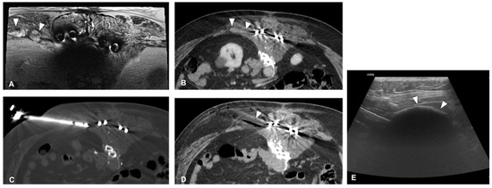
Desmoid tumors are rare benign tumors originating from musculoaponeurotic structures [62][85]. Despite their benign nature, they can display local aggressiveness, causing disability and sometimes pain. The ESMO advises initial observation and subsequent medical treatment for progressing tumors. Cryoablation, an interventional radiology technique, is recommended for desmoid tumor patients due to its ability to induce cell death through multiple cycles of freezing [63][64][65][86,87,88] (Figure 45).
Figure 45. A 46-year-old patient with desmoid tumor of the abdominal wall showed in axial T2 fat sat MR image ((D)—arrowhead). Axial (A) and sagittal oblique (B) CT images showing the cryoprobe inside the lesion and the post-procedure ice ball containing the whole lesion (C). Axial T2 fat sat MR image 3 months after treatment shows an increase in T2 signal inside the lesion, suggesting necrosis (E), and the control with 6 months shows no residual tumor (F).
Given the high recurrence rate, surgery’s role in treating desmoid tumors is restricted only to selected cases [66][89]. However, percutaneous cryoablation has recently come into the spotlight as a promising treatment option, demonstrating positive results in safety, effectiveness, and symptom alleviation [67][68][69][90,91,92]

5.1. Cryoablation for Disease Control

Mandel et al. [70][98] conducted a study aimed at comparing the outcomes of patients with primary and recurrent extra-abdominal desmoid tumors undergoing percutaneous cryoablation with those undergoing surgical treatment. In this study, 22 patients treated with cryoablation were compared with 33 surgical patients. The median monitoring periods were 16.3 months for cryoablation and 14.9 months for surgery. Local recurrence-free survival (LRFS) rates for two years stood at 59% for cryoablation and 71% for surgery, although the median LRFS for surgery was not met. Two-year disease control reached 85% for all patients, with no significant difference between the cryoablation and surgical groups.

5.2. Ablation Margin

Although technically benign tumors, desmoid tumors have a high potential for local recurrence and local invasion. For this reason, they should be treated as malignant lesions, and to obtain a curative effect, all the lesions should be covered by the ice ball, leaving some margins if possible.

5.3. Pain Reduction

One of the main symptoms of a desmoid tumor is local pain and discomfort. Bouhamama et al. [71][99] focused on cryoablation’s analgesic efficacy. This study emphasizes the analgesic effect of cryoablation in the treatment of desmoid tumors, reporting a significant reduction in pain post-procedure. It also reports a disease-free survival rate of 42.2% at 3 years, providing a more complete picture of the long-term efficacy of cryoablation.

5.4. Patient Selection

In contrast, Testa et al. [72][97] highlighted the trend toward active monitoring as the first line of treatment for desmoid tumors (DTs), reserving systemic and local ablative therapies for cases where the disease is advancing or causing symptoms. This study suggested that determining which patients are better suited for a primary non-interventionist approach as opposed to a direct interventional treatment like cryoablation remains uncertain.

6. Technical Consideration

6.1. Planning and Approach

Cryoablation planning requires pre-procedural imaging, ideally within a month prior to the procedure. Thin-section CT is recommended for bone lesions, while MRI with dynamic contrast enhancement and high spatial resolution techniques offer higher sensitivity and specificity for soft tissue lesions [73][100].

6.2. Needle Placement and Hydro Dissection

When the lesion is not deeply located and is outside the bone, as in soft tissue tumors, the ultrasound-guided placement of the needles may expedite the process and reduce patient exposure to ionizing radiation [73][74][75][100,101,102]. Hydrodissection is a valuable technique, used to create a safe margin between the tumor and adjacent critical structures. To enhance visibility during control CT scans, it is recommended to use iodinated water for hydro dissection. This allows the operator to clearly delineate the dissected area [76][58].

6.3. Ablation and Monitoring

During the ablation phase, real-time visualization of the ice ball using ultrasound can assist in limiting the number of CT scans. However, the complete ice ball cannot be fully visualized due to shadowing; therefore, visualization through CT is essential [77][103]. When performing cryoablation near neural structures, such as the spine or peripheral nerves, continuous monitoring of evoked potentials is necessary.

7. Conclusions

Percutaneous cryoablation is a reliable and successful method for treating a range of bone and soft tissue tumors, being increasingly incorporated into the multidisciplinary decision-making process of tumor boards. The procedure’s benefits encompass prompt alleviation of pain, enhancement of life quality, and limited unwanted side effects. Precision in needle placement and ice ball visualization, facilitated by imaging guidance, further enhances the safety and accuracy of the procedure, making it a viable option even for lesions located near critical structures. Despite these benefits, the decision to perform cryoablation should consider tumor location, size, and aggressiveness. Cryoablation has demonstrated substantial potential in handling a variety of musculoskeletal tumors, from retroperitoneal soft tissue sarcomas to sacral chordomas, bone metastases, and benign bone and soft tissue tumors, underlining its adaptability. However, more research is required to fully understand the extent of cryoablation’s capabilities.
ScholarVision Creations