p38γ MAPK in Physiology and Disease: Comparison
Please note this is a comparison between Version 2 by Jessie Wu and Version 1 by Guan Chen.

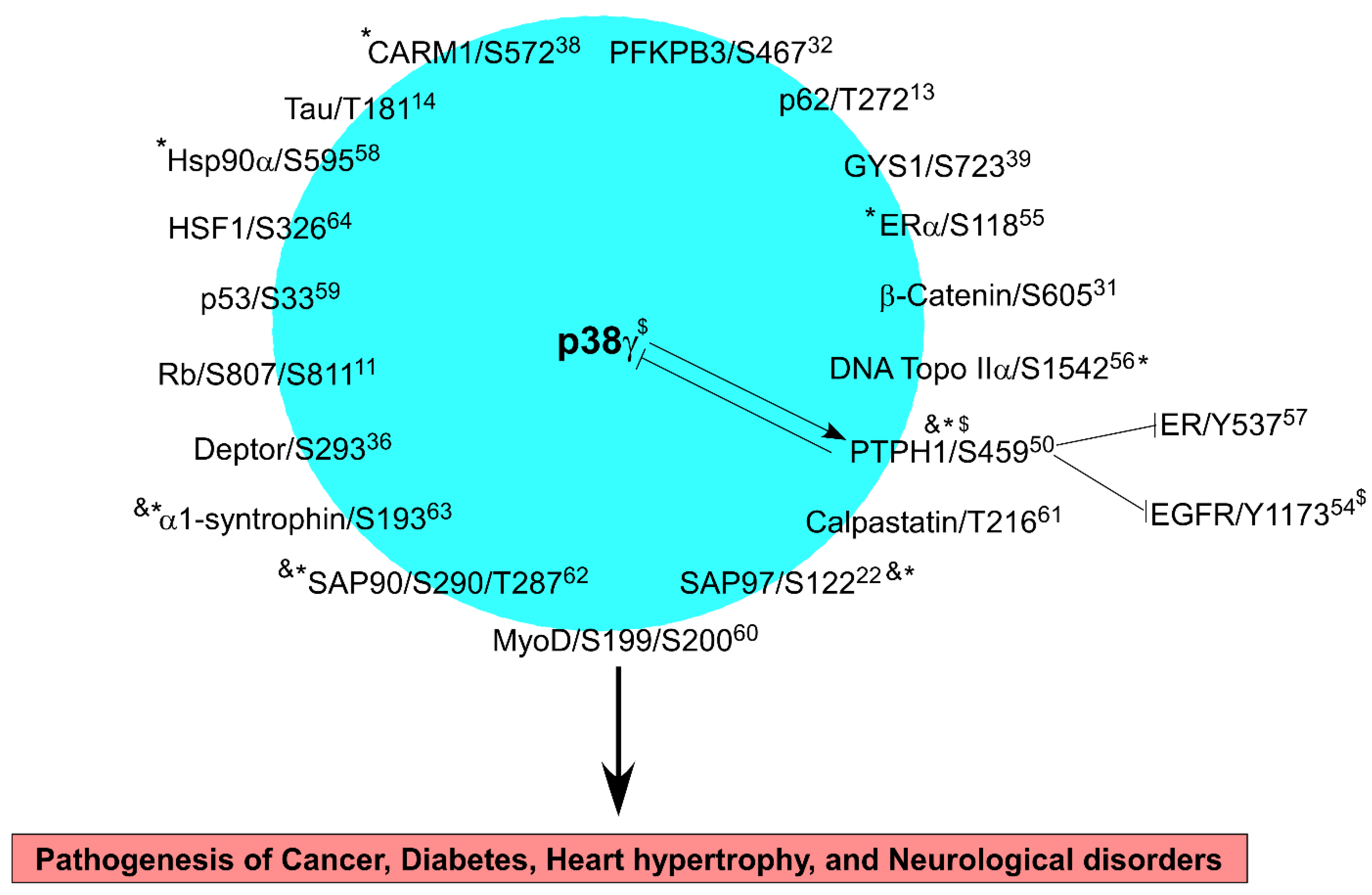
p38γ MAPK (also called ERK6 or SAPK3) is a family member of stress-activated MAPKs and has common and specific roles as compared to other p38 proteins in signal transduction. In addition to inflammation, p38γ metabolic signaling is involved in physiological exercise and in pathogenesis of cancer, diabetes, and Alzheimer’s disease, indicating its potential as a therapeutic target. p38γphosphorylates at least 19 substrates through which p38γ activity is further modified to regulate life-important cellular processes such as proliferation, differentiation, cell death, and transformation, thereby impacting biological outcomes of p38γ-driven pathogenesis. P38γ signaling is characterized by its unique reciprocal regulation with its specific phosphatase PTPH1 and by its direct binding to promoter DNAs, leading to transcriptional activation of targets including cancer-like stem cell drivers.

  • p38γ
  • signal transduction
  • cancer
  • diseases

1. Introduction

p38γ mitogen-activated protein kinase (MAPK) is an isoform of p38 family proteins (p38α, β, γ, and δ) that are encoded by four different genes in different chromosomes. p38α (Gene name: MAPK14) and p38β (MAPK11) are expressed ubiquitously, whereas p38γ (MAPK12) and p38δ (MAPK13) are expressed in certain tissues (for example, p38γ in skeletal muscle; and p38δ in the salivary, pituitary, and adrenal glands) [1]. All p38s contain a conserved Thr–Gly–Tyr (TGY) dual-phosphorylation motif within the kinase activation loop, and both Thr and Tyr phosphorylation are necessary to fully activate the kinase. p38 MAPKs are phosphorylated and activated by MAPK kinase kinase 3 (MKK3) and/or MKK6, which in turn phosphorylate a substrate containing an ST/P motif [1]. While p38α and p38β can directly phosphorylate more than 100 substrates [2], p38γ and p38δ have specific and non-overlapping substrates [3,4][3][4]. In response to stress and inflammatory stimuli, p38α is most frequently activated, while other p38 family members are phosphorylated by a mechanism depending on cell type and/or stimuli. Although these isoform-specific activations are important for different cellular outcomes, the mechanisms involved are largely unknown [2,5,6][2][5][6]. Isoform-specific and tissue-dependent effects of p38 MAPKs in inflammation and inflammation-associated oncogenesis have been recently reviewed [7]. This mreseanuscriptrch will focus on recent discoveries regarding p38γ inflammatory and metabolic signaling in physiology and diseases.
p38γ was cloned in 1996 [8,9,10][8][9][10] and is about 60% homologous to p38α. A three-dimensional (3D) search of active CDK1 and CDK2 revealed a higher degree of structural similarity with p38γ than with other stress kinases [11]. P38γ is unique among MAPKs due to its C-terminal sequence, called the PDZ motif, which can bind to PDZ domain proteins, which may be the determinant for its distinct activity as compared to its close family member p38δ. This allows p38γ to form PDZ-dependent protein complexes essential for its specific activities.

2. p38γ Signaling to Its Substrates in Physiology and Diseases

p38γ is a serine/threonine kinase and both its expression and phosphorylation are important for its inflammatory, metabolic, and oncogenic signaling (Figure 1). In response to the KRAS oncogene, p38γ is induced in rat intestinal epithelial cells (IEC-6) but not in mouse NIH3T3 fibroblasts [44,45][12][13]. In pancreatic epithelial HPNE cells, however, KRAS transformation increases both p38γ protein expression and phosphorylation. These results suggest that the KRAS oncogene activates p38γ through increased expression and elevated phosphorylation by a mechanism depending on cell types and tissue origin. Because p38γ phosphorylation is decreased by transient KRAS co-transfection in IEC-6 cells [44][12] and p38γ is the only MAPK that contains a PDZ-domain binding motif at its C terminus [46][14], wresearchers sought to search for its specific phosphatase using a PDZ-based two-hybrid screening. WeResearchers found that wild-type p38γ, but not its PDZ-deleted mutant, interacts with a PDZ-domain containing protein tyrosine phosphatase H1 (PTPH1), which decreases p38γ phosphorylation in vitro and in vivo [47][15]. Through PDZ binding, p38γ was previously shown to phosphorylate SAP97/S122 [22][16], which is implicated in ethanol-activated and p38γ-dependent stimulation of the cancer-like stem cell (CSC) population and breast cancer growth [48][17]. Of interest, PDZ binding is required for p38γ [44,47,49][12][15][18] and PTPH1 [50,51,52][19][20][21] oncogenic activity [53][22], as expression of their PDZ-binding-deficient mutants by stable transfection or application of peptide to disrupt the endogenous p38γ/PTPH1 interaction inhibits cancer cell growth [47][15]. The same PDZ binding also enables p38γ phosphorylation of PTPH1 at S459 in vitro and in vivo, whereas p38α lacks this activity, which is required for PTPH1 phosphatase and oncogenic activity [50][19]. These results indicate a critical role of the PDZ complex in oncogenesis, which requires phosphorylation/de-phosphorylation of p38γ and PTPH1. Consistent with this speculation, after tetracycline-inducible KRAS expression (tet-on system), upregulated p38γ is dominant and persistent, whereas the resultant p-PTPH1/S459 is delayed and transient [54][23]. This leads to increased EGFR protein expression (due to p38γ) and enhanced EGFR de-phosphorylation (due to PTPH1), resulting in a phenotype of increased levels of un-phosphorylated EGFR proteins in KRAS-mutant colon cancer cells [54][23]. Because KRAS induces the protein expression of p38γ and PTPH1, this reciprocal p38γ dephosphorylation and PTPH1 phosphorylation may be not only important for transformation but for certain phenotypes of KRAS-dependent tumors such as those resistant to EGFR inhibitors [12][24].
Figure 1. p38γ signaling network in diseases. The number after each protein indicates the reference number in which the phosphorylation was demonstrated. The reciprocal effects of p38γ and PTPH1 were illustrated with PTPH1 substrates (EGFR and ER) also listed. p38γ may cooperate with one or more substrates to impact pathogenesis of a disease (highlighted inside the box below) and one phosphorylation event may be involved in several diseases. * indicates a specific phosphorylation by p38γ and not by its family member p38α, whereas & shows a PDZ-dependent reaction. $ indicates that EGFR depends on p38γ PDZ motif and phosphorylation to form a complex with both p38γ and PTPH1.
In cancer cells, p38γ phosphorylates additional substrates. In breast cancer cells p38γ phosphorylates estrogen receptor α (ER) at S118 and inhibits its proteasome-dependent degradation [55][25]. This phosphorylation event at ER/S118 enables p38γ to cooperate with c-Jun in binding the cyclin D1 promoter, leading to increased cyclin D1 expression and decreased breast cancer sensitivity to anti-estrogen Tamoxifen (TAM) [55][25]. Additionally, p38γ phosphorylates DNA Topo IIα/S1542 in vitro and in vivo, thereby increasing Topo II stability and catalytic activity as well as breast cancer cell sensitivity to Topo II inhibitors [56][26]. In addition, p38γ-phosphorylated protein phosphatase PTPH1 can dephosphorylate ER at Y539 and increase nuclear ER accumulation and enhance breast cancer sensitivity to anti-estrogens [57][27]. Moreover, p38γ can cooperate with c-Jun to stimulate epidermal growth factor receptor (EGFR) transcription [54][23]. PTPH1 can also dephosphorylate EGFR in breast cancer cells and disrupt EGFR interaction with ER in the cytoplasmic membrane and thereby sensitize breast cancer cells to EGFR inhibitor lapatinib [52][21]. Thus, p38γ can directly phosphorylate substrates in cancer cells to impact cellular outcomes and indirectly regulate cancer cell growth via phosphorylating its substrate PTPH1 that is required for de-phosphorylation of proliferative proteins (Figure 1).
Several studies showed that p38γ can phosphorylate several oncogenic proteins in cancer cells and/or tissues, and that is implicated in tumor-growth in mice. Proteomic screening identified that p38γ specifically interacts with heat shock protein 90 α (HSP90α) in KRAS-mutated but not KRAS wild-type colon cancer cells, and phosphorylates HSP90α at S595, thus preventing mutant KRAS protein from degradation and conferring sensitivity of KRAS mutant colon cancer cells to HSP90 inhibitor 17-alllylaminogeldanamycin (17-AAG) [58][28]. Application of the p38γ inhibitor pirfenidone (PFD) blocked p38γ-induced HSP90/S595 phosphorylation, promoted mutant KRAS degradation, and inhibited colon cancer xenograft growth in nude mice [58][28]. β-catenin is a key member of the Wnt pathway and studies by Yin et al. found that intestinal epithelial cell (IEC)-specific knockout of p38γ suppresses the β-catenin/Wnt pathway [31][29]. Mechanistic analysis further showed that p38γ binds and phosphorylates β-catenin at S605, which is important for β-catenin stability, and that PFD requires epithelial p38γ to inhibit phosphorylation of several oncogenic substrates including PTPH1, Hsp90, and β-catenin to inhibit colon tumorigenesis [31][29]. These results indicate that p38γ may cooperate with several substrates to promote colon cancer development and growth through phosphorylation (Figure 1). p38γ-induced SAP97/S122 may be also involved in mammary tumor development and growth in vitro [22,48][16][17]. In pancreatic cancer, p38γ binds phosphofructokinase-2/fructose-2,6-bisphosphatase 3 (PFKFB3) in KRAS-transformed cells and phosphorylates PFKFB3 at S467 in human pancreatic cancer cells and in mouse KPC tumors [32][30]. Moreover, p-PFKFB3 protein levels were only completely depleted in p38γ-expressing KPC tumor cells by a combination of the p38γ inhibitor PFD with the PFKFB3 inhibitor PFK15, and expression of the phosphorylation mutant PFKFB3/S467A in KPC tumors decreased the tumor growth in nude mice, as compared to the tumor expressing wild-type PFKFB3. These results demonstrate that epithelial p38γ may be a systemic target for therapeutic intervention through phosphorylating its substrates.
Studies further showed that p38γ can phosphorylate tumor suppressors and thereby regulate tumorigenesis. p38γ phosphorylates the retinoblastoma (Rb) at S807/S811 through which p38γ compensates for the loss of CDK1 or CDK2, and both p38γ conditional knockout (KO) and the application of PFD significantly prevent carcinogen-induced liver tumor tumorigenesis [11]. Moreover, p38γ, as well as p38α, can phosphorylate p53/S33 and mediate RAS oncogene-induced senescence, but only p38γ, but not p38α, is essential for oncogenic Ras-induced transcriptional activity of p53 [59][31]. Whether p38γ phosphorylating p53 impacts transformation and tumor growth in murine models has not been demonstrated.
p38γ also cooperates with its substrates to impact other pathological processes. In a study of neurological disorders, p38γ was found to phosphorylate tau protein at T181, which together with extracellular amyloid-β (AB), orchestrates neuronal dysfunction in Alzheimer’s disease, whereas a site-specific tau phosphorylation disrupts the PSD-95/tau/Fyn interaction and inhibits Aβ toxicity [14][32]. In a study of muscle stem cell commitment, p38γ phosphorylates coactivator-associated arginine methyltransferase 1 Carm1 at S572 to prevent its nuclear translocation, which functions to oppose the activity of p38α and stimulates self-renewal rather than differentiation [38][33]. In a study of heart hypertrophy, however, p38γ and p38δ phosphorylate mTOR complex 1 (mTORC1) and mTOR complex 2 (mTORC2) inhibitor DEPTOR at S145, S244, and S265, promoting its downregulation [36][34]. Furthermore, p38γ phosphorylates MyoD at S199/S200, thereby enhancing MyoD occupancy on the Myogenin promoter to form a repressive transcriptional complex [60][35].
Further, p38γ can phosphorylate insulin receptor substrate p62 at T269/S272, which may contribute to type 2 diabetes [13][36]. During heart development, p38γ and p38δ contribute to the cardiac metabolic switch through inhibitory phosphorylation of glycogen synthase 1 (GYS1) at S723, S727, and T278, leading to glycogen metabolism inactivation [39][37]. Calpastation is also phosphorylated by p38γ at T216/S219, resulting in its reduced activity to inhibit the protease calpain in ventricular remodeling [61][38]. SAP90 is phosphorylated by p38γ and ERK2 at T287 and S290, which may be important for their co-localization in neurons [62][39]. In addition, p38γ phosphorylates α1-syntrophin at S193/S201 though PDZ binding to regulate its localization and substrate specificity [63][40]. While several p38 MAPKs can regulate heat shock factor 1 (HSF1), p38γ is the principal isoform responsible for its phosphorylation at S326 in cells, which affects the extent and duration of the heat shock response [64][41]. These results together indicate that p38γ may cooperate with its specific and common substrates and other p38s and/or MAPKs to form a signaling network through phosphorylation to regulate oncogenesis, diabetes, heart hypertrophy, and neurological disorders (Figure 1).

References

  1. Ono, K.; Han, J. The p38 signal transduction pathway. Activation and function. Cell. Sign. 2000, 12, 1–13.
  2. Canovas, B.; Nebreda, A.R. Diversity and versatility of p38 kinase signalling in health and disease. Nat. Rev. Mol. Cell Biol. 2021, 22, 1–21.
  3. Qi, X.M.; Wang, F.; Chen, G. p38 Gamma MAPK Encyclopedia of Signaling Molecules, 2nd ed.; Choi, S., Ed.; Springer: Berlin/Heidelberg, Germany, 2018; pp. 3718–3727.
  4. Cuenda, A.; Sanz-Ezquerro, J. p38g and p38d: From spectators to key physiological players. Trends Biochem. Sci. 2017, 42, 431–442.
  5. Pramanik, R.; Qi, X.; Borowicz, S.; Choubey, D.; Schultz, R.M.; Han, J.; Chen, G. p38 isoforms have opposite effects on AP-1-dependent transcription through regulation of c-Jun: The determinant role of the isoforms in the p38 MAPK signal specificity. J. Biol. Chem. 2003, 278, 4831–4839.
  6. Loesch, M.; Chen, G. The p38 MAPK stress pathway as a tumor suppressor or more? Front. Biosci. 2008, 13, 3581–3593.
  7. Qin, J.; Xin, H.; Qi, X.; Chen, G. Isoform-specific and cell/tissue-dependent effects of p38 MAPKs in regulating inflammation and inflammation-associated oncogenesis. Front. Biosci. Landmark 2022, 27, 031.
  8. Li, Z.; Jiang, Y.; Ulevitch, R.J.; Han, J. The primary structure of p38g: A new member of p38 group of MAP kinases. Biochem. Biophys. Res. Commun. 1996, 228, 334–340.
  9. Lechner, C.; Zahalka, M.A.; Giot, J.; Moller, N.P.; Ullrich, A. ERK6, a mitogen-activated protein kinase involved in C2C12 myoblast differentiation. Proc. Natl. Acad. Sci. USA 1996, 93, 4355–4359.
  10. Mertens, S.; Craxton, M.; Goedert, M. SAP kinase-3, a new member of the family of mammalian stress-activated protein kinases. FEBS Lett. 1996, 383, 273–276.
  11. Tomas-Loba, A.; Manieri, E.; Gonzalez-Teran, B.; Mora, A.; Leiva-Vega, L.; Santamans, A.M. p38g is essential for cell cycle progression and liver tumorigenesis. Nature 2019, 568, 557–560.
  12. Tang, J.; Qi, X.; Mercola, D.; Han, J.; Chen, G. Essential role of p38g in K-Ras transformation independent of phosphorylation. J. Biol. Chem. 2005, 280, 23910–23917.
  13. Pohl, N.M.; Qi, X.; Loesch, M.; Tang, J.; Li, Q.; Chen, G. Tissue-specific roles of p38g MAPK in Ras transformation. AACR Proc. 2006, 47, 2534.
  14. Wagner, E.F.; Nebreda, A.R. Signal integration by JNK and p38 MAPK pathways in cancer development. Nat. Rev. Cancer 2009, 9, 537–549.
  15. Hou, S.W.; Zhi, H.Y.; Pohl, N.; Loesch, M.; Qi, X.M.; Li, R.S. PTPH1 Dephosphorylates and Cooperates with p38γ MAPK to Increase Ras Oncogenesis through PDZ-Mediated Interaction. Cancer Res. 2010, 70, 2910.
  16. Sabio, G.; Simon, J.; Arthur, C.; Kuma, Y.; Peggie, M.; Carr, J.; Murray-Tait, V.; Centeno, F.; Goebeler, M.; Morrice, N.; et al. p38g regulates the localisation of SAP97 in the cytoskeleton by modulating its interaction with GKAP. EMBO J. 2005, 24, 1134–1145.
  17. Xu, M.; Ren, Z.; Wang, X.; Comer, A.; Frank, J.A.; Ke, Z. ErB2 and p38g MAPK mediate alcohol-induced increase in breast cancer stem cells. Mol. Cancer 2016, 15, 52.
  18. Hou, S.; Lepp, A.; Chen, G. p38 gamma MAP kinase. UCSD-Nat. Mol. Pages 2010.
  19. Hou, S.W.; Suresh, P.S.; Qi, X.; Lepp, A.; Mirza, S.; Chen, G. p38g Mitogen-activated Protein Kinase Signals through Phosphorylating Its Phosphatase PTPH1 in Regulating Ras Protein Oncogenesis and Stress Response. J. Biol. Chem. 2012, 287, 27895–27905.
  20. Zhi, H.; Hou, S.W.; Li, R.; Basir, Z.; Xiang, A.; Szabo, A. PTPH1 cooperates with vitamin D receptor to stimulate breast cancer growth through their mutual stabilization. Oncogene 2011, 30, 1706–1715.
  21. Ma, S.; Yin, N.; Qi, X.; Pfister, S.L.; Zhang, M.; Ma, R.; Chen, G. Tyrosine dephosphorylation enhances the therapeutic target activity of epidermal growth factor receptor (EGFR) by disrupting its interaction with estrogen receptor (ER). Oncotarget 2015, 6, 13320–13333.
  22. Chen, K.; Lin, S.; Wu, M.; Ho, M.; Santhanam, A.; Chou, C.; Meng, T.; Wang, A. Reciprocal allosteric regulation of p38g and PTPN3 involves a PDZ domain-modulated complex formation. Sci. Signal. 2014, 7, ra98.
  23. Yin, N.; Lepp, A.; Ji, Y.; Mortensen, M.; Hou, S.; Qi, X. The K-Ras effector p38g MAPK confers intrinsic resistance to tyrosine kinase inhibitors by stimulating EGFR transcription and EGFR dephosphorylation. J. Biol. Chem. 2017, 292, 15070–15079.
  24. Qi, X.; Wang, F.; Mortensen, M.; Wertz, R.; Chen, G. Targeting an oncogenic kinase/phosphatase signaling network for cancer therapy. Acta Pharm. Sin. B 2018, 8, 511–517.
  25. Qi, X.; Zhi, H.; Lepp, A.; Wang, P.; Huang, J.; Basir, Z. p38g mitogen-activated protein kinase (MAPK) confers breast cancer hormone sensitivity by switching estrogen receptor (ER) signaling from classical to nonclassical pathway via stimulating ER phosphorylation and c-Jun transcription. J. Biol. Chem. 2012, 287, 14681–14691.
  26. Qi, X.; Hou, S.; Lepp, A.; Li, R.; Basir, Z.; Lou, Z.; Chen, G. Phosphorylation and stabilization of topoisomerase IIa protein by p38g mitogen-activated protein kinase sensitize breast cancer cells to its poisons. J. Biol. Chem. 2011, 286, 35883–35890.
  27. Suresh, P.S.; Ma, S.; Migliaccio, A.; Chen, G. Protein-tyrosine phosphatase H1 increases breast cancer sensitivity to antiestrogens by dephosphorylating estrogen receptor at tyr537. Mol. Cancer Ther. 2014, 13, 230–238.
  28. Qi, X.M.; Xie, C.; Hou, S.; Li, G.; Yin, N.; Dong, L.; Lepp, A.; Chesnik, M.A.; Mirza, S.P.; Szabo, A.; et al. Identification of a ternary protein-complex as a therapeutic target for K-Ras-dependent colon cancer. Oncotarget 2014, 5, 4269–4282.
  29. Yin, N.; Qi, X.; Tsai, S.; Lu, Y.; Basir, Z.; Oshima, K. p38g MAPK is required for inflammation-associated colon tumorigenesis. Oncogene 2016, 35, 1039–1048.
  30. Wang, F.; Qi, X.; Wertz, R.; Mortensen, M.; Hagen, C.; Evans, J. p38g MAPK is essential for aerobic glycolysis and pancreatic tumorigenesis. Cancer Res. 2020, 80, 3251–3264.
  31. Kwong, J.; Hong, L.; Liao, R.; Deng, Q.; Han, J.; Sun, P. p38a and p38g mediates oncogenic ras-induced senescence through different mechanisms. J. Biol. Chem. 2009, 284, 11237–11246.
  32. Ittner, A.; Chua, S.W.; Bertz, J.; Volkerling, A.; van der Hoven, J.; Gladbach, A. Site-specific phosphorylation of tau inhibits amyloid-b toxicity in alzheimer’s mice. Science 2016, 354, 904–908.
  33. Chang, N.; Sincennes, M.; Chevalier, F.P.; Brun, C.E.; Lacaria, M.; Segales, J. The dystrophin glycoprotein complex regulates the epigenetic activation of muscle stem cell commitment. Cell Metab. 2019, 22, 755–768.
  34. Gonzalez-Teran, B.; Lopez, J.A.; Rodriguez, E.; Leiva, L.; Martinez-Martinez, S.; Bernal, J.A. p38g and d promote heart hypertrophy by targeting the mTOR-inhibitory protein DEPTOR for degradation. Nat. Commun. 2016, 7, 10477.
  35. Gillespie, M.A.; Grand, F.L.; Scime, A.; Kuang, S.; von Maltzahn, J.; Seale, V.; Cuenda, A.; Ranish, J.A.; Rudnicki, M.A. p38g-dependent gene silencing restricts entry into the myogenic differentiation program. J. Cell Biol. 2009, 187, 991–1005.
  36. Koh, A.; Molinaro, A.; Stahlman, M.; Khan, M.T.; Schmidt, C.; Manneras-Holm, L. Microbially produced imidazole propionate impairs insulin signaling through mTORC1. Cell 2018, 175, 947–961.
  37. Santamans, A.M.; Montalvo-Romeral, V.; Mora, A.; Lopez, J.A.; Gonzalez-Romero, F.; Jimenez-Blasco, D. p38γ and p38δ regulate postnatal cardiac metabolism through glycogen synthase 1. PLoS Biol. 2021, 19, e3001447.
  38. Loonat, A.A.; Martin, E.D.; Sarafraz-Shekary, N.; Tilgner, K.; Hertz, N.T.; Levin, R. p38g MAPK contributes to left ventricular remodeling after pathologic stress and disinhibits calpain through phosphorylation of calpastatin. FASEB J. 2019, 33, 13131–13144.
  39. Sabio, G.; Reuver, S.; Feijoo, C.; Hasegawa, M.; Thomas, G.M.; Centeno, F.; Kuhlendahl, S.; Leal-Ortiz, S.; Goedert, M.; Garner, C.; et al. Stress- and mitogen-induced phosphorylation of the synapse-associated protein SAP90/PSD-95 by activation of SAPK3/p38g and ERK1/ERK2. Biochem. J. 2004, 380, 19–30.
  40. Hasegawa, M.; Cuenda, A.; Spillantini, M.G.; Thomas, G.M.; Buee-Scherrer, V.; Cohen, P.; Goedert, M. Stress-activated protein kinase-3 interacts with the PDZ domain of a1-syntrophin: A mechanism for specific substrate recognition. J. Biol. Chem. 1999, 274, 12626–12631.
  41. Naidu, S.D.; Sutherland, C.; Zhang, Y.; Risco, A.; Vega, L.D.L.; Caunt, C.J. Heat shock factor 1 is a substrate for p38 mitogen-activated protein kinases. Mol. Cell. Biol. 2016, 36, 2403–2417.
More
Video Production Service