Your browser does not fully support modern features. Please upgrade for a smoother experience.
Please note this is a comparison between Version 1 by Mara Schvarzstein and Version 2 by Wendy Huang.
Whole genome duplication (WGD) or polyploidization can occur at the cellular, tissue, and organismal levels. At the cellular level, tetraploidization has been proposed as a driver of aneuploidy and genome instability and correlates strongly with cancer progression, metastasis, and the development of drug resistance. WGD is also a key developmental strategy for regulating cell size, metabolism, and cellular function. In specific tissues, WGD is involved in normal development (e.g., organogenesis), tissue homeostasis, wound healing, and regeneration. At the organismal level, WGD propels evolutionary processes such as adaptation, speciation, and crop domestication.
- whole genome duplication
- polyploidization
- polyploidy
- aneuploidy
- adaptation
1. Introduction
Whole genome duplication (WGD) or polyploidization is vital for normal development [1], tissue homeostasis [2][3][2,3], and regeneration [4] (Figure 1). It is also a driver of evolutionary processes [5] including adaptation [6], speciation [7], and crop domestication [8]. In addition, WGD can drive pathological conditions, especially oncogenesis [1][9][10][1,9,10]. In nature, WGD exists at the cellular, tissue, organ, and organismal levels. Polyploid cells are normally found in most multicellular diploid organisms [11][12][11,12]. In humans, certain cells from the placenta [13], mammary glands [14], liver [15], heart [16], skin [17], and bone marrow must become polyploid to perform specialized functions [18], for tissue homeostasis [3], or as part of the wound healing process [19]. For instance, during development, megakaryocytes acquire 16 copies of the genome and thus become giant cells that are hypermetabolic. This polyploidization is a basic requirement to produce platelets in the blood, as platelets are produced by fragmenting the cytoplasm of megakaryocytes [20][21][22][20,21,22]. In addition, WGD is involved in oncogenesis, metastasis, and the development of resistance to chemotherapeutic drugs, and polyploid cells may accumulate in patients with neurodegenerative disorders, cardiovascular disease, and diabetes [23][24][25][26][23,24,25,26].
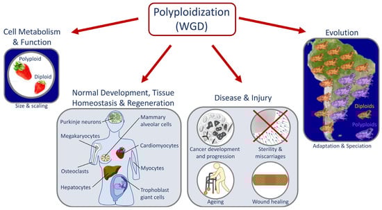
Figure 1. Roles of whole genome duplication (WGD). Diagram depicting key examples of processes affected by polyploidization/WGD. Polyploidy can occur at the cellular, tissue, or organismal levels. It is a key step in important biological processes affecting cellular metabolism and function, normal development, tissue homeostasis, regeneration, wound healing, organismal adaptation to the environment, and speciation. In addition, polyploidization can cause and/or prevent disease and repair injuries.
Polyploid cells can arise by cell fusion resulting in larger multinucleate cells, by endoduplication (alternating cycles between G and S phases without undergoing mitosis or cytokinesis) resulting in mononucleated cells, or by endomitosis (the cell enters mitosis but does not complete cell division) to generate either mononucleated giant cells or multinucleated cells [27]. In some cyanobacteria, polyploidy is established and maintained by misregulation of the replisome machinery [28][29][30][28,29,30].
2. Causes and Downstream Effects of WGD
The outcomes of WGD are diverse in nature—ranging from changes in gene expression to altered cell size and scaling and to adaptation to stress [31][32][31,32] (Figure 2). The effects of polyploidization are surprisingly similar in different biological contexts (e.g., in cells, tissues, and organisms) [4][11][33][34][4,11,33,34] and regardless of the biological process (e.g., during development, evolution, regeneration, and oncogenesis) [1][3][35][36][37][38][39][40][41][1,3,35,36,37,38,39,40,41] where WGD takes place (Figure 2). A common feature of WGD is that it is induced by stress, and it provides the changes that result in plasticity and thus enhances adaptation to stress in normal and pathological conditions. For instance, environmental stresses induce WGD in tissues (e.g., wounding or viral infection) [42][43][44][42,43,44] or organisms (e.g., large changes in temperature or water availability and salinity) [45][46][47][48][49][50][45,46,47,48,49,50]. Furthermore, stress-induced WGD is a platform for genomic diversity and versatility that provides the means for rapid adaptability (Figure 1 and Figure 2). Whereas in whole organisms, this can elicit a competitive advantage in a changing environment [32][51][32,51]; in cancer, it elicits an adaptive advantage that promotes oncogenesis, metastasis, and the development of drug resistance [32][51][32,51]. The latter is similar to how the pathogenic yeast Candida abdicans becomes resistant to antifungal drugs [6]. Therefore, it is widely expected that queries about mechanisms that give rise to WGD and its effects in both sub-organismal and organismal systems will likely reveal additional commonalities across systems and processes.
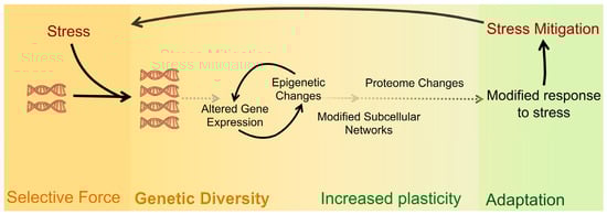
Figure 2. Shared causes and downstream effects of WGD across biological settings. Many forms of stress can lead an organism to undergo WGD. Whether as a response to the stress itself or as a way to adapt to the changing environment and/or different selective pressures, gene and epigenome changes lead to an altered proteome that supports an adaptive response to the initial stress exposure.
Polyploidy is often confused with aneuploidy, in part because, in humans, both phenomena are hallmarks of cancer [1][39][40][52][1,39,40,52]. Observations from numerous studies identify WGD in about 30% of human cancers [53][54][53,54] and as a precursor for many malignancies in cancer evolution [39][55][56][57][58][59][39,55,56,57,58,59]. Although WGD does not seem to require a preexisting cancer driver mutation; it most often emerges after oncogenic mutations, such as lesions in the TP53 genes [53][56][53,56]. WGD promotes diversification of copy number alterations (CNAs) and is permissive for other chromosomal aberrations and genome instability associated with poor cancer patient prognosis [39][58][59][39,58,59]. Interestingly, a recent study revealed a mechanism by which WGD enhances cancer-promoting alterations in DNA organization and gene expression [58]. Induction of WGD can reduce the levels of proteins involved in chromatin packaging, resulting in reorganization of the 3D DNA compartments and domains. Over time, loss of this domain structure predisposes the cell to additional cancer-promoting alterations and expression of oncogenic genes [58]. Polyploidization can also be protective, as in the liver where it both provides the basis for genetic adaptation to hepatotoxic stress and also protects from malignant transformations [21][60][21,60].
Organismal WGD is common in extant plants [7][32][61][62][63][7,32,61,62,63] and is also observed in protists [64], fungi [51][65][66][51,65,66], bacteria, and archaebacteria [67][68][67,68] and in several animal clades [2][12][35][36][40][41][69][70][2,12,35,36,40,41,69,70]. In both prokaryotes and eukaryotes, the effects of WGD are comparable with increased resistance to UV irradiation, cell size, adaptability to environmental changes, and evolvability [28][71][28,71]. Evidence for ancestral WGD events is found in the genomes of organisms of most clades, including vertebrates [36][72][73][36,72,73]. Polyploid metazoans arise due to errors during meiosis that result in the formation of diploid gametes that when fertilized give rise to polyploid organisms. Fertilization between diploid (2n) and haploid (n) gametes results in a triploid (3n) organism; fertilization between two diploid (2n) gametes results in a tetraploid (4n) [33][35][33,35]. When the diploid gametes of two related species fuse, the resulting organism is said to be an allopolyploid, whereas when diploid gametes from the same species fuse the result is said to be an autopolyploid [73]. Polyploidization from the cell to the organismal level most often results in lethality [32][74][32,74]. For instance, human triploid and tetraploid embryos die during gestation or soon after delivery, but triploid and tetraploid mosaic children may survive after birth [75][76][75,76]. This lethality may be caused by errors partitioning the additional chromosomes during cell division, by WGD-driven increases in cell size that interfere with tissues or organ scaling, and by epigenetic changes and gene expression changes that affect dose-dependent processes and complexes [73]. The latter may be especially true in human triploids and tetraploids which may have difficulty equalizing expression between the silenced X chromosomes and the extra sets of autosomes since variable numbers of Barr bodies may be found in these cells [75]. A newly formed polyploid may very occasionally overcome these obstacles, e.g., by establishing balanced genome expression. Then, if polyploidy provides a competitive advantage—as in a changing environment—polyploid lineages may outgrow their diploid counterparts and become established [32][41][73][32,41,73]. The established polyploid population will continue to evolve and then may “diploidize” to become a new species that has an adaptive advantage compared to the parental diploid species [32].
3. Laboratory Multicellular Organism Models for WGD
A major impediment to studying WGD and its impacts on the physiology and function of cells, tissues, and organs is the scarcity of laboratory models that permit comparison between isogenic organisms with differing ploidy [33]. Many laboratory models are sterile or embryonic lethal when polyploid [77][78][79][77,78,79]. Generation of autopolyploid laboratory model organisms that are not sterile (e.g., plants) frequently involves the use of chemicals such as colchicine, known to cause aneuploidy and genetic mutations. These polyploids, therefore, may not be truly isogenic with the parental diploid strains they were derived from [51][73][51,73]. The recently developed method for generating tetraploids in Caenorhabditis utilizes transient knock-down of a meiosis-specific cohesin (i.e., rec-8) by RNA interference to produce diploid gametes [80]. Therefore, the derived tetraploids are unlikely to differ significantly from the diploid strains from which they were derived (see Figure 3A for an overview of the method) [80].
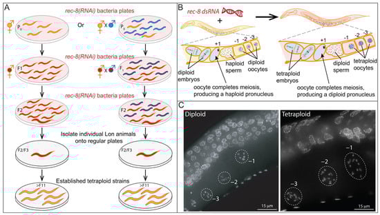
Figure 3. Method for rapid isolation of C. elegans tetraploids. (A). Diagram of the published method for generating tetraploid strains from diploids. This protocol relies on the transient knockdown of the meiosis-specific cohesin rec-8 which results in the production of diploid instead of haploid gametes. Thus, upon fertilization, tetraploid progeny (which are longer and bigger) are produced. The method works both during the selfing of the hermaphrodites (left) and during outcrossing (right). Crossing allows for the generation of tetraploids with two different genetic backgrounds. After two to three generations of exposure to rec-8 dsRNA, the larger hermaphrodites are individually plated on normal growth media. Strains that consistently produce large (Lon) hermaphrodites are then screened to confirm that these strains are tetraploids. (B). Diploid hermaphrodites (left) produce haploid gametes. Haploid sperm are stored in the spermatheca. Oocytes prior to the meiotic divisions have six pairs of connected homologous chromosomes that can be visualized by DAPI in the diakinesis-stage oocytes (Figure 3C, below) just adjacent to the spermatheca (−1, −2, and −3 oocytes based on position). The oocyte moves through the spermatheca and is fertilized and completes the meiotic divisions to produce a haploid pronucleus. The tetraploid worms derived from exposure to rec-8 dsRNA, (right) produce diploid gametes with 12 DAPI bodies. (C) Examples of fixed hermaphrodites stained with DAPI to screen for ploidy by counting chromosome pairs in the −1, −2, and −3 diakinesis oocytes.