Bimetallic Au–Ag Nanoparticles: Comparison
Please note this is a comparison between Version 1 by Muthu M.S. and Version 2 by Peter Tang.

Bimetallic nanoparticles (NPs) with two separate metals have been found to have stronger antibacterial potential than their monometallic versions. This enhanced antibacterial efficiency of bimetallic nanoparticles is due to the synergistic effect of their participating monometallic counterparts. To distinguish between bacteria and mammals, the existence of diverse metal transport systems and metalloproteins is necessary for the use of bimetallic Au–Ag NPs, just like any other metal NPs. Due to their very low toxicity toward human cells, these bimetallic NPs, particularly gold–silver NPs, might prove to be an effective weapon in the arsenal to beat emerging drug-resistant bacteria. The cellular mechanism of bimetallic nanoparticles for antibacterial activity consists of cell membrane degradation, disturbance in homeostasis, oxidative stress, and the production of reactive oxygen species. The synthesis of bimetallic nanoparticles can be performed by a bottom-up and top-down strategy. The bottom-up technique generally includes sol-gel, chemical vapor deposition, green synthesis, and co-precipitation methods, whereas the top-down technique includes the laser ablation method.

  • antibacterial
  • bimetallic
  • gold–silver
  • multidrug resistance
  • nanoparticles
  • wound healing

1. Introduction

Infections caused by microbes have turned out to be a significant source of morbidity and death worldwide [1]. The World Health Organization (WHO) reported that antimicrobial resistance (AMR) is among the top 10 health threats confronting humanity in 2021. According to the most thorough report published in The Lancet, more than 1.2 million people have died due to antibiotic-resistant bacterial infections [2]. If current trends continue, it is estimated that over 10 million people will die each year from drug-resistant diseases by 2050. The misuse and abuse of antimicrobials has mostly caused the development of drug-resistant bacteria. Microbial drug-resistant based infections are a worldwide issue that requires immediate action from health authorities and policymakers to avert avoidable deaths. Nanoparticles (NPs) are commonly employed as an alternative to antibiotics to target microorganisms. Nanomaterials exhibit broad-spectrum antibacterial properties. Noble metal NPs (Cu, Ag, and Au) have been used in food preservatives, medical device coatings, and medical equipment because they have been demonstrated to have powerful and long-lasting antibacterial effects against a wide range of microbes [3]. Recently, because of their unique physical and chemical characteristics, bimetal nanoparticles have been proposed as a viable antibiotic replacement. Bimetallic gold–silver nanoparticles have opened a new field of research due to their broad-spectrum antibacterial capabilities. Because of the synergy between the metals that make up their composition, they have outstanding characteristics and a wide range of uses in the biomedical field [4][5][4,5]. Silver nanoparticles have shown the highest surface plasmon resonance (SPR), whereas gold nanoparticles, on the other hand, have great chemical stability. Bimetallic gold–silver nanoparticles demonstrated SPR bands that can be exploited in the phototherapy of cancer and catalytic biosensors [6][7][8][6,7,8]. Recently, AlZaban et al., synthesized bimetallic gold–silver (Au–Ag) core–shell NPs for the catalysis of the trans-esterification process in a fungal isolate (Fusarium solani) and to enhance biodiesel production [9]. In another study, Renones et al., investigated the reducing property of Au–Ag NPs loaded with TiO2, that photocatalyzed the conversion of CO2 with water [10]. Additionally, Aazam et al., employed bimetallic Au–Ag NPs for the elimination of chromium (VI) as an adsorbent in a liquid solution [11]. The microgeometry and electrical structure of the initial nano-sized single metal were both altered when two distinct metal atoms were combined. This combination caused an impact of synergy that enhanced the catalytic functioning of the new nanoalloy by improving their selectivity, activity, and stability [12][13][14][12,13,14]. To date, the antibacterial properties of different metallic nanoparticles have been studied, and there is consensus regarding silver nanoparticles as them being the most effective against bacteria [15][16][17][15,16,17]. Gao et al., synthesized Ag NPs with diverse morphologies and evaluated their antibacterial properties under controlled settings. Their results revealed that triangular Ag nanoplates have superior antibacterial capabilities than Ag nanospheres [18]. The findings supported that the size and shape of nanoparticles significantly affect their antibacterial properties [19]. However, the exact mechanisms by which silver nanoparticles eradicate pathogens are mostly unknown. It was expected that the Ag+ ions are released from the nanoparticles that are mainly responsible for their antibacterial activity. The low stability and reported toxicity of Ag NPs to mammalian cells limit their antibacterial applications [17][18][19][20][21][17,18,19,20,21]. Humans generally come into contact with Ag NPs by skin contact, inhalation, oral intake, or blood circulation. Numerous studies reporting on the in vivo and in vitro toxicity of AgNO3 are based on their amounts, size, and exposure length to the mammalian cells or tissue [22][23][22,23]. In an in vitro study, it was observed that Ag NPs are toxic to mammalian cells originating from the integumentary, hepatic, pulmonary, neurological, and circulatory systems as well as the sexual organs [24]. Similarly, the in vivo toxicity of Ag NPs administered via inhalation, ingestion, or intravenous/IP injection has been reported, and they are detectable in blood and cause harm to several organs, including the lungs, abdominal organs, and nervous system [25]. In a study, Li et al., performed a study on D. magna, a freshwater filter-feeding crustacean to test the comparative toxicity of characterized Au, Ag, and bimetallic Ag–Au NPs. The results revealed that all of the nanoparticles examined had dose-dependent toxicological effects on D. magna. The LC50 of Au was found to be 70 mg/L while Ag had 30 μg/L (Au is approximately 1000-fold less toxic than Ag), whereas for Au–Ag NPs, the LC50 value was found to be 12–15 μg/L (depending on the composition of Au and Ag) [26]. Additionally, the liver and lungs are the primary destinations or targets for prolonged Ag NP exposure [27]. To overcome the current restrictions, Ag NPs must be prepared with various compositions to increase their characteristics and suitability for therapeutic applications.
Among all metallic nanoparticles, silver nanoparticles have incomparable antimicrobial potential, while gold nanoparticles (Au-NPs) have good antibacterial activity, high biocompatibility, and the ability to change surfaces in many ways [28][29][28,29]. Recently, Ni et al., showed how to quickly and easily make uniform porous hydroxylapatite-decorated Ag nanocomposites that have excellent antibacterial properties against Escherichia coli (E. coli), Pseudomonas aeruginosa (P. aeruginosa), and Staphylococcus aureus (S. aureus) [30]. Similarly, Zhang et al., synthesized polyvinyl alcohol (PVA) nanofibers with embedded silver nanoparticles that were used as antibacterial agents [31].
Some small organic molecules (such as specific amines, pharmaceutical intermediates, and indole derivatives) can complement Au NPs and have proved to be effective even against MDR strains [5][32][33][5,32,33]. Many bimetallic NPs (Au–Pd, Au–Pt, and Mn–Fe, etc.) were effective against MDR bacteria [34][35][34,35]. In a study, Bahrami et al., reported that the efficacy of antibiotic drugs improved after combining them with an Ag–Au alloy. Bimetallic Au–Ag NPs and their antibacterial effects are based on the biological activities of bimetallic Au–Ag NPs, not on monometallic NPs (Au–NPs or Ag–NPs) [36]. Over the last few decades, many workers have reported the synthesis of bimetallic Au–Ag NPs using diverse techniques [35][37][38][39][40][41][35,37,38,39,40,41]. There are two classes of bimetallic nanostructures: the first are a mixed type and the second are isolated ones. Further, they can be divided into alloys, intermetallic, subclusters, and core–shell based on the configuration of the atoms (Figure 1). It is possible to synthesize bimetallic core–shell NPs made of an Ag/Au core or Au/Ag shell. Mohsin et al., synthesized bimetallic Ag(core)/Au(shell) and Au (core)/Ag (shell) core–shell nanoparticles successfully in the aqueous phase by using the citrate reduction method (changes in salt concentration, the temperature ranged from 25 to 100 °C, and the pH was 2 to 12 of salt solutions for both sets of reaction conditions during the production of the shell structure) [42].
Figure 1. Various arrangements of bimetallic atoms in the nanoparticles. (A) Alloy, (B) intermetallic, (C) subclusters, (D) core–shell, (E) multishell core–shell, and (F) multiple-core materials coated by a single shell and (G) disordered alloy. (Different metal atoms are depicted in different colors).

2. The Antibacterial Mode of Action of Au–Ag NPs

The invasion of silver ions into the bacterial cell is thought to be the antibacterial action of silver complexes. Silver salts, e.g., AgNO3, Na3C6H5O7, etc., are not the only source of metal complexes and can affect the target sites [43][44][45][43,44,45]. Bimetallic Au–Ag NPs, just like any other metal NPs, can differentiate bacterial cells from mammalian cells due to the presence of various metal transport systems and metalloproteins. As illustrated in Figure 2, bimetallic Au–Ag NPs are predicted to have antibacterial activity in a variety of ways.
Figure 2. Different antibacterial modes of action displayed by bimetallic Au–Ag NPs. (1) Impede electron transport chain, (2) DNA damage, (3) Protein degradation, (4) Ribosomal bifurcation, (5) Enzyme deactivation, (6) Reduces ROS generation, (7) Impairment of proton efflux pump function, (8) Leakage of cellular content (bacteria), (9) Disruption of the cell membrane and (10) Prevention of biofilm formation.
Au–Ag NPs have certain advantages over traditional organic antimicrobials because the target is not limited to the biochemical processes of bacteria (replication, transcription, and translation). However, it also affects other molecular targets, which primarily include the alteration in cell membrane structure due to genotoxicity, protein, and enzyme damage. Additionally, signal transduction inhibition controls bacterial growth due to altered electrostatic interactions, disturbed homeostasis via protein binding, ROS production, oxidative stress, and protein and enzyme damage [46]. The possible mechanism of Au–Ag bimetallic NPs for antibacterial activity is described below:

2.1. Cell Membrane Degradation

The bacterial cell surface and spores are negatively charged due to the acid functional groups in proteins (at a physiological pH). Gram-negative bacteria have a more significant negative charge than Gram-positive bacteria, resulting in a higher charge/surface area in the lipopolysaccharide lipid bilayer than other phospholipids [47]. Electrostatic communication begins at the NPs surface when positively charged NPs come into contact with negatively charged bacterial cell membranes. The electrostatic force of attraction increases as the surface area increases. The rise in the ratio of exterior surface area/unit of mass in NPs makes this impact more apparent than in bulk equivalents. The interaction between bacteria and nanoparticles changes the structure and permeability of bacterial cell membranes, leading to oxidative stress and the amplification of bacterial protein damage [48][49][48,49].
After cell wall breakage, the water component of cytosol comes out, which causes the cell to discharge its cytosolic components. Cells try to counterbalance this situation through bacterial electron transport systems and proton efflux pumps [50]. The blockage of cellular respiration disrupts vitality transmission, and cell death arises from the ensuing ion shortage and the suppression of membrane stability caused by cell wall cracking [51]. Due to the participation of metal-based NPs, these sorts of occurrences have been seen (silver, gold, magnesium oxide, titanium oxide, and zinc oxide). Metallic NPs, such as silver, have been shown to interact with the sulfurous components of the cell membrane, causing the ion generation to block the cell wall arrangement [52]. Apart from this, metallic NPs can upset cellular respiration and influence cell division, as well as affect DNA synthesis [53].

2.2. Disturbance in Homeostasis

The exposure of bacteria to metal or metal ions creates an interruption in the normal metabolic function of bacteria. Metallic NPs’ antibacterial activity is revealed when they attach to cytosolic proteins, DNA, and enzymes. The negative charge on a lipopolysaccharide gets neutralized by positively charged metal ions that make the outer membrane further permeable. Therefore, the buildup of metal ions inside bacterial cells is due to the electrostatic interaction of metallic ions and bacterial cells. The proliferation of bacterial cells is regulated by the disorganized bacterial cell membrane. Upon the internalization of NPs into the bacterial cell, the respiratory and metabolic functions, as well as ATP generation, begin to be disrupted. Metallic ions bind to the peptidoglycan layer’s SH groups, causing the cell wall to break down [54]. In particular, silver obstructs the replication machinery and different cell division stages by binding with cellular respiration and DNA enzymes. Similarly, Au NPs do this by binding with DNA and regulating the genes within the cell [55][56][55,56].

2.3. Oxidative Stress and the Production of Reactive Oxygen Species (ROS)

ROS (superoxide anions (O2), hydroxyl radicals (OH.), hydrogen peroxide (H2O2), and organic hydroperoxides) are produced when metallic NPs cluster on the bacteria’s outer cell membrane [57]. The produced ROS may prove to be lethal for microbes [58], as they can break down the bioorganic molecules (amino acids, proteins, carbohydrates, lipids, and nucleic acids, which are macronutrients) of the bacteria. ROS production requires the functioning of the redox cycle, functional moieties containing oxygen groups on the nanoparticle surface, and cell–particle interactions. ROS is produced on nanoparticles mostly as a result of changes in surface electrical characteristics and a reduction in molecular size. Superoxide anion (O2) production is facilitated by electron donor/acceptor contacts and interactions with molecular oxygen [59][60][59,60].

3. Synthesis Routes of BIMETALLIC Au–Ag Nanoparticles

The synthesis and growth mechanism of nanostructures and nanomaterials is one of the most important variables in their use and applications in numerous fields. Although a nanostructured material may have been a promising choice in one application, it may be more useful in another. The technique of growth and synthesis is also important. When it comes to the synthesis of metallic nanoparticles, there are two different methods [61]. The first is known as the top-down strategy, while the second is known as the bottom-up strategy (Figure 3A,B). The top-down technique is useful for producing long-range technical structures and connecting macroscopic devices, and the bottom-up approach is better for producing and arranging short-range orders at the nanoscale. While the former is concerned with shrinking the size of current technological devices, the latter is concerned with the construction of ever more complicated molecular devices on an atomic scale [62]. A detailed description of the synthesis techniques of bimetallic nanoparticles is outlined in detail below:
Figure 3.
Synthesis methods of nanoparticles: (
A
) bottom-up, (
B
) top-down approaches.

3.1. Bottom-Up Method

The bottom-up method is referred to as the constructive technique. The bottom-up approach is opposed to the top-down approach. Nanomaterials (NMs) of the desired structure, size, and chemical content can be obtained by the growth and self-assembly of atoms and molecules as their building blocks [63]. In a study, Fujimoto et al., synthesized bismuth and platinum (Bi–Pt) bimetallic nanoparticles using a bottom-up technique [64]. Another work was reported by Kawai et al., about the synthesis of bimetallic Au–TiO2 (gold–titanium) by the bottom-up technique [65].

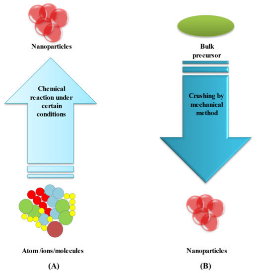
3.2. Top-Down Method

This method is used to transform bulk material into tiny nanoparticles. Top-down techniques are straightforward to employ. However, they are unsuccessful when producing irregularly shaped and very small particles. The main disadvantage of the top-down approach is the difficulty in acquiring a suitable particle size and shape [66][67][95,96]. Laser ablation is the most controllable top-down approach. Bulk material is treated with a laser beam (in this case, a bimetallic Au–Ag alloy). Under optimum conditions, well-dispersed bimetallic Au–Ag NPs can be synthesized, which can then be fractionated and surface-functionalized. A two-step synthesis, which involves laser irradiating a combination of silver and gold nanoparticles, is another alternative [68][97].

4. Antibacterial Properties of Bimetallic Au–Ag NPs

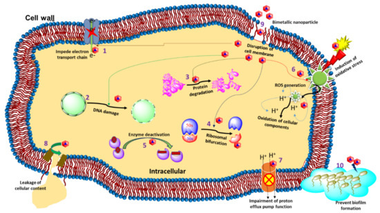
Controlling the invasion of new bacterial infections, their increasing proliferative powers, and antibacterial resistance, all of which have major public health implications, necessitates the use of extremely potent antimicrobial agents. Due to their synergistic effects, broad spectrum of physiochemical properties, and various mechanisms of action, bimetallic nanoparticles synthesized by combining two distinct metals have recently emerged as having a promising antibacterial efficiency exceeding those of their monometallic counterparts. Consequently, Au–Ag bimetallic nanoparticles are of great importance in imaging, biomedical devices, and nanomedicine [69][107]. In a study, Ding et al., synthesized Au–Ag core–shell NPs via a chemical route and investigated their antimicrobial efficacy. In this study, they reported the aggregation of Au–Ag core–shell NPs onto the bacterial surface, which led to improved imaging because of the improved two-photon photoluminescence. These nanoparticles were found to have antibacterial action against S. aureus while being less harmful to human dermal fibroblasts [70][81]. On the other hand, Bankura et al., reported the use of dextran as a reducing agent for the synthesis of Au–Ag alloy NPs and investigated their antimicrobial efficacy. The antibacterial activity of a 0.1 mg/mL concentration of Ag-Au alloy NPs was found to be significant against bacteria (B. subtilis, B. cereus, E. coli, and P. aeruginosa) with zones of inhibition of 24, 21, 17, and 20 mm [71][108]. In another report, the author followed a photosynthetic route to synthesize Au–Ag alloy nanoparticles for the first time. The bioreduction material in the study was essential oil from Coleus aromaticus. Gram-negative E. coli and Gram-positive S. aureus were used to test the antibacterial efficacy of the photosynthesized Au–Ag alloy nanoparticles. An inhibitory zone of 28 mm for the alloy nanoparticles (synthesized with 150 µL essential oil) demonstrated their strong bactericidal activity against E. coli. An in vitro antioxidant assay of the herbal-deduced nanoparticles also exhibited intense free radical (superoxide, hydroxyl, and nitric oxide radicals) scavenging activity [72][109]. Similarly, Amina et al., prepared Au–Ag alloy nanoparticles by using a microwave-assisted technique that utilized an extract of Asparagus racemosus root. In addition, the green-synthesized bimetallic alloy nanoparticles were tested against five different bacterial strains (Bacillus subtilis (ATCC 6633), Escherichia coli (ATCC 25922), Klebsiella pneumonia (Urine), Pseudomonas aeruginosa (ATCC 27853), and Staphylococcus aureus (ATCC 25923). It was reported that P. aeruginosa and S. aureus strains were the most susceptible (highest zone of inhibition) towards Au–Ag alloy nanoparticles versus single metal nanoparticles synthesized with plant extract [73][110]. Additionally, Gopinath et al., synthesized green bimetallic (Au–Ag) nanoparticles by using Gloriosa superba aqueous leaf extract. It was demonstrated that the developed nanoparticles had higher antibacterial as well as antibiofilm activities against Gram-positive and Gram-negative bacteria. The authors found a significant zone of inhibition at 6.33 ± 0.33 mm and 5.33 ± 0.33 mm for B. subtilis and E. coli, respectively [74][111]. In another study, recently developed biosynthesized Au–Ag NPs without the incorporation of a surfactant or stabilizing agent. It was observed that when the pH of a solution with E. coli and Au ions was raised, Au nanomaterials were formed. Core–shell Au–Ag nanostructures were generated in an ordered manner after Ag ions combined with the Au core. The spectroscopic and microscopic analyses confirmed the structural composition of the biosynthetic bimetallic Au–Ag nanoparticles [75][121]. In a similar study, Liu et al., reported that their bimetallic NPs showed stronger application possibilities in the superfast colorimetric monitoring of H2O2, photothermal treatments, and antimicrobial therapy. Without using 3,3′,5,5′-tetramethylbenzidine or peroxidase, their bimetallic Au–Cu NPs were able to sense H2O2 quickly and calorimetrically [76][122]. Furthermore, Au–Ag NPs could improve antibacterial activity without increasing cytotoxicity, ensuring that silver could be used in clinical settings [69][107]. In recent studies, Kalwar et al., created Au–Ag-NP-decorated cellulose nanofibers. Cellulose acetate nanofibers were made by electrospinning, and alkaline hydrolysis was used to deacetylate them. The Au–Ag NPs were coated on the surface of cellulose nanofibers using a dipping process to create an excellent wound dressing material. Furthermore, their antibacterial activity against E. coli and S. aureus was tested, and the Au–Ag NPs/cellulose was found to be a good antimicrobial material [77][123]. Villalobos-Noriega et al., synthesized bimetallic core–shell Au–Ag NPs by a green approach. Root extract of Rumex hymenosepalus containing catechins and stilbenes acted as a reducing agent in the NPs synthesis. The growth kinetics of microorganisms was analyzed by the Gompertz model. The findings suggested that silver NPs and bimetallic Au–Ag NPs had a dose-dependent effect on the lag phase and growth rate of E. coli and Candida albicans, with the Au–Ag NPs having a better response [78][79][120,124].

5. Bimetallic Nanoparticles Targeting Multidrug-Resistant Bacteria

Multidrug-resistant (MDR) bacteria are widely recognized as one of the most serious current public health issues, killing an estimated 700,000 people each year throughout the world [80][125]. Furthermore, treating MDR bacteria with ineffective antibiotics promotes the expansion of bacterial tolerance. For example, almost more than 50% of S. aureus strains obtained from several US hospitals are methicillin-resistant, with some strains also being resistant to vancomycin and carbapenems [81][126]. MDR microorganisms are frequently linked to nosocomial infection. Some MDR bacteria, on the other hand, have become common sources of community-acquired illnesses. This is a significant breakthrough since community-wide MDR bacteria dissemination leads to a significant rise in the population at risk and causes an increase in the number of MDR-bacteria-related diseases. When the incidence of resistance patterns in bacteria causing community-acquired infections exceeds a certain threshold, broad-spectrum antibacterial and/or combination antibacterial therapy is indicated for the empiric treatment of community-acquired disorders. Efforts to combat drug-resistant diseases are being hampered by the sluggish discovery of new antibiotics. It is anticipated that there will be no effective antibiotics available by 2050 if no new antibiotics are discovered [82][127]. Due to the lack of effective antibiotics against MDR bacteria, developing nanoparticles has been used as a substitute. It has also been observed that bimetallic NPs are efficient against bacteria, including MDR bacteria [83][128]. Several studies have shown bimetallic NPs to be effective against MDR bacteria. When monometallic counterparts were joined to form bimetallic NPs, the antibacterial activity was increased [84][129]. In a study, Wang et al., reported that Au NPs and mercaptophenylboronic acid (MBA) are incapable of acting as antibiotics separately. However, when MBA was coupled with Au NPs, the Au–MBA NPs showed significant antibacterial activity against Gram-positive MDR clinical isolates (e.g., MDR Staphyloccocus aureus and MDR Staphyloccocus epidermidis) [85][130]. In a similar study, Zhao and his collaborators synthesized bimetallic NPs by combining two salt solutions in an aqueous phase and reducing them with sodium borohydride. Monometallic NPs were also synthesized by the same method as the corresponding salt for comparison. The antibacterial NPs’ MIC (minimal inhibitory concentration) against E. coli and S. aureus were determined. Out of the nine different synthesized bimetallic NPs screened, two types of bimetallic NPs, namely the AuRh and AuRu NPs, showed MICs of 7 and 20 µg/mL, respectively, against E. coli and S. aureus. All of these bimetallic NPs were ineffective against S. aureus, with the MIC for S. aureus exceeding 128 µg/mL [86][131]. Kumar and his colleagues developed carbohydrate-coated bimetallic Au–Ag NPs, which were more effective against MDR strains than their monometallic counterparts (i.e., Ag NPs and Au NPs). The Au–Ag NPs were significantly more capable against Gram-negative MDR E. coli and Enterobacter cloacae than standard antibiotics. An in vivo study also exhibited that bimetallic Au–Ag NPs were almost 11,000 times more effective than Gentamicin at killing MDR MRSA infecting mice skin wounds. The Au–Ag NPs could heal and regenerate the infected wounds faster and without scarring. The in vivo results showed that Au–Ag NPs are an effective antibacterial agent against MDR strains with no adverse side effects [87][82]. Other forms of Au–Ag bimetallic NPs have been investigated and their antibacterial activity studied, although mostly as coating agents rather than as a delivery method [88][132].

6. Gold, Silver, and Gold–Silver Nanomaterials for Wound Healing

Wound healing is a complex biological process involving a series of cellular and molecular interactions targeted at repairing the injured tissue and restoring its protective function. The wound healing process occurs simultaneously in four different steps: hemostasis, inflammation, proliferation, and remodeling, all of which occur simultaneously. Various medications are now available on the market that can aid with wound healing. For wound healing, drugs that target blood coagulation, inflammatory reactions, platelet function, and cell proliferation are often employed. Glucocorticoids, nonsteroidal anti-inflammatory drugs, and chemotherapeutic agents are examples of these medications [87][89][90][82,133,134]. Bimetallic Au–Ag NPs are attractive candidates for wound dressing integration due to their high antibacterial potential and reduced toxicity profile compared to monometallic silver and gold NPs. According to Mârza et al., the antibacterial characteristics of silver can impact the healing process of skin regeneration, and in the meantime, the antibacterial properties of silver can assist an open wound by avoiding bacterial infection [91][135].
The skin healing and regeneration ability of bioactive glass with spherical gold nanocages in Vaseline ointments were examined in vivo in this researchtudy, which compared bioactive-glass–Vaseline and bioactive glass with spherical-gold–Vaseline ointments. Because the spherical gold nanocages were supported by silver, they had a high antibacterial activity. The findings indicated that the presence of silver in a wound affected the healing process. Jiang et al., developed a green synthetic method for bimetallic Au–Ag NPs without using any surfactants and stabilizers. When E. coli and Au ions were retained in the same solution, Au nanoparticles were formed first by raising the pH. Core–shell Au–Ag nanoparticles were generated in an ordered manner when the Ag ions combined.
Video Production Service