Recently, scientists have successfully developed a number of specific recognition fluorescent probes using different strategies, for example, through binding different identifying groups or changing the molecular structure by reaction with the target
[18][19][18,19]. The organic semiconductor with high biocompatibility is widely used in biosensors
[20][21][20,21], such as AIE (aggregation-induced emission) based probes. The small size of the organic semiconductor probes makes it easy to penetrate cells
[22]. In addition, the outstanding diffusion properties of organic semiconductor probes help improve the resolution of imaging
[23]. However, there are still some problems that restrict the development of organic semiconductor probes. On the one hand, organic semiconductor probes usually have poor photostability
[24][25][24,25]. On the other hand, their toxic effects and metabolism in organisms remain a challenge for organic semiconductor probes application. To solve these problems, researchers have constructed OSNs (organic semiconductor nanoparticles) by attaching or wrapping organic semiconductors in inorganic matrices
[26], Metal-organic frameworks (MOFs)
[27][28][29][27,28,29], polymers
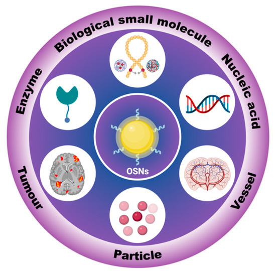
[30], etc. Due to their excellent properties, OSNs are widely used in biosensors (
Figure 1).
OSNs are widely used as biosensors for high-resolution tracking in vitro and in vivo
[31][32][31,32]. Compared to inorganic fluorescent materials, OSNs can be designed with various photosensitive properties and high biocompatibility through different design strategies
[33][34][33,34]. However, to construct high-performance OSNs for bioimaging, the materials must not only exhibit intense fluorescence, but they must also be designed in a comprehensive manner. For instance, the attachment and wrapping between OSNs and nanoparticles should be stable to prevent detachment
[35][36][35,36]. Additionally, toxicity is a crucial factor in bioimaging, as some reacting OSNs may be non-toxic before reacting with the target, but they could become toxic when combined with it
[37]. Furthermore, the sensitivity and effectiveness of OSN sensors still require further improvements for practical applications.
2. In Vitro Tracking
2.1. Biological Small Molecules Tracking
Biological small molecules in the body can reveal health conditions, and their detection and tracing are of great importance for biomedical research. Glutathione (GSH), the essential endogenous antioxidant, is the most abundant intracellular nonprotein thiol in mammalian and eukaryotic cells
[38][39][38,39]. The disturbances in GSH content are generally considered to be associated with various human diseases, such as psoriasis, human immunodeficiency virus (HIV), liver damage, and diabetes
[40][41][42][40,41,42]. Given its importance in medicine, achieving highly sensitive detection of GSH, as well as tracking of GSH in living cells, is urgent. Moreover, the tracking of GSH in organisms is favorable for biological investigations and early disease diagnoses
[43].
2.2. Enzyme Concentration Measurement
Enzymes are widely present in the body and act as biocatalysts. The International Union of Biochemistry and Molecular Biology (IUBMB) classifies enzymes into seven categories, including oxidoreductases, transferases, hydrolases, lyases, isomerases, ligases, and translocases
[44][47]. They all play decisive roles in the activity of the organism. If the activity of enzymes is weakened due to certain defects caused by various factors, it can lead to abnormal reactions, disorders of substance metabolism, and even development of clinical diseases. As a consequence, tracking the concentration and activity of enzymes is of major importance.
2.3. Nucleic Acid Concentration Measurement
Nucleic acid concludes deoxyribonucleic acid (DNA) and ribonucleic acid (RNA). It is a biological macromolecular compound polymerized by many nucleotides and is one of the most basic substances of life
[45][46][47][62,63,64]. DNA is the main basis for storing, replicating, and transmitting genetic information. Meanwhile, RNA plays an important role in protein synthesis. Thus, facile and reliable methods for the detection of DNA are of vital importance to the medical field, such as medical diagnosis, mutational analysis, gene therapy, biological studies, and specific genomic techniques
[48][49][65,66].
3. In Vivo Tracking
Different from the biosensors used for in vitro imaging, the influencing factors for in vivo tracking are more complex. Factors, such as water solubility, diffusion resistance, blood circulation, pH, and temperature, all need to be systematically considered during probe design. Moreover, the probes may be non-toxic to the target but toxic to other cells or organs. Therefore, before applying probe technology for in vivo tracking, in vitro experiments should first be performed to ensure its biosafety. Additionally, the probe should have different metabolism speeds corresponding to different tracking periods. In addition, stability and biocompatibility both are decisive factors for OSNs probes in in vivo tracking
[50][82]. Reasonable molecular designing can improve the stability and biocompatibility of OSNs, such as designing a stable molecular skeleton, reducing halogen atom content, and so on
[51][52][53][54][83,84,85,86]. Moreover, changes in molecular polarity due to changes in molecular structure may change the toxicity of the system.
3.1. Tumor Localization
Today, cancer is a major public health problem and the major cause of death globally
[55][56][87,88]. At present, the main diagnostic methods of cancer include ultrasound imaging (US)
[57][89], single photon emission computed tomography (SPECT)
[58][59][60][90,91,92], positron emission tomography (PET)
[61][62][63][93,94,95], electronic computed tomography (CT)
[64][96], magnetic resonance imaging (MRI)
[65][66][67][97,98,99], and optical imaging
[68][69][70][100,101,102]. Among them, radiological imaging has not only unavoidable radiological risks but also certain deficiencies in specificity, sensitivity, resolution, etc.
[71][72][73][74][103,104,105,106]. As a non-invasive technique, fluorescence imaging has the advantages of low risk of harm to humans, high sensitivity, and short response time, thus receiving increasing attention from researchers.
3.2. Blood Vessel Imaging
In the biomedical field, fluorescence imaging, as a highly sensitive non-invasive imaging technique, poses less risk of harm to the human body and has a shorter response time. It has great potential in organ tracking, especially for deep-tissue diagnosis
[75][116]. X-ray radiography is a common method for gastrointestinal disease diagnosis
[76][77][78][117,118,119]. Before the X-ray radiography, the patient needs to take the contrast agent orally, such as in the form of a barium meal. The barium meal will be excreted out of the body by defecation
[79][80][81][82][83][120,121,122,123,124]. In short-period medical examinations, metabolism is crucial for the contrast medium.
3.3. Particles Tracking
Air pollution is an important health concern globally. According to data published by the World Health Organization (WHO), upwards of four million people die early each year due to outdoor air pollution, and the main cause of this is particulate matter pollution
[84][85][133,134]. Therefore, exploring the deposition of particulate matter in the body and developing imaging techniques for it is a crucial area of cutting-edge scientific research. With the rapid development of nanotechnology, an increasing number of studies are applying biosensors and nanotechnology for tracing particulate matter and in vivo imaging. OSNs, consisting of fluorescently stained polystyrene nanoparticles, are now widely used to model atmospheric particulate matter. In one study, this OSN was successfully used to observe the dynamic deposition of particles in the lungs of mice in real-time using a two-photon microscope
[86][135].
4. Conclusions
Organic semiconductor nanoparticles (OSNs) offer several advantages for biosensing applications, including strong fluorescence emission, adjustable emission wavelength, and high biocompatibility, among others. Furthermore, by employing different design strategies,
scholarswe can develop a wide range of functional probes to meet various biosensing requirements. The development of OSNs has the potential to advance the fields of biology and medical science in several ways. Firstly, OSNs can serve as a platform for researchers to monitor the circulation of ions or biological molecules in organisms, thus deepening our understanding of their underlying mechanisms. Secondly, OSNs can be utilized for in vivo tracking to enable faster and more accurate disease diagnosis. Overall, the development of OSNs has the potential to significantly enhance medical science.