Submitted Successfully!
To reward your contribution, here is a gift for you: A free trial for our video production service.
Thank you for your contribution! You can also upload a video entry or images related to this topic.
Version Summary Created by Modification Content Size Created at Operation
1 + 3276 word(s) 3276 2021-04-27 06:00:16 |
2 Edited redundant sentence in the Introduction. -19 word(s) 3257 2021-05-02 16:59:06 |

Video Upload Options

Do you have a full video?

Confirm

Are you sure to Delete?
Cite
If you have any further questions, please contact Encyclopedia Editorial Office.
Larocca, D. Uncoupling Aging from Chronological Time. Encyclopedia. Available online: https://encyclopedia.pub/entry/9261 (accessed on 15 May 2024).
Larocca D. Uncoupling Aging from Chronological Time. Encyclopedia. Available at: https://encyclopedia.pub/entry/9261. Accessed May 15, 2024.
Larocca, Dana. "Uncoupling Aging from Chronological Time" Encyclopedia, https://encyclopedia.pub/entry/9261 (accessed May 15, 2024).
Larocca, D. (2021, May 02). Uncoupling Aging from Chronological Time. In Encyclopedia. https://encyclopedia.pub/entry/9261
Larocca, Dana. "Uncoupling Aging from Chronological Time." Encyclopedia. Web. 02 May, 2021.
Uncoupling Aging from Chronological Time
Edit

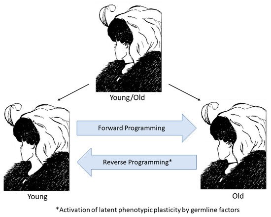
Cellular life evolved from simple unicellular organisms that could replicate indefinitely, being essentially ageless. At this point, life split into two fundamentally different cell types: the immortal germline representing an unbroken lineage of cell division with no intrinsic endpoint and the mortal soma, which ages and dies. We consider aging as a process not fixed to the pace of chronological time but one that can speed up or slow down depending on the rate of intrinsic cellular clocks. Moreover germline factor reprogramming might be used to slow the rate of aging and potentially reverse it by causing the clocks to tick backward. Therefore, reprogramming may eventually lead to therapeutic strategies to treat degenerative diseases by altering aging itself, the one condition common to us all.

aging cellular clocks reprogramming development epigenetics DNA methylation telomeres transposable elements longevity regeneration

1. Introduction

In the late 19th century, Weismann accurately described the dual nature of life, separating it into the immortal germline and mortal soma. The germline can forever renew itself, giving birth to young organisms at each generation, and thus, we refer to germline cells as clock-free and somatic cells as clock-bound. Indeed, we can trace an unbroken chain of cell division from all life on earth going back 4 billion years to converge at the base of the evolutionary tree to a single cell, LUCA, the “last universal common ancestor” [1]. In contrast, somatic cells lose their robustness with time exhibiting the hallmarks of aging [2], which eventually lead to organismal degeneration and an exponential increase in the probability of death with the passage of time [3]. Three cellular aging clocks that bind the soma to time include the telomeric clock [4], DNA methylation (DNAm) clock, also known as the epigenetic or Horvath clock [5], and the transposable element (TE) clock [6], which are likely primary because they directly affect the genome either by telomere shortening, chemical modification of DNA, or transposition induced double stranded breaks (DSB)[7]. Many more cellular aging clocks such as transcriptomic, proteomic, and metabolomic clocks [8][9] likely exist. The molecular basis of the immortal germline extending from the first unicellular organisms through to the current multitude of lifeforms would not be understood until the mid-20th century with the discovery of DNA. Although the information stored in DNA changes through mutation and reassortment of genetic alleles as a driver of evolution, the DNA nevertheless retains all the information needed to make a new body, so each generation is born young. As Weismann expressed in a letter to Nature does not exist, but only immortal forms of activity of organized matter” [10]. We may now extend this to “immortal information encoded in DNA of organized matter.” As we have described, the germline either escapes or resets the aging clocks so that the genomic information is preserved. Thus, the germline preserves the information needed to perpetuate itself indefinitely and to make a new body, even though the body that carries it ages. The natural or engineered application of germline strategies to the soma may slow down or even reverse aging, thus uncoupling aging from chronological time.

2. Plasticity of Aging and Development

Unlike the immortal germline, the aging soma follows a predictable course that is coupled to time yet flexible enough to accelerate or decelerate depending on environmental and evolutionary pressure. Indeed, we see a spectrum of aging rates leading to diverse lifespans among animal species from the very rapid aging in the mayfly (hours), to the fruit fly (days), or dwarf pygmy goby (weeks), to gradual aging in most mammals (year/decades), to very slow aging as in the Aldabra giant tortoise, bowhead whale, Greenland shark (centuries).  At the very extreme end are examples of indefinite lifespans with no apparent intrinsic aging as in hydra, planaria, sponges, and mussels[11]. Remarkably, it is now possible to reverse the cellular clocks of development and aging, winding them back from an aged somatic cell to the earliest stage of life. We have evidence from cloning (somatic cell nuclear transfer (SCNT)) experiments first performed in frogs and later in mammals, including humans [12][13][14] that while somatic cells age and accumulate the key hallmarks of aging with time [2], the information to make a young cell is preserved in the DNA. During SCNT, exposure of a somatic nucleus to germline factors in the egg resets the aging clock to zero, such that cloned animals are born young as if they were conceived from two germ cells [15][16][17]. Moreover, Yamanaka demonstrated over a decade ago that forced expression of as few as four germline factors, OCT4, KLF4, SOX2, and c-MYC (OKSM), alone could be used to reprogram development in vitro, thus reverting a differentiated cell back to an induced pluripotent stem cell (iPSC) [18]. However, it was initially unclear whether the reprogramming of a differentiated cell would also result in reverting the hallmarks of aging to a younger state. Initial studies suggested that iPSC lines were potentially limited in their differentiation capacity because of short telomeres [19][20]. However, later studies have demonstrated that reprogramming of aged and senescent cultured fibroblasts and even in fibroblasts obtained from centenarian donors restores telomeres to embryonic length [21][22][23]. Indeed, reprogramming has even been shown to reset telomere length in cells from a donor near the current limit of human aging (114 years) [24].  The telomere clock ticks rapidly in patients with premature aging syndromes, but telomeres in these cells can also be reset to embryonic length by reprogramming [24][25][26]. In addition, epigenetic markers of aging such as senescence-associated heterochromatin foci (SAHF) and heterochromatin protein-1α (HP1α) are restored to young levels by reprogramming cells from prematurely aged as well as naturally aged donors [22][27]. Other hallmarks of aging, including mitochondrial fitness, have been shown to be reversed by reprogramming [22][28]. The rejuvenating effect of reprogramming has also been demonstrated at the epigenetic and transcriptomic level in iPSC lines that were differentiated to their respective cell type of origin, such as mesenchymal stromal cells, hematopoietic stem cells, and fibroblasts [29][30]. Moreover, iPSC from old mice can be differentiated, in vivo, into heart cells that closely resemble their younger counterparts [31]. Taken together, these studies strongly support the reversal of both developmental stage and aging by germline factor reprogramming to iPSCs.
Interestingly, examples of reversal of developmental stage can be found in nature as an adaptation to environmental conditions as observed in the expression of embryonic pathways in the regenerating limbs of urodeles or in oncogenic transformation. One of the most profound natural reversals, noted by Weismann in 1883, is seen in the hydrozoan, Torritopsis dohrnii, which normally develops into a medusa from a benthic polyp form but undergoes a reversal from medusa to polyp via transformation to a cyst intermediate under adverse conditions such as injury, starvation or senescence [32]. Comparative transcriptome analysis of the cyst versus medusa and polyp forms shows enrichment in gene pathways such as DNA integration, transposition, repair, and telomere maintenance suggesting germline-like maintenance of genome integrity in the intermediate cyst stage [33]. However, it remains to be determined whether there is suppression of TEs at the cyst stage, as in the hydra [34] and long-lived termite reproductives [35]. In contrast, somatic cell-associated pathways such as cell signaling, aging, and differentiation were downregulated in the cyst stage [33]. Another example of developmental reversal occurs in the process of sex transition in a marine fish, the bluehead wrasse, which remarkably involves some of the same germline factors used for cell reprogramming. The female wrasse undergoes a rapid behavioral and complete physical transformation to sperm-producing terminal phase (TP) male in 8–10 days in response to the loss of the dominant TP male and the subsequent increase in cortisol levels. Mutually antagonistic male and female gene networks determine and maintain gonadal fate in fishes and thus account for the retention of a feature of embryonic development, bipotentiality of sex, into adulthood. The mechanism of transformation in the bluehead wrasse has been recently elucidated at the molecular level [36]. Transcriptomic and methylomic analysis of gonads at various stages from the female through TP male reveals that the gonads pass through an undifferentiated midway stage that resembles mammalian pluripotent stem cells (PSC) and primordial germ cells (PGC) rather than transdifferentiation from the female to male state [36]. For example, Polycomb group members of PRC2, which are responsible for tri-methylation of lysine 27 on histone H3 (H3K27me3) and are downregulated in PSC, are also downregulated during the midway stage of the female to male transition. The variant histone H2A.2, which is also low in PSC, shows a similar pattern. In addition, writers and erasers of histone acetylation are expressed dynamically during the transition. Evidence of extensive reprogramming of DNA methylation was also observed with upregulation of ten-eleven translocation demethylase (TET) (as in PSC and PGC) midway through transition leading to a shift from female to male pattern of methyltransferases. Genome-wide DNA methylation changes from the female to male pattern were found to occur. Notably, like the transcriptome, the midway methylome state represents a developmental shift rather than an intermediate differentiated state. Indeed, it would be interesting to determine whether the DNAm age of the gonad regresses during the transition if an epigenetic aging clock can be created for marine fishes. These data illustrate remarkable plasticity in a normally committed developmental process of sex determination via epigenetic reprogramming involving transition through an earlier developmental state. Remarkably mutually antagonistic gene networks that both determine and maintain sex in adulthood have also been found in mice indicating conservation of some degree of plasticity even in mammals [37][38]. Given the relationship of aging to developmental processes and examples of the maintenance of developmental plasticity into adulthood, could epigenetic reprogramming uncover latent phenotypic plasticity in mammals to return aging adults to a younger epigenetic state and thereby reverse aging?

3. Reversal of Aging In Vitro

Given the astonishing degree of phenotypic plasticity observed in the forward and reverse programming of development and aging in nature and in the laboratory, we may perhaps view young and old organisms as two epigenetic states of the same genome with the young state contained (as information) within the old and the old state contained in the young as a potentiality that becomes actualized by the ticking of the cellular clocks with the passage of time (Figure 3). If aging is indeed a continuation of development as suggested by the behavior of cellular aging clocks, then it is not surprising that reprogramming can reverse the forward programming of both development and aging. However, it also raises the intriguing possibility of reversing aging without loss of developmental status if reprogramming is capable of turning back development and aging in the reverse order in which they occurred. A study of transcriptomic and methylomic data from a time course of fibroblasts undergoing reprogramming suggests that this may indeed be the case and that reprogramming is reverse programming of aging first followed by a reversal of developmental state (Figure 4) [39]. The study shows a linear decrease in DNAm age during the early phase of reprogramming (partial reprogramming; day 3–11) when fibroblast identity has not been lost. At this early stage, the cells undergoing reprogramming have a high propensity for spontaneous reversion to their initial fully differentiated state. Importantly, early and late markers of pluripotency (LIN28DNMT3AZIC3TERT) are not induced, and the fibroblast defining genes are not fully repressed during the initial age reversal/partial reprogramming phase before the DNAm age has reached zero (day 20). Similarly, genes we have identified as fetal/adult-specific (post-embryonic to fetal transition (EFT)), such as COX7A1 and ADIRF, are reduced but not to minimal levels during partial reprogramming, and embryonic-specific genes (pre-EFT) such as PCDHB2 are not expressed until day 20 (Figure 4) [40]. Another study has recently reported rejuvenation of transcriptomic and DNAm age by as much as 30 years in transiently reprogrammed fibroblasts from middle-aged donors [41]. It will be important to obtain single-cell analysis to determine the dynamics of these changes within subpopulations of the reprogramming factor-treated cells. The data suggest, however, that reprogramming first runs the epigenetic aging steps in the reverse order that they occurred during the life history followed by a reversal of developmental state. Therefore, these data indicate the potential to apply reprogramming methods therapeutically for rejuvenating aged cells, tissues, and perhaps whole organisms without loss of cell identity.
Figure 3. The two faces of epigenetic aging. The top illustration is an illusion designed so that the viewer will see either a young woman or an old woman. However, both are present in the same figure, just as the young and old woman are present in the same person as the genome’s epigenetic potential. Forward programming takes the young woman through a series of epigenetic states to an old woman as the DNAm, and other cellular clocks tick forward. Reverse programming can potentially be achieved using germline factors to revert the genome to an earlier epigenetic age.
Figure 4. Age reversal precedes reversion of developmental state during reprogramming of fibroblasts using OKSM germline factors. Post-EFT genes (COX7A1, ADIRF) are rapidly reduced during early (partial) reprogramming (day 3–20) while a linear decrease in DNAm age is occurring. Pre-EFT genes are turned on at day 20 when DNAm age has gone to zero, and their expression increases as cells reverse their developmental state to full pluripotency (≥28 days). The data suggest the possibility of age reprogramming without permanent loss of cell identity. Figure is adapted from Olova et al [39].

4. Reversal of Aging In Vivo

Initial attempts at in vivo reprogramming using 4-factor (OKSM) reprogramming were not encouraging because they resulted in extensive tumor formation [42][43]. However, Ocampo et al. later reported reversal of epigenetic aging markers with no signs of tumor development in transgenic OKSM mice when the factors are induced at a lower dose and on a cyclic schedule of 2 days on and 5 days off [44]. Instead of tumor formation, cyclic OKSM induction led to amelioration of the premature aging phenotype and extension of lifespan in a mouse model of HGPS. They further demonstrated that cyclic OKSM induction resulted in greater resistance to metabolic disease following pancreatic injury and increased repair of skeletal muscle damage in physiologically aged mice. Additional in vivo studies have shown reduced scar formation of skin wounds using viral vector-mediated partial reprogramming and protection against liver damage using a small molecule approach [45][46]. The question remained, however, as to whether partial reprogramming in vivo reversed the DNAm clock. Two recent studies provide evidence suggesting that a reversal of the DNAm clock during in vivo reprogramming may be possible [47][48]. In one study, transient expression of six reprogramming factors, OCT4, SOX2, KLF4, LIN28, c-MYC, and NANOG (OSKLMN), was obtained using mRNA transfection, which resulted in a reversion of fibroblasts and endothelial cells to a younger phenotype as measured by epigenomic markers (tri-methylation of lysine 9 on histone 3 (H3K9me3), HP1γ, and lamina-associated polypeptide 2α (LAP2α)), mitochondrial fitness, autophagy, transcriptome, and DNAm clock [48]. Transient expression of OSKLMN mitigated the inflammatory phenotype of osteoarthritic chondrocytes in culture, and implants of ex vivo OSKLMN treated aged mouse or human skeletal muscle stem cells resulted in rejuvenated muscle regenerative response following injury in physiologically aged mice with no signs of tumor formation [48]. In another in vivo study, only three factors (OKS) were delivered to mice using an inducible adeno-associated virus vector (AAV). The results indicate that induction of OKS resulted in the protection of retinal ganglia cells and regeneration of axons in an optic nerve crush injury model, and the same treatment restored vision in a mouse model of glaucoma [47]. Importantly, the DNAm age was reversed by the treatment in both models, and the regenerative effects were Tet1 and Tet2 dependent in the optic nerve model, indicating that these reprogramming factors may be “reverse programming” the epigenetic clock to an earlier state (DNAm age) with an enhanced regenerative capacity [47]. The introduction of germline factors into somatic cells, in vivo, reveals a potential for phenotypic plasticity in adult mammals that was previously observed only in lower life forms.
The prospect of reversing organismic aging could potentially reduce or eliminate many age-associated degenerative diseases such as heart disease, cancer, diabetes, osteoporosis, sarcopenia, neurodegenerative disease, and skin aging. Thus, rejuvenation therapies could potentially alleviate many of the social and economic costs that we face globally because of an ever-increasing demographic shift to an older population. However, critical barriers remain to develop rejuvenating therapies that involve germline factor reprogramming. For example, more studies are needed on naturally aged experimental animals to determine the effect of such treatments on lifespan and health span. Moreover, careful control of the timing and dosage of reprogramming factors will need to be developed to minimize the risk of tumor formation. The further development of epigenetic aging clocks in experimental animals and tissue culture will be helpful for screening reprogramming agents and delivery strategies. Strategies for transient expression of reprogramming factors include AAV mediated inducible gene delivery, RNA transfer, and small molecules [46][47][48]. The use of extracellular vesicles such as exosomes to deliver reprogramming RNAs is also a promising approach because of their long half-life in vivo and low immunogenicity [49]. Pre-clinical studies have shown the efficacy of engineered exosomes for the treatment of pancreatic cancer, and they can be effectively scaled using standard bioreactors [50]. In addition to their potential as a delivery vehicle, exosomes produced by young mesenchymal stem cells (MSC) may, on their own, be an effective way to slow or reverse some aspects of aging [51]. The development of reprogramming therapies may even benefit from the considerable progress in RNA delivery and manufacturing that has resulted from the accelerated efforts to develop COVID-19 vaccines [52]. Careful attention will be needed to assess the appropriate indications and regulatory considerations when developing clinical studies. Initial proof of concept studies may involve specific indications such as arthritic joint disease; however, later studies may be possible using resistance to age-related disease as an outcome as in the proposed TAME study [53].

5. Concluding Remarks

The intimate relationship between aging and developmental clocks suggests that the adaptation of developmental clock rate to environmental pressure might contribute to the wide variation in lifespan observed between species. For example, humans and mole-rats exhibit neoteny (retention of embryonic or juvenile characteristics through adulthood), where slowing the rate of development correlates with an extension of lifespan. The application of germline strategies in somatic stem cells has resulted in the remarkable regenerative capacity of lower life forms that are capable of indefinite lifespans, such as sponges, planarians, and hydra. This regenerative capacity has become increasingly restricted as more complex life forms evolved, being confined to the pre-EFT period in mammals. However, retention of extensive capacity for regeneration is observed in lower vertebrates, including fishes, amphibians, and reptiles, which also exhibit remarkable phenotypic plasticity in their capacity for metamorphosis and in certain cases of remarkable reversals of developmental stage and sexual development. Finally, reprogramming using germline factors can uncover a similar but latent phenotypic plasticity in mammals by reverting both the developmental state and cellular age. Indeed, both natural phenotypic plasticity in the blue wrasse and partial reprogramming involve the repression of DNA methyl transferases (DNMT) and induction of demethylases (TET) which, by a yet-to-be-determined mechanism, may enable the DNAm clock to tick backward. The discovery that partial reprogramming can reverse the aging clock without permanent alteration of cellular identity has led to initial studies that demonstrate the potential to reverse organismic aging. Although there are many challenges ahead, our current understanding of cellular clocks and our ability to reprogram them using germline factors opens the door to many promising therapeutic approaches to slowing down, preventing, or reversing aging itself and thus treating the many age-related diseases that burden society. Indeed, if these approaches can be made practical and scalable, we may find ourselves in a future in which we have no time to age.

References

  1. Weiss, M.C.; Preiner, M.; Xavier, J.C.; Zimorski, V.; Martin, W.F.; The last universal common ancestor between ancient Earth chemistry and the onset of genetics.. PLoS Genet. 2018, 14, e1007518.
  2. Carlos López-Otín; Maria A. Blasco; Linda Partridge; Manuel Serrano; Guido Kroemer; The Hallmarks of Aging. Cell 2013, 153, 1194-1217, 10.1016/j.cell.2013.05.039.
  3. Strehler, B.L.; Mildvan, A.S.; General theory of mortality and aging. Science 1960, 132, 14-21.
  4. Calvin B. Harley; A. Bruce Futcher; Carol W. Greider; Telomeres shorten during ageing of human fibroblasts. Nature 1990, 345, 458-460, 10.1038/345458a0.
  5. Steve Horvath; DNA methylation age of human tissues and cell types. Genome Biology 2012, 14, R115-R115, 10.1186/gb-2013-14-10-r115.
  6. John M. Sedivy; Jill A. Kreiling; Nicola Neretti; Marco De Cecco; Steven W. Criscione; Jeffrey W. Hofmann; Xiaoai Zhao; Takahiro Ito; Abigail L. Peterson; Death by transposition – the enemy within?. BioEssays 2013, 35, 1035-1043, 10.1002/bies.201300097.
  7. Dana Larocca; Jieun Lee; Michael West; Ivan Labat; Hal Sternberg; No Time to Age: Uncoupling Aging from Chronological Time. Genes 2021, 12, 611, 10.3390/genes12050611.
  8. Juulia Jylhävä; Nancy L. Pedersen; Sara Hägg; Biological Age Predictors. EBioMedicine 2017, 21, 29-36, 10.1016/j.ebiom.2017.03.046.
  9. David H. Meyer; Björn Schumacher; BiT age: A transcriptome‐based aging clock near the theoretical limit of accuracy. Aging Cell 2021, 20, e13320, 10.1111/acel.13320.
  10. Thomas B. L. Kirkwood; Thomas Cremer; Cytogerontology since 1881: A reappraisal of August Weismann and a review of modern progress. Quality of Life Research 1982, 60, 101-121, 10.1007/bf00569695.
  11. Finch, C.E.. Longevity, Senescence, and the Genome; University of Chicago Press: London, UK, 1994; pp. 43-247.
  12. J B Gurdon; The developmental capacity of nuclei taken from intestinal epithelium cells of feeding tadpoles.. Journal of embryology and experimental morphology 1962, 10, 622-640.
  13. K. H. S. Campbell; J. McWhir; W. A. Ritchie; I. Wilmut; Sheep cloned by nuclear transfer from a cultured cell line. Nature 1996, 380, 64-66, 10.1038/380064a0.
  14. Masahito Tachibana; Paula Amato; Michelle Sparman; Nuria Marti Gutierrez; Rebecca Tippner-Hedges; Hong Ma; Eunju Kang; Alimujiang Fulati; Hyo-Sang Lee; Hathaitip Sritanaudomchai; et al.Keith MastersonJanine LarsonDeborah EatonKaren Sadler-FreddDavid BattagliaDavid LeeDiana WuJeffrey JensenPhillip PattonSumita GokhaleRichard L. StoufferN WolfShoukhrat Mitalipov Human Embryonic Stem Cells Derived by Somatic Cell Nuclear Transfer. Cell 2013, 154, 465-466, 10.1016/j.cell.2013.06.042.
  15. Dean H. Betts; Vilceu Bordignon; Jonathan R. Hill; Quinton Winger; Mark E. Westhusin; Lawrence C. Smith; W. Allan King; Reprogramming of telomerase activity and rebuilding of telomere length in cloned cattle. Proceedings of the National Academy of Sciences 2001, 98, 1077-1082, 10.1073/pnas.031559298.
  16. Robert P. Lanza; Jose B. Cibelli; Catherine Blackwell; Vincent J. Cristofalo; Mary Kay Francis; Gabriela M. Baerlocher; Jennifer Mak; Michael Schertzer; Elizabeth A. Chavez; Nancy Sawyer; et al.Peter M. LansdorpMichael D. West Extension of Cell Life-Span and Telomere Length in Animals Cloned from Senescent Somatic Cells. Science 2000, 288, 665-669, 10.1126/science.288.5466.665.
  17. R. P. Lanza; Cloned Cattle Can Be Healthy and Normal. Science 2001, 294, 1893-1894, 10.1126/science.1063440.
  18. Kazutoshi Takahashi; Koji Tanabe; Mari Ohnuki; Megumi Narita; Tomoko Ichisaka; Kiichiro Tomoda; Shinya Yamanaka; Induction of Pluripotent Stem Cells from Adult Human Fibroblasts by Defined Factors. Cell 2007, 131, 861-872, 10.1016/j.cell.2007.11.019.
  19. Bao-Yang Hu; Jason P. Weick; Junying Yu; Li-Xiang Ma; Xiao-Qing Zhang; James A. Thomson; Su-Chun Zhang; Neural differentiation of human induced pluripotent stem cells follows developmental principles but with variable potency. Proceedings of the National Academy of Sciences 2010, 107, 4335-4340, 10.1073/pnas.0910012107.
  20. Qiang Feng; Irina Klimanskaya; Ignatius Gomes; Hoon Kim; Young Chung; George R. Honig; Kwang-Soo Kim; Robert Lanza; Shi-Jiang Lu; Hemangioblastic Derivatives from Human Induced Pluripotent Stem Cells Exhibit Limited Expansion and Early Senescence. STEM CELLS 2010, 28, 704-712, 10.1002/stem.321.
  21. H Vaziri; Kb Chapman; A Guigova; J Teichroeb; M D Lacher; H Sternberg; I Singec; L Briggs; J Wheeler; J Sampathkumar; et al.R GonzalezD LaroccaJ MuraiE SnyderWh AndrewsWd FunkM D West Spontaneous reversal of the developmental aging of normal human cells following transcriptional reprogramming. Regenerative Medicine 2010, 5, 345-363, 10.2217/rme.10.21.
  22. Laure Lapasset; Ollivier Milhavet; Alexandre Prieur; Emilie Besnard; Amelie Babled; Nafissa Aït-Hamou; Julia Leschik; Franck Pellestor; Jean-Marie Ramirez; John De Vos; et al.Sylvain LehmannJean-Marc Lemaitre Rejuvenating senescent and centenarian human cells by reprogramming through the pluripotent state. Genes & Development 2011, 25, 2248-2253, 10.1101/gad.173922.111.
  23. Takuya Yagi; Arifumi Kosakai; Daisuke Ito; Yohei Okada; Wado Akamatsu; Yoshihiro Nihei; Akira Nabetani; Fuyuki Ishikawa; Yasumichi Arai; Nobuyoshi Hirose; et al.Hideyuki OkanoNorihiro Suzuki Establishment of Induced Pluripotent Stem Cells from Centenarians for Neurodegenerative Disease Research. PLOS ONE 2012, 7, e41572, 10.1371/journal.pone.0041572.
  24. Jieun Lee; Paola A. Bignone; L.S. Coles; Yang Liu; Evan Snyder; Dana Larocca; Induced pluripotency and spontaneous reversal of cellular aging in supercentenarian donor cells. Biochemical and Biophysical Research Communications 2020, 525, 563-569, 10.1016/j.bbrc.2020.02.092.
  25. Guang-Hui Liu; Basam Z. Barkho; Sergio Zamudio Ruiz; Dinh Diep; Jing Qu; Sheng-Lian Yang; Athanasia D. Panopoulos; Keiichiro Suzuki; Leo Kurian; Christopher T Walsh; et al.James ThompsonStephanie BoueHo Lim FungIgnacio Sancho-MartinezKun ZhangJohn Yates IiiJuan Carlos Izpisua Belmonte Recapitulation of premature ageing with iPSCs from Hutchinson–Gilford progeria syndrome. Nature 2011, 472, 221-225, 10.1038/nature09879.
  26. Hoi-Hung Cheung; Xiaozhuo Liu; Lucile Canterel-Thouennon; Lu Li; Catherine Edmonson; Owen M. Rennert; Telomerase Protects Werner Syndrome Lineage-Specific Stem Cells from Premature Aging. Stem Cell Reports 2014, 2, 534-546, 10.1016/j.stemcr.2014.02.006.
  27. Guang-Hui Liu; Basam Z. Barkho; Sergio Zamudio Ruiz; Dinh Diep; Jing Qu; Sheng-Lian Yang; Athanasia D. Panopoulos; Keiichiro Suzuki; Leo Kurian; Christopher T Walsh; et al.James ThompsonStephanie BoueHo Lim FungIgnacio Sancho-MartinezKun ZhangJohn Yates IiiJuan Carlos Izpisua Belmonte Recapitulation of premature ageing with iPSCs from Hutchinson–Gilford progeria syndrome. Nature 2011, 472, 221-225, 10.1038/nature09879.
  28. Steven T. Suhr; Eun Ah Chang; Jonathan Tjong; Nathan Alcasid; Guy A. Perkins; Marcelo D. Goissis; Mark H. Ellisman; Gloria I. Perez; Jose B. Cibelli; Mitochondrial Rejuvenation After Induced Pluripotency. PLOS ONE 2010, 5, e14095, 10.1371/journal.pone.0014095.
  29. Joana Frobel; Hatim Hemeda; Michael Lenz; Giulio Abagnale; Sylvia Joussen; Bernd Denecke; Tomo Šarić; Martin Zenke; Wolfgang Wagner; Epigenetic Rejuvenation of Mesenchymal Stromal Cells Derived from Induced Pluripotent Stem Cells. Stem Cell Reports 2014, 3, 414-422, 10.1016/j.stemcr.2014.07.003.
  30. Martin Wahlestedt; Eva Erlandsson; Trine Kristiansen; Rong Lu; Cord Brakebusch; Irving L. Weissman; Joan Yuan; Javier Martin-Gonzalez; David Bryder; Clonal reversal of ageing-associated stem cell lineage bias via a pluripotent intermediate. Nature Communications 2017, 8, 14533, 10.1038/ncomms14533.
  31. Zhao Cheng; Hong-Ling Peng; Rong Zhang; Xian-Ming Fu; Guang-Sen Zhang; Rejuvenation of Cardiac Tissue Developed from Reprogrammed Aged Somatic Cells. Rejuvenation Research 2017, 20, 389-400, 10.1089/rej.2017.1930.
  32. Weissman, A. Die Entstehung der Sexualzellen Bei Den Hydromedusen: Zugleich Ein Betrag zur Kenntniss des Baues und der Lebenserscheinungen Dieser Gruppe; Gustav Fischer Verlag: Jena, Germany, 1883.
  33. Matsumoto, Y.; Piraino, S.; Miglietta, M.P. Transcriptome Characterization of Reverse Development in Turritopsis dohrnii (Hydrozoa, Cnidaria). G3 Genes Genomes Genet. 2019, 9, 4127–4138.
  34. Bryan B. Teefy; Stefan Siebert; Jack F. Cazet; Haifan Lin; Celina E. Juliano; PIWI–piRNA pathway-mediated transposable element repression in Hydra somatic stem cells. RNA 2020, 26, 550-563, 10.1261/rna.072835.119.
  35. Daniel Elsner; Karen Meusemann; Judith Korb; Longevity and transposon defense, the case of termite reproductives. Proceedings of the National Academy of Sciences 2018, 115, 5504-5509, 10.1073/pnas.1804046115.
  36. Todd, E.V.; Ortega-Recalde, O.; Liu, H.; Lamm, M.S.; Rutherford, K.M.; Cross, H.; Black, M.A.; Kardailsky, O.; Graves, J.A.M.; Hore, T.A.; et al. Stress, novel sex genes, and epigenetic reprogramming orchestrate socially controlled sex change. Sci. Adv. 2019, 5, eaaw7006.
  37. Uhlenhaut, N.H.; Jakob, S.; Anlag, K.; Eisenberger, T.; Sekido, R.; Kress, J.; Treier, A.C.; Klugmann, C.; Klasen, C.; Holter, N.I.; et al. Somatic sex reprogramming of adult ovaries to testes by FOXL2 ablation. Cell 2009, 139, 1130–1142.
  38. Matson, C.K.; Murphy, M.W.; Sarver, A.L.; Griswold, M.D.; Bardwell, V.J.; Zarkower, D. DMRT1 prevents female reprogramming in the postnatal mammalian testis. Nature 2011, 476, 101–104.
  39. Olova, N.; Simpson, D.J.; Marioni, R.E.; Chandra, T. Partial reprogramming induces a steady decline in epigenetic age before loss of somatic identity. Aging Cell 2019, 18, e12877.
  40. West, M.D.; Labat, I.; Sternberg, H.; Larocca, D.; Nasonkin, I.; Chapman, K.B.; Singh, R.; Makarev, E.; Aliper, A.; Kazennov, A.; et al. Use of deep neural network ensembles to identify embryonic-fetal transition markers: Repression of COX7A1 in embryonic and cancer cells. Oncotarget 2018, 9, 7796–7811.
  41. Gill, D.; Parry, A.; Santos, F.; Hernando-Herraez, I.; Stubbs, T.M.; Milagre, I.; Reik, W. Multi-omic rejuvenation of human cells by maturation phase transient reprogramming. bioRxiv 2021.
  42. Ohnishi, K.; Semi, K.; Yamamoto, T.; Shimizu, M.; Tanaka, A.; Mitsunaga, K.; Okita, K.; Osafune, K.; Arioka, Y.; Maeda, T.; et al. Premature termination of reprogramming in vivo leads to cancer development through altered epigenetic regulation. Cell 2014, 156, 663–677.
  43. Abad, M.; Mosteiro, L.; Pantoja, C.; Canamero, M.; Rayon, T.; Ors, I.; Grana, O.; Megias, D.; Dominguez, O.; Martinez, D.; et al. Reprogramming in vivo produces teratomas and iPS cells with totipotency features. Nature 2013, 502, 340–345.
  44. Ocampo, A.; Reddy, P.; Martinez-Redondo, P.; Platero-Luengo, A.; Hatanaka, F.; Hishida, T.; Li, M.; Lam, D.; Kurita, M.; Beyret, E.; et al. In Vivo Amelioration of Age-Associated Hallmarks by Partial Reprogramming. Cell 2016, 167, 1719–1733.e1712.
  45. Doeser, M.C.; Scholer, H.R.; Wu, G. Reduction of Fibrosis and Scar Formation by Partial Reprogramming In Vivo. Stem Cells 2018, 36, 1216–1225.
  46. Tang, Y.; Cheng, L. Cocktail of chemical compounds robustly promoting cell reprogramming protects liver against acute injury. Protein Cell 2017, 8, 273–283.
  47. Lu, Y.; Brommer, B.; Tian, X.; Krishnan, A.; Meer, M.; Wang, C.; Vera, D.L.; Zeng, Q.; Yu, D.; Bonkowski, M.S.; et al. Reprogramming to recover youthful epigenetic information and restore vision. Nature 2020, 588, 124–129.
  48. Sarkar, T.J.; Quarta, M.; Mukherjee, S.; Colville, A.; Paine, P.; Doan, L.; Tran, C.M.; Chu, C.R.; Horvath, S.; Qi, L.S.; et al. Transient non-integrative expression of nuclear reprogramming factors promotes multifaceted amelioration of aging in human cells. Nat. Commun. 2020, 11, 1545.
  49. Agarwal, S.; Agarwal, V.; Agarwal, M.; Singh, M. Exosomes: Structure, Biogenesis, Types and Application in Diagnosis and Gene and Drug Delivery. Curr. Gene Ther. 2020, 20, 195–206.
  50. Mendt, M.; Kamerkar, S.; Sugimoto, H.; McAndrews, K.M.; Wu, C.C.; Gagea, M.; Yang, S.; Blanko EV, R.; Peng, Q.; Ma, X.; et al. Generation and testing of clinical-grade exosomes for pancreatic cancer. JCI Insight 2018, 3, e99263.
  51. Dorronsoro, A.; Santiago, F.E.; Grassi, D.; Zhang, T.; Lai, R.C.; McGowan, S.J.; Angelini, L.; Lavasani, M.; Corbo, L.; Lu, A.; et al. Mesenchymal stem cell-derived extracellular vesicles reduce senescence and extend health span in mouse models of aging. Aging Cell 2021, 20, e13337.
  52. Walsh, E.E.; Frenck, R.W., Jr.; Falsey, A.R.; Kitchin, N.; Absalon, J.; Gurtman, A.; Lockhart, S.; Neuzil, K.; Mulligan, M.J.; Bailey, R.; et al. Safety and Immunogenicity of Two RNA-Based Covid-19 Vaccine Candidates. N. Engl. J. Med. 2020, 383, 2439–2450.
  53. Justice, J.N.; Niedernhofer, L.; Robbins, P.D.; Aroda, V.R.; Espeland, M.A.; Kritchevsky, S.B.; Kuchel, G.A.; Barzilai, N. Development of Clinical Trials to Extend Healthy Lifespan. Cardiovasc. Endocrinol. Metab. 2018, 7, 80–83.
More
Information
Contributor MDPI registered users' name will be linked to their SciProfiles pages. To register with us, please refer to https://encyclopedia.pub/register :
View Times: 469
Revisions: 2 times (View History)
Update Date: 02 May 2021
1000/1000