Submitted Successfully!
To reward your contribution, here is a gift for you: A free trial for our video production service.
Thank you for your contribution! You can also upload a video entry or images related to this topic.
Version Summary Created by Modification Content Size Created at Operation
1 -- 6353 2024-03-14 07:31:53 |
2 Txet format revised Meta information modification 6353 2024-04-15 07:19:34 |

Video Upload Options

Do you have a full video?

Confirm

Are you sure to Delete?
Cite
If you have any further questions, please contact Encyclopedia Editorial Office.
Ornoy, A.; Echefu, B.; Becker, M. Valproic Acid in Pregnancy Revisited. Encyclopedia. Available online: https://encyclopedia.pub/entry/56240 (accessed on 29 April 2024).
Ornoy A, Echefu B, Becker M. Valproic Acid in Pregnancy Revisited. Encyclopedia. Available at: https://encyclopedia.pub/entry/56240. Accessed April 29, 2024.
Ornoy, Asher, Boniface Echefu, Maria Becker. "Valproic Acid in Pregnancy Revisited" Encyclopedia, https://encyclopedia.pub/entry/56240 (accessed April 29, 2024).
Ornoy, A., Echefu, B., & Becker, M. (2024, March 14). Valproic Acid in Pregnancy Revisited. In Encyclopedia. https://encyclopedia.pub/entry/56240
Ornoy, Asher, et al. "Valproic Acid in Pregnancy Revisited." Encyclopedia. Web. 14 March, 2024.
Valproic Acid in Pregnancy Revisited
Edit

Valproic acid (VPA) is a very effective anticonvulsant and mood stabilizer with relatively few side effects. Being an epigenetic modulator, it undergoes clinical trials for the treatment of advanced prostatic and breast cancer. However, in pregnancy, it seems to be the most teratogenic antiepileptic drug. Among the proven effects are congenital malformations in about 10%. The more common congenital malformations are neural tube defects, cardiac anomalies, urogenital malformations including hypospadias, skeletal malformations and orofacial clefts. These effects are dose related; daily doses below 600 mg have a limited teratogenic potential. VPA, when added to other anti-seizure medications, increases the malformations rate. It induces malformations even when taken for indications other than epilepsy, adding to the data that epilepsy is not responsible for the teratogenic effects.

valproic acid teratogenic antiepileptic drug neural tube defects autism

1. Introduction

Although the most sensitive period to teratogens is during active organogenesis at post conception weeks 3–8, there are several organs (i.e., teeth, external genitalia, brain) that continue to be very active developmentally beyond that period and may therefore still be affected by teratogens. Moreover, many drugs, especially antiepileptic drugs (AEDs) and drugs affecting mood are to be taken throughout pregnancy, thus possibly affecting the conceptus before and after the period of active organogenesis [1][2][3][4]. Therefore, teratogenic drugs that are taken for long periods of time are generally more hazardous compared to drugs administered for a limited time.
AEDs are used to control various types of convulsive disorders; some of them are administered as mood stabilizers or are sometimes administered for migraine and neuralgia treatment [3][4][5]. In addition to the anatomic and functional damage that can be caused by many of these drugs, AEDs also have the potential to induce in the offspring neurological, behavioral and cognitive effects.
In the majority of epileptic women planning a pregnancy, antiepileptic drugs cannot be discontinued because of the risk of seizures during pregnancy, which can be harmful to both mother and child [4][6]. Due to variations in the rate and degree of the teratogenic potential of the different AEDs, it is generally advised to use the less teratogenic drugs in the minimal effective dose during pregnancy.
There seems to be sufficient data to imply that valproic acid (VPA) is a highly teratogenic drug, apparently the most teratogenic antiepileptic drug [6][7][8][9][10][11]. There are also studies showing that the different types of epilepsy (if untreated) are not teratogenic [6][12]. This is further demonstrated by the use of some non-teratogenic antiepileptic drugs (i.e., lamotrigine) that do not increase the rate of congenital malformations, indicating that the teratogenic effects of the AEDs result from the direct effects of the drugs on the developing embryo and fetus. Moreover, VPA administered for the treatment of bipolar disorder also resulted in increased malformation rates [6][7], inducing a significant change in the guidelines for VPA prescription [13]. Many of these drugs may also cause withdrawal symptoms in the newborn infant.
The contrasting faces of VPA, many therapeutic benefits as opposed to the high percent of rather severe abnormalities if taken during pregnancy, remind us of the well—known Novella written by Robert Louis Stevenson and published in 1886, “Strange Case of Dr. Jekyll and Mr. Hyde”, in which Dr. Jekyll is the good and perfect man (a very effective drug) but may become Mr. Hyde (if taken during pregnancy), the ultimate evil and a murderer.

2. The Therapeutic Effects of VPA

Valproic acid (2-propylpentanoic acid) is a short-chain fatty acid and is one of the substances in the group of histone deacetylase inhibitors (HDACi).
VPA has been on the market as an anticonvulsant since 1974, and is used in many countries because of its efficiency against several types of epilepsy and as a mood stabilizer. One of its main actions is the increase in the level of gamma amino butyric acid (GABA) in the brain. GABA, an inhibitory neurotransmitter, is an important inhibitor of seizures, as a reduction in GABA levels may potentiate seizures. For seizure control, the daily doses range between 300 mg and 2 gr, aiming to achieve therapeutic plasma levels of 50–100 microgram/mL. The lower doses are usually administered in the treatment of bipolar disorder, for manic patients, and in the treatment of migraines.
VPA has several additional and important beneficial effects. It is a potent epigenetic modulator and as such is used as an anticancer therapeutic agent in advanced prostatic cancer and in breast cancer [6][14][15]. Moreover, VPA also has reno-protective effects in diabetic nephropathy [15], protects the kidneys from acute renal ischemia reperfusion damage, has neuroprotective effects enhancing neuronal repair after stroke, and even has some antifungal and antimicrobial effects.

3. Possible Damage and Major Congenital Malformations (MCMs) Caused by VPA in Pregnancy

In humans, several Major Congenital Malformations (MCMs) are attributed to VPA teratogenicity and have been encoded under the phrase, “Valproate syndrome” that was mainly defined due to typical craniofacial dysmorphism [6]. There were arguments as to whether these malformations are VPA-induced or due to confounding factors, as pregnant patients tend to use multiple AEDs, and secondly, because these types of MCMs were similar to those previously reported in carbamazepine and phenytoin monotherapies [2][4][6][16][17]. Neural Tube Defects (NTD) were observed in 1–2% of VPA-exposed infants [18][19][20]. Cardiovascular, craniofacial and orofacial malformations, skeletal malformations and limb reduction abnormalities, rib and phalangeal malformations, tracheomalacia, urogenital malformations, hypospadias and visual and hearing deficits were all described [7][10][18][21][22][23]. In addition, a high rate of spontaneous abortions [24] and reduced neonatal weight [7] were reported. Enlarged cerebral ventricles, hypoplasia of the corpus callosum and an abnormal septum pellucidum are only part of the brain malformations. This may often cause severe neurodevelopmental problems, but neurodevelopmental abnormalities are often observed without any distinct malformations in the brain or other organs (i.e., ASD, ADHD intellectual impairment ext.) [8][19][24][25][26][27][28][29].
Many, but not all, of these malformations have been found in experimental animal studies as well. In rodents, as detailed later, it is mainly associated with exencephaly alongside malformations of various organs.

4. VPA and Dose-Related MCMs

Eadie and Vajda [30] reported in a study from the Australian antiepileptic drugs registry that in 172 pregnancies exposed in utero to VPA monotherapy, the rate of MCMs was as high as 15.1%. The rate of malformations increased steeply with daily doses above 1400 mg. Campbell et al. [31] assessed the dose response of prenatal VPA teratogenicity and found that in daily doses of less than 600 mg, the rate of malformations was 5.0%, in daily doses of 600–1000 mg the rate increased to 6.1% but in doses above 1000 mg it was 10.4%. Such a dose response of VPA teratogenicity was also reported by other investigators with an increasing rate of congenital abnormalities in doses above 1000 mg/day [7][8][32][33].

5. VPA and Polytherapy

VPA Polytherapy also increases the rate of MCMs compared to monotherapy. When an antiepileptic medication is given in combination with other AEDs it may be difficult to point exactly to the drug with the major contribution to the abnormal outcome. This may also relate to the combinations of VPA with other AEDs, raising the question as to which is the major contributor. However, there are a number of studies demonstrating that the contribution of VPA to abnormal outcomes in pregnancies treated with several AEDs is a major one [34][35][36].
Holmes et al. [37] found that with the addition of VPA to lamotrigine, the rate of congenital malformations increased to 9.1% as compared to lamotrigine alone, which was only 2.9%. However, the rate was not increased when other AEDs, except VPA, were added to lamotrigine treatment. Similarly, the risk of malformations in children exposed to VPA and carbamazepine was 15.4%, while carbamazepine with any other AED was only 2.5%. Thus, VPA seems to be the main drug that has synergistic effects with other AEDs.

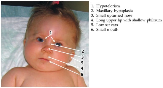
6. Valproic Acid Syndrome (Valproic Acid Spectrum Disorder)

A specific set of facial dysmorphic features related to the effects of VPA on the developing embryo and fetus was first described by DiLiberti et al. in 1984, in seven infants [38]. This syndrome was later corroborated by many authors describing additional children exposed in utero to VPA and exhibiting similar facial features [11][39][40][41][42][43]. The main clinical findings include intrauterine growth retardation (IUGR), a long and thin upper lip, shallow philtrum, epicanthal folds and mid face hypoplasia manifested by a flat nasal bridge, small upturned nose and downturned angles of the mouth (Figure 1). Many of the children with “valproate syndrome” also have other congenital anomalies, developmental delay and neurological impairment [40]. A similar set of anomalies was also described in the offspring of women using other AEDs in pregnancy, with definitions representing quite similar facial dysmorphic features following in utero exposure to phenytoin, carbamazepine, phenobarbital, trimethadione and primidone [19][42]. Thus, it is accepted that these dysmorphic features constitute the “Antiepileptic Drugs Syndrome”.
Figure 1. Typical facial changes in VPA syndrome.
Dean et al. [44] proposed a set of clinical findings that might be of help in diagnosing the “fetal antiepileptic drug syndrome” and they also included facial dysmorphic features that seem to be common to all antiepileptic drugs. Kini et al. [11] found that among fifty-six children exposed in utero to VPA, only five (9%) had normal facial appearance while twenty-four (42%) had moderate to severe facial dysmorphic features similar to those described for the VPA syndrome. A reduction in verbal intelligence was common among these children. Based on many studies, it seems that the “fetal valproate syndrome” is not significantly distinct from the “antiepileptic drug syndrome. It is difficult to diagnose VPA embryopathy from the facial appearance only, as similar facial features can be observed with other AEDs and also in a small percentage of normal non-exposed children, without a history of intrauterine VPA exposure. The specific facial features, often accompanied by other major anomalies and/or developmental delay, are a helpful tool in the diagnosis of VPA embryopathy [11][19][42][44].

7. VPA and Neurodevelopmental Problems

7.1. Impaired Neurodevelopment

Impaired neurodevelopment induced by prenatal VPA is based on prospective and retrospective neurodevelopmental studies of a relatively large number of children prenatally exposed to VPA. Neurodevelopmental deficits, including delayed motor skills, reduced cognitive functions, language impairment, specific neurodevelopmental syndromes (i.e., ASD and ADHD) and behavioral deficits were all described [6][9][45][46][47]. There seems to be a strong correlation between the dysmorphic facial features constituting the valproate syndrome and neurodevelopmental/cognitive impairment.
While evaluating the results of developmental studies, it is important to remember that there may be many confounding factors that should also be considered. Some of them, like alcohol or drug abuse, are not reported. Hence, the presence of the typical facial features of valproate syndrome is an important marker for the involvement of VPA in neurodevelopmental problems.

7.2. Intellectual and Learning Disabilities

As of today, the intellectual abilities of hundreds of children exposed in utero to VPA have been studied [11][26][42][44][45][46][47][48][49][50][51][52][53][54][55]. Koch et al. [46] studied 40 children exposed in utero to a single antiepileptic drug: phenobarbital, phenytoin or VPA. The VPA-exposed neonates exhibited high excitability and the degree of excitation correlated with the VPA serum concentrations at birth. Later, at six years of age, the neurological dysfunction of these children further correlated with their neonatal VPA blood levels [46]. Kini et al. [11] studied 63 children, 0.5–16 years of age who were exposed in utero to VPA, and found that 14% had major anomalies.
Shallcross et al. [56], in the UK, studied the neurodevelopment of 44 VPA-exposed infants of less than 24 months of age in comparison to 51 infants born to mothers treated with levetiracetam and 97 controls using the Griffith Mental Development Scales. They found normal development in infants prenatally exposed to levetiracetam, but reduced mental abilities in those born to VPA-treated mothers. [56]

7.3. VPA and ADHD

During the last years, information has accumulated that prenatal VPA not only increases the rate of ASD among the offspring, but also significantly increases the prevalence of ADHD. In the study [45] on 30 children exposed prenatally to VPA compared to 42 children exposed to lamotrigine and 52 non-exposed children [57] to assess preschool ADHD. Higher scores imply a higher probability of ADHD. The global score of the parents Conners questionnaire was significantly higher (p = 0.01) than that of lamotrigine-exposed children or of controls. The ADHD score of the teacher’s questionnaire was also higher, but the difference was insignificant. Similarly, visual–motor integration, visual perception and motor control were significantly lower. None of the VPA-exposed children had facial dysmorphic features.
Cohen et al., 2013 studied the rate of inattentive and combined types of ADHD in 45, six-year-old children prenatally exposed to VPA in comparison to children exposed to other AEDs [58]. They found that 10 of the children (22%) had either inattentive or combined types of ADHD according to the parents’ BASC questionnaire and 11 (26%) had either inattentive or combined types of ADHD according to the teachers’ BASC questionnaire.

7.4. VPA and Autism Spectrum Disorder (ASD)

A possible association between in utero VPA exposure and ASD was apparently first observed by Christianson et al. who described four children exposed in utero to VPA; all demonstrated developmental delay and one of these children also had ASD [48]. Later, Williams and Hesh [59] and Rasalam et al. [60] described additional children with the typical facial features of VPA embryopathy who also developed the typical findings of autism as outlined in the DSM IV. Moore et al. [42] found that among 57 children affected by antiepileptic drugs, four had ASD and two had Asperger syndrome. Five of the affected children (10.8% of 46 exposed to VPA) were exposed to VPA alone or combined with carbamazepine or phenytoin.
Other investigators, generally using the ICD 9 criteria, found a slightly lower rate of ASD, but one still significantly higher than in the general population. For example, Christensen et al. conducted a population-based study evaluating 655,615 children born from 1996 to 2006 in Denmark, with 508 prenatally exposed to VPA [61]. They found a clear association between prenatal exposure to VPA and an increased risk of childhood autism. However, the prevalence of ASD (ASD and childhood autism) was only about 4.42% with a hazard ratio of 2.9. Later, several studies reported similar associations of prenatal VPA treatment and ASD [62][63][64][65][66]

8. VPA and Folic Acid Administration in Human

VPA and many antiepileptic drugs (phenytoin, barbiturates, carbamazepine and lamotrigine) may interfere with folic acid absorption or metabolism, possibly an additional cause for their induction of congenital anomalies [6][24]. It is therefore recommended to treat women on AEDs at preconception and in the first 2–3 months of pregnancy with folic acid, which protects humans from NTD and possibly cardiac and oro-facial malformations. Although the use of folic acid supplementation has been shown to generally decrease the incidence of NTD in humans, there is disagreement as to the benefit of folic acid in reducing the rate of AED-induced congenital malformations and NTD, especially following VPA intake [16][20][22][23][24][25]. Despite the uncertainty of effectiveness, it is recommended for women on AED therapy to take 4–5 mg/day of folic acid prior to any planned pregnancy.

9. VPA Transplacental Passage and Secretion into Milk and Semen

A basic principle for the action of any agent on the embryo and fetus is its ability to cross the placenta. VPA is known to cross the human placenta, and the clearance of VPA is increased during pregnancy [67][68]. Valproic acid levels in cord serum are often higher than in the mother and may be up to five times higher than the levels in maternal serum at term, with mean ratios of 1.4–2.4 and a very large variation [69]. These increased concentrations in the fetus have been attributed to a better binding in the fetal compartment than in the maternal. The high, possibly toxic concentrations in the fetus, may partially explain the high teratogenicity of this drug.
VPA is excreted into human milk in relatively low concentrations [70][71][72]. Available reports suggest that a suckling infant may ingest less than 5% of the weight-adjusted maternal daily dose [68]. Kacirova et al. [70], in a study carried out in 2021 women, examined the VPA concentrations in breast milk and newborn serum and found that the infant serum concentrations and maternal concentrations in milk are low, ranging from 0.01 to 1.61, with an average of 0.51. Hence, lactation by VPA-treated mothers is permissible.

10. Malformations in Children of Untreated Epileptic Women

Since many of the antiepileptic drugs are teratogenic in man and in experimental animals, the question often asked is whether epilepsy may cause an increase in the rate of MCMs without any relation to treatment. This is in spite of the fact that there are several AEDs that are not teratogenic and do not increase the rate of MCMs in prenatally exposed children (i.e., Lamotrigine, Levetiracetam). Moreover, the studies carried out in experimental animals, which demonstrate AED teratogenicity, are performed on non-epileptic animals. That epilepsy might play an important role in the teratogenicity of AEDs was shown by Shapiro et al. in 1976 [73], who analyzed the data from two cohorts of children born to treated and untreated epileptic mothers—the Finnish and American data that did not include women treated by VPA. The authors raised the possibility that the increased rate of malformations found in the offspring of epileptic mothers treated by phenytoin stems from maternal epilepsy since a similar rate of malformations (apparently major and minor malformations) was similar in children born to non-treated epileptic women. If the father had epilepsy, the rate of anomalies was intermediate between that of the epileptic and control mothers.
Kaaja et al. [74] prospectively studied the outcome of pregnancies in 988 epileptic women, of which 239 were not exposed during the first trimester of pregnancy to AEDs while the others were treated with various AEDs. They found at birth a 3.8% rate of MCMs among the offspring of the treated women and a rate of only 0.8% among the offspring of untreated epileptic women, with no correlation between the number of seizures during pregnancy and the outcome. 
In addition, several studies have found that when AEDs are administered for psychiatric indications (i.e., VPA or carbamazepine for the treatment of bipolar disorder) they are still teratogenic, with the rate of malformations being similar to that in children of treated epileptic mothers [7].

11. Animal Studies on VPA-Induced Teratogenicity

11.1. VPA-Induced MCMs–General

Since the confirmation of VPA as a potent human teratogen, many studies employing different laboratory animal species (mice, rats, zebra fish, hamsters, drosophila, chicken, calf, rabbits and monkeys) of various strains have made extensive efforts to recapitulate the adverse effects of VPA reported in humans, in order to understand the mechanisms by which this drug mediates teratogenicity [75][76][77][78][79][80][81][82][83][84][85].
Similar to its effects in humans, VPA has exhibited dose-related and developmental-age-dependent teratogenic effects in various forms including, but not limited to, increased mortality rate, intrauterine growth retardation, gross craniofacial and skeletal anomalies, epigenetic aberrations and neurodevelopmental disorders, including ASD-like behaviors, in all animal species studied [86][87][88][89][90][91].

11.2. VPA-Induced General (Microscopic) Effects on the Nervous Tissue

VPA teratogenicity is manifested in brain tissue in different forms and magnitude, in a dose- and sex-related fashion. Mowery et al. [92] reported related abnormalities in the cerebellar nuclei of rodents prenatally exposed to VPA, with interpositus, fastigial and dentate nuclei of the cerebellum mostly affected. Hara et al., demonstrated in female mice offspring that exposure to VPA at GD 12.5 significantly decreased the number of Nissl-positive cells in the prefrontal cortex, supporting the dimorphism of VPA-induced social and communication deficits, and morphological changes in the somatosensory and prefrontal cortex leading to memory deficits [93]. In the parietal and occipital lobes of mouse cerebral cortex, VPA induced a characteristic asymmetry of parvalbumin (PV) cell reduction across hemispheres [94].

11.3. VPA-Induced Microscopic Pathological Changes in the Liver and Heart

VPA may induce hepatotoxicity and increased liver oxidative stress [95]. Hence, it is not surprising that it may causes hepatic damage/malformations in the VPA-exposed fetuses. One of the very few studies on the teratogenic effect of VPA on the liver reported that a single intraperitoneal injection of VPA (200 mg/kg) on GD 8 in pregnant mice resulted in offspring with a smaller liver size, whereby microscopic studies revealed a dilation of the central vein of liver lobules, breakage of the endothelial lining of the liver’s central veins, edematous swelling and a distortion of the normal architecture of the liver parenchyma [96]. Electron microscopic studies of the kidneys of mice prenatally exposed to VPA showed irregular glomerular basal membranes and marked deletions of the foot process [97][98]. Prenatal VPA induced the upregulation of thrombospondin-1 and activin proteins, inhibiting angiogenesis and vasculogenesis (manifested as failed endothelial cell tube formation) in a dose-related manner [99].

11.4. VPA-Induced Congenital Malformations in Animals

VPA administration during pregnancy alone or in combination with other drugs, induces a high number of anomalies, resorption and increased mortality rate. Most of the studies reported on exencephaly and spina bifida occulta as well as spina bifida aperta, thereby mimicking NTD as found in humans [6][28][76][87][88][89][100][101][102][103][104][105]. As expected, in addition to the dose relationship of VPA in the pathophysiology of NTD, gestational age is a crucial factor in determining what type of NTD is induced by VPA exposure. For example, Nau and co-workers treated mice with multiple high doses of VPA on gestation day 9 (GD) but not GD 8, and successfully produced posterior neural tube failure coinciding with spina bifida, unlike preceding studies where VPA on day 8 only produced cranial NTD (exencephaly) [100]. The rate of NTD was significantly reduced by high doses of folic or folinic acid [106]. Methionine, if given to pregnant mice a short time prior to VPA injection, significantly reduced embryonic VPA damage [107].

12. VPA-Induced Autistic-like Behavior in Animals

Rodents exposed to VPA during prenatal development exhibit behavioral deficits that resemble the characteristics observed in individuals with autism [108][109][110][111], a fact that strengthens the possible association in man between in utero VPA exposure and ASD. Prenatal VPA exposure in animals has been proposed as a model of autism that has both construct and face validity due to similarity in disease etiology and resemblance to human symptoms [109][112].
Rodier et al. [113] found that an injection of 350 mg/kg body weight to rats during days 11.5–12.5 of pregnancy, at the time of neural tube closure, reduced the number of neurons in the motor nuclei of cranial nerves within the brainstem, without any other morphological changes in the brain. This was similar to the postmortem changes observed in the brain of an autistic child [113]. Later, the same group also found a reduction in the size of the cerebellar hemispheres and in the number of cerebellar Purkinje cells following an injection of 600 mg/kg VPA on day 12.5 of gestation in rats [114]. These findings were consistent with those observed in ASD in humans, such as a reduction in Purkinje cell numbers in the cerebellum and anomalies of the inferior olive in the brainstem and the deep cerebellar nuclei [115][116].
ASD-like behavior was observed in mice injected with a single dose of 600 mg/Kg VPA on postnatal day 4. In addition to these behavioral studies, scholars also observed on day 60 changes in the expression of many genes, several of them already found to be associated with ASD in the SFARI list of genes [91][117]. This implies a possible epigenetic mechanism underlying the effects of VPA, probably related to the well-known inhibitory effects of VPA on histone deacetylase. There was also increased oxidative stress in the prefrontal cortex of VPA-treated pups. All effects including the changes in gene expression and behavior were sex-related [118].
Similar findings were observed following the injection of VPA on day 12 of gestation. Additionally, these mouse models showed evidence of abnormal but typical autistic-like calls on ultrasonic vocalization, abnormal EEG recordings and an imbalance between excitatory and inhibitory neurotransmission with repetitive and abnormal social behaviors typical to ASD patients reported and reviewed in [14][91][118][119][120][121]. The suggested mechanisms of action in the brain include increased glutamatergic neural density which leads to an excitatory/inhibitory imbalance, altered monoamines brain turnover, increased reactive oxygen species and epigenetic modifications [91][122].

13. VPA-Induced Changes in Gene Expression in the Brain of Animals

Studies utilizing animal models have shown that prenatal exposure to VPA can lead to changes in the expression of genes related to brain development, synaptic function and neurotransmitter signaling pathways, which are associated with ASD risk genes [91][117][123][124][125][126][127].
Feleke et al. [124] investigated how prenatal exposure to VPA influenced gene expression in term, day 21 fetuses. They studied fetuses from epileptic (Genetic Absence Epilepsy Rats from Strasbourg) and non-epileptic control rats to understand the impact of the genetic background. The analyses of genome-wide gene expression data showed that the majority of genes differentially expressed by VPA in epileptic pups exhibited similar changes to those observed in non-epileptic pups [124]. The investigators concluded that the altered pattern of gene expression triggered by VPA is independent of the genetic epilepsy background. Pathway enrichment analysis revealed that genes downregulated by VPA exposure exhibited enrichment for functional processes associated with the modulation of synaptic function and neuronal processes such as genes related to the glutamate receptor complex and neurotransmitter receptor activity, including genes for the regulation of insulin secretion [124].
Huang et al. [126] investigated the gene expressing analysis using a microarray in 50-day-old male rat hippocampi exposed to 600 mg/kg VPA on day 12.5. The majority of the 721 genes that were differently expressed were downregulated. The enrichment analysis of differentially-expressed genes in VPA-exposed rat hippocampi revealed associations with the plasma membrane, G-protein signaling, amine binding, and calcium signaling.

14. The Suggested Mechanism of the Teratogenic Action of VPA

14.1. Folate One-Carbon Metabolism (OCM) and Folate Deficiency

Theories on the mechanism of action of AED-induced malformations with anti-folate activity focused on the reduction in folic acid levels that often occurs after treatment with VPA and other AEDs. Indeed, VPA acts specifically as an antimetabolite to folic acid [24][111][128]. Reduced embryonic folic acid may disrupt gene expression, increase embryonic oxidative stress and induce changes in protein synthesis [106][128]. As folic acid antagonists generally produce birth defects [16], it would be consistent for anticonvulsant-mediated folic acid deficiency to result in fetal anomalies of the type observed in the offspring of VPA-treated women, i.e., NTD, cardiac anomalies and neurodevelopmental delay [28].
Through this pathway, VPA causes alterations in the glycine cleavage system, and blocks the dihydrofolate reductase and other enzymes involved in folate and SAMe cycles [107][129][130][131]. These effects of VPA may prevent the transfer of a one-carbon unit for the relevant cycles to progress, leading to abnormal methylation, phospholipid, polyamines, protein and nucleic acid synthesis [132][133]. It is reported that VPA, via the OCM, may also increase the oxidative stress burden, which will further induce major and minor congenital malformations [130][134][135][136][137]. Pregnant Wistar rats treated with different doses of VPA in mid and late gestational periods revealed alterations in several genes and enzymes of the OCM and folate transporter (folR1) [138]. Reynold and co-workers [139][140] demonstrated that VPA alters the expression of methylenetetrahydrofolate reductase and methyltetrahydrofolate reductase, and formyl hydrofolate reductase and dihydrofolate reductase. Alterations of the former are mediated in homocysteinuria while alterations of the later are involved in anti-tumorigenesis. VPA’s effect on formyl hydrofolate reductase inhibits the purine pathway responsible for protein synthesis [140]. The question of whether folic acid supplementation in pregnancy is effective in reducing the VPA teratogenicity is in debate.
The fact that folic acid administration, even in large doses, does not seem to prevent VPA-induced MCM may suggest that the above proposed mechanism of VPA teratogenic effects is of minor importance. Indeed, recent findings have continued to demonstrate the possibility that VPA teratogenicity is not primarily caused by its interference with folic acid metabolism, but by the alteration of several other pathways and mechanisms that play different significant roles in the OCM [6][14][140][141][142][143][144][145][146]. These current observations are evoking attention towards other components, including the important rate-limiting factors of OCM via which folic acid exerts its influence against the pathophysiology of VPA. (Figure 2).
Figure 2. Suggested mechanism of the teratogenic action of VPA-inducing epigenetic changes. Several biological pathways by which VPA causes congenital malformations and neurodevelopmental abnormalities have been proposed. Among them, one-carbon metabolism (OCM) and inhibition of HDAC enzymes seem to be the main contributors to VPA teratogenicity. VPA blocks the dihydrofolate reductase and methyltransferases (MTases) enzymes that are involved in folate and SAMe cycles in one-carbon metabolism (OCM). Thus, VPA interferences in OCM resulted in depleted endogenous folate levels, SAM: SAH ratio imbalance and higher plasma concentrations of SAH with lower concentrations of plasma SAMe that were indeed observed in women with NTD-affected pregnancies. The decline in SAMe production, a critical methyl donor, contributes to a decrease in DNA methylation levels, subsequently increasing gene expression. VPA’s disruption of folate-dependent pathways and the subsequent impact on SAMe levels, in turn, influences DNA methylation, and gene expression, and potentially contributes to the observed developmental abnormalities and congenital malformations associated with VPA exposure during pregnancy. Similarly, inhibition of HDAC enzymes leads to enhanced histone acetylation and formation of loose chromatin, and therefore an increase in gene expression.

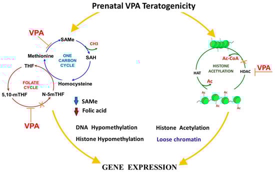
14.2. Alterations in the SAMe Cycle and VPA-Induced Malformations

S-Adenosine Methionine (SAMe), an FDA-approved food additive sold over the counter, has been found to have antagonistic actions against VPA. Being the primary and major biological methyl donor synthetized by all living organisms, it donates its methyl group to nucleic acid, proteins, and many other molecules for normal cell functioning [14]. SAMe serves as a precursor for the synthesis of polyamines, lipids, nucleotides, proteins, monoamine neurotransmitters, and glutathione (GSH) via three main interconnected metabolic pathways: polyamine synthesis, transmethylation, and transsulfuration [14]. The reaction between methionine and ATP catalyzed by methyltransferase (MTases) produces SAMe which is further catalyzed by other MTases, as a result of which S-adenosylhomocysteine (SAH) is produced. SAH is a competitive inhibitor of MTases and is essentially hydrolyzed to homocysteine and adenosine by adenosylhomocysteinase (AHCY). The ratio of SAM:SAH is used in determining cellular methylation potential whereby imbalance in this ratio will result in failures in methionine, SAMe, and reduced glutathione (GSH) synthesis, leading to homocysteinuria [131][147]. This represents the hallmark of aberrant methylation origin where VPA is a major culprit [148][149][150]. Women with NTD-affected pregnancies possess higher plasma concentrations of SAH with lower concentrations of plasma SAMe. Such metabolic arrangement corresponds significantly with reduced methylation capacity in consistently affected pregnancies [151]. Semmler and co-workers [130] showed that VPA altered the SAM:SAH ratio apparently leading to reduced hippocampal cell numbers, reduced brainstem volume, and impaired memory and learning.

14.3. The Inhibition of Histone Deacetylases (HDAC)

There is growing interest and several clinical trials using VPA (and other HDAC inhibitors), especially in the field of cancer research, on the premise that HDACs are promising targets for therapeutic interventions intended to reverse aberrant histone acetylation states. Acetylation is tightly governed by the opposing actions of two large families of enzymes: histone acetyltransferases (HATs) and histone deacetylases (HDACs) [6][120][152]. The proper regulation of these two opposing enzymatic activities is of paramount importance for the normal control of development, the failure of which will result in disease conditions.
Another mechanism of VPA teratogenicity is this observed inhibition of HDACs leading to changes in gene expression induced in the neonate when VPA was administered on postnatal day 4, and in the fetus when given on prenatal day 12 of pregnancy [90]. Paradoxically, the epigenetic involvement of histone acetylation and deacetylation in numerous pathophysiological pathways implies that HDAC inhibition is capable of leading to the adverse effect of an aberrant global regulation of gene expression [152] (Figure 2).
The first report of VPA as an HDAC inhibitor was apparently by Menegola et al. [90] who, in HeLa cell lines, demonstrated VPA’s inhibitory activities on HDAC, its capability of halting the cell cycle to induce growth arrest and apoptosis. This inhibitory effect of VPA on proliferating cells presumably explains its teratogenicity affecting neural tube closure [90]. Histological analysis and RT-PCR assays to examine the expression of myocardial cell-related genes in mice exposed to VPA revealed that the transcriptional levels of heart development-related genes (CHF1, Tbx5 and MEF2), are significantly increased in the hearts of VPA-exposed mouse fetuses [153].
Interestingly, the RNA sequencing analysis of human cortical neurons treated with VPA revealed that the genes showing differential expression were notably associated with functions such as mRNA splicing, mRNA processing, histone modification, and metabolism-related gene sets [154]. The analysis of differential transcript usage (DTU) revealed that VPA exposure brings about significant changes in DTU isoforms within the genes that are crucial for neurodevelopment and coincide with identified ASD-risk genes [154].

14.4. Increased Oxidative Stress

Several AEDs, similar to diabetes and lead (Pb) teratogenicity cause a heightened oxidative stress burden on the developing fetus [4][155][156][157][158] which is implied as one of VPA’s mechanisms of teratogenic action [6]. Generally, the developing embryo lacks a fully functional antioxidant defense system in the early gestational age, and a consequent irreversible embryonic and fetal damage may result from the abnormal elevation of reactive oxygen-nitrogen species. The brain particularly lags behind other fetal organs considering the development of the body’s antioxidant system, and therefore is found to be more adversely affected in instances of oxidative stress [159]. VPA, when injected intraperitoneally on GD 8 to pregnant ICR mice induced marked fetal malformations and evidence of the formation of peroxynitrite and S-nitrosylation in addition to an altered GSH level that induced the apoptosis of neural tube cells and macrophages [84].

14.5. Mitochondrial Dysfunction

The dysregulation of mitochondrial metabolism has been implicated as a possible pathophysiology of abnormalities caused by VPA treatment [14][103]. Salimi reported a collapse of mitochondrial membrane potential that would undermine synaptic plasticity [81]. Investigations on the effects of VPA in undermining the functional integrity of mitochondria revealed various outcomes, including the conformational alteration of the inner membrane and transport chain proteins, ATP depletion and increased reactive oxygen species (ROS) [160][161]. A dose-dependent VPA-induced cristae distortion in pyramidal neurons that altered mitochondrial ultrastructure was suggested as a sign of an impairment of synaptic plasticity and neurogenesis in treated rodents [162][163]. It was also observed in pigs that VPA inhibits mitochondrial respiratory chain complexes I, leading to the asymmetrical distribution and fragmentation of cristae, raised oxidative stress and the enlargement of the mitochondrial matrix [164][165][166].

14.6. Inositol Depletion

Inositol plays a crucial role in major signaling pathways that influence diverse cellular functions and offers an interface between membranes and the cytosol, the coordination of endocytosis and vesicle trafficking. The depletion of inositol reportedly causes cranial NTDs in mouse embryos, in a scenario that folic acid deficiency did not- and this was explained by the fact that inositol is a key factor in cell shaping and rearrangement. Therefore, depletion in the levels of inositol may have severe adverse developmental consequences [167][168][169][170].

15. The Prevention of VPA Teratogenicity in Animals in Relation to Congenital Malformations

15.1. Attempts to Minimize VPA Teratogenicity

Generally, if a substance is a suspected or proven teratogen, the advice is, whenever possible, to refrain from exposure. Often, this exposure cannot be prevented as the pregnant woman needs to continue her exposure to the teratogen for her or her conceptus’ health. A vivid example is the need to continue the treatment with anti-seizure medications in spite of their possible teratogenicity. Hence, the possibility to prevent the undesirable effects of a teratogen is of utmost importance [171]. A vivid example for such an approach is the prevention of NTD and other malformations by folic acid that has the backing of many animal and human studies.
Several investigators have made attempts to improve the efficacy and usage of VPA, by introducing different changes to the chemical structure of VPA in order to minimize the teratogenic effects while simultaneously retaining its therapeutic activity and potency [87][172][173][174][175][176][177]. So far, most attempts were not successful. However, with several VPA analogs now in clinical trial, there are possibilities that a potent and less teratogenic VPA congener will soon emerge.

15.2. VPA and Folic Acid (FA) Administration in Animals

As mentioned above, the prevention of VPA-induced malformations in human by folic acid administration has relatively little effect. Controversial findings were also reported in animals. Several investigators reported that folinic acid, regardless of the route of administration, failed to reduce the incidence of VPA-induced exencephaly in rodents. Neither did it alter the teratogenicity of VPA on the brain, liver, or kidney and other embryonic tissues, suggesting that folate probably has no significance in the pathophysiology of VPA-induced congenital malformations [129].
Furugen et al. [138] reported that VPA altered mRNA levels of major carriers for folate, glucose, choline, SAMe and some hormones. They concluded that VPA targets the folate receptor FOLR1, and possibly other folate receptors, leading to the direct inhibition of placental folate uptake. Earlier, Fathe et al. [178], utilizing cell culture modeling, found that VPA is a noncompetitive inhibitor of high affinity folate receptors such as folate receptorα (FRα). Hence, VPA may interfere with folate metabolism by inhibiting glutamate formyl transferase, an enzyme that produces 5-formyltetrahydrofolate (folinic acid), thereby inhibiting methionine, choline and SAMe syntheses, leading to adverse epigenetic changes [139][140].

16. The Prevention of the VPA Induction of ASD in Preclinical Animal Models

Preventing or treating VPA-induced ASD-like behavior in animal models has been a subject of experimental research, primarily to better understand the mechanisms involved and to explore potential preventive measures. Some strategies and findings from experimental studies include supplementation with antioxidants [179][180], epigenetic modulator molecules [14][120], environmental enrichment [181], pharmacological [182] and genetic interventions [183].
The timing of pharmacological treatments in studies significantly impacts their effects and outcomes. Treating VPA-exposed offspring at different stages of development, whether it is prenatally, early postnatally, during adolescence or adulthood, can yield different results and treatment efficacy. Most reported pharmacological studies based on the strategy of giving treatments to VPA-exposed offspring at adolescence or adulthood reported that the beneficial effects of treatments were generally transient.
Kumar and Sharma [182] investigated the effects of minocycline, a known modulator of retinoic acid signaling, in an ASD-like model induced by 500 mg/kg VPA given to pregnant rats on day 12.5.of gestation. Oral treatment with 25 and 50 mg/kg minocycline from postnatal days 21–50 normalized social interaction, spontaneous alteration, exploratory activity, intestinal motility, serotonin levels and prefrontal cortex mitochondrial complex activity.

17. Conclusions

Valproic acid seems to be the highest teratogenic drug among the AEDs and therefore, if possible, should be avoided during childbearing age, especially in pregnancy. Because NTD may be induced in the third week post fertilization, it may be too late to stop the medication when pregnancy is first diagnosed. Therefore, VPA-treated women at childbearing age should use contraceptives and stop the medication before any planned pregnancy. Moreover, due to its neurobehavioral negative effects even late in pregnancy (i.e., intellectual disability, ASD, ADHD and other neurodevelopmental disabilities), it seems advisable to change VPA to a different antiepileptic drug even during pregnancy if the alternative drug is effective. VPA significantly increases the teratogenic potential of other antiepileptic drugs. If there is no alternative to VPA treatment, the pregnancy should be considered high risk and must have proper follow-up, including appropriate antenatal diagnosis. It is also advised to use the lowest effective amount divided into three daily doses to minimize fluctuations of serum levels of VPA. Daily doses lower than 600 mg seem to have a low teratogenic effect. The addition of 4–5 mg/day of folic acid should also be considered, although its efficacy is somewhat in doubt.

References

  1. Ornoy, A. The impact of intrauterine exposure versus postnatal environment in neurodevelopmental toxicity: Long-term neurobehavioral studies in children at risk for developmental disorders. Toxicol. Lett. 2003, 140–141, 171–181.
  2. Arnon, J.; Shechtman, S.; Ornoy, A. The use of psychiatric drugs in pregnancy and lactation. Isr. J. Psychiatry Relat. Sci. 2000, 37, 205–222.
  3. Jentink, J.; Loane, M.A.; Dolk, H.; Barisic, I.; Garne, E.; Morris, J.K.; de Jong-van den Berg, L.T. Valproic acid monotherapy in pregnancy and major congenital malformations. New Engl. J. Med. 2010, 362, 2185–2193.
  4. Ornoy, A. Neuroteratogens in man: An overview with special emphasis on the teratogenicity of antiepileptic drugs in pregnancy. Reprod. Toxicol. 2006, 22, 214–226.
  5. Kultima, K.; Nyström, A.-M.; Scholz, B.; Gustafson, A.-L.; Dencker, L.; Stigson, M. Valproic Acid Teratogenicity: A Toxicogenomics Approach. Environ. Health Perspect. 2004, 112, 1225–1235.
  6. Ornoy, A. Valproic acid in pregnancy: How much are we endangering the embryo and fetus? Reprod. Toxicol. 2009, 28, 1–10.
  7. Diav-Citrin, O.; Shechtman, S.; Bar-Oz, B.; Cantrell, D.; Arnon, J.; Ornoy, A. Pregnancy outcome after in utero exposure to valproate: Evidence of dose relationship in teratogenic effect. CNS Drugs 2008, 22, 325–334.
  8. Morrow, J.; Russell, A.; Guthrie, E.; Parsons, L.; Robertson, I.; Waddell, R.; Irwin, B.; McGivern, R.C.; Morrison, P.J.; Craig, J. Malformation risks of antiepileptic drugs in pregnancy: A prospective study from the UK Epilepsy and Pregnancy Register. J. Neurol. Neurosurg. Psychiatry 2006, 77, 193–198.
  9. Bromley, R.; Weston, J.; Adab, N.; Greenhalgh, J.; Sanniti, A.; McKay, A.J. Treatment for epilepsy in pregnancy: Neurodevelopmental outcomes in the child. Cochrane Database Syst. Rev. 2014, 10, CD010236.
  10. Weston, J.; Bromley, R.; Jackson, C.F.; Adab, N.; Clayton-Smith, J.; Greenhalgh, J.; Hounsome, J.; McKay, A.J.; Tudur Smith, C.; Marson, A.G. Monotherapy treatment of epilepsy in pregnancy: Congenital malformation outcomes in the child. Cochrane Database Syst. Rev. 2016, 11, CD010224.
  11. Kini, U.; Adab, N.; Vinten, J.; Fryer, A.; Clayton-Smith, J. Dysmorphic features: An important clue to the diagnosis and severity of fetal anticonvulsant syndromes. Arch. Dis. Child. Fetal Neonatal Ed. 2006, 91, F90–F95.
  12. Fried, S.; Kozer, E.; Nulman, I.; Einarson, T.R.; Koren, G. Malformation rates in children of women with untreated epilepsy: A meta-analysis. Drug Saf. 2004, 27, 197–202.
  13. Anmella, G.; Pacchiarotti, I.; Cubała, W.J.; Dudek, D.; Maina, G.; Thomas, P.; Vieta, E. Expert advice on the management of valproate in women with bipolar disorder at childbearing age. Eur. Neuropsychopharmacol. J. Eur. Coll. Neuropsychopharmacol. 2019, 29, 1199–1212.
  14. Ornoy, A.; Becker, M.; Weinstein-Fudim, L.; Ergaz, Z. S-Adenosine Methionine (SAMe) and Valproic Acid (VPA) as Epigenetic Modulators: Special Emphasis on their Interactions Affecting Nervous Tissue during Pregnancy. Int. J. Mol. Sci. 2020, 21, 3721.
  15. Singh, D.; Gupta, S.; Verma, I.; Morsy, M.A.; Nair, A.B.; Ahmed, A.F. Hidden pharmacological activities of valproic acid: A new insight. Biomed. Pharmacother. 2021, 142, 112021.
  16. Ornoy, A.; Arnon, J. Clinical teratology. West. J. Med. 1993, 159, 382.
  17. Philbrook, N.A.; Nikolovska, A.; Maciver, R.D.; Belanger, C.L.; Winn, L.M. Characterizing the effects of in utero exposure to valproic acid on murine fetal heart development. Birth Defects Res. 2019, 111, 1551–1560.
  18. Bromley, R.; Adab, N.; Bluett-Duncan, M.; Clayton-Smith, J.; Christensen, J.; Edwards, K.; Greenhalgh, J.; Hill, R.A.; Jackson, C.F.; Khanom, S.; et al. Monotherapy treatment of epilepsy in pregnancy: Congenital malformation outcomes in the child. Cochrane Database Syst. Rev. 2023, 8, CD010224.
  19. Yerby, M.S. Pregnancy, teratogenesis, and epilepsy. Neurol. Clin. 1994, 12, 749–771.
  20. Bjerkedal, T.; Czeizel, A.; Goujard, J.; Kallen, B.; Mastroiacova, P.; Nevin, N.; Oakley, G., Jr.; Robert, E. Valproic acid and spina bifida. Lancet 1982, 2, 1096.
  21. Verloes, A.; Frikiche, A.; Gremillet, C.; Paquay, T.; Decortis, T.; Rigo, J.; Senterre, J. Proximal phocomelia and radial ray aplasia in fetal valproic syndrome. Eur. J. Pediatr. 1990, 149, 266–267.
  22. Kim, H.H.; Laufer, M.R. Developmental abnormalities of the female reproductive tract. Curr. Opin. Obstet. Gynecol. 1994, 6, 518–525.
  23. Okada, A.; Kurihara, H.; Aoki, Y.; Bialer, M.; Fujiwara, M. Amidic modification of valproic acid reduces skeletal teratogenicity in mice. Birth Defects Res. Part B Dev. Reprod. Toxicol. 2004, 71, 47–53.
  24. Yerby, M.S. Management issues for women with epilepsy: Neural tube defects and folic acid supplementation. Neurology 2003, 61, S23–S26.
  25. Koren, G.; Nava-Ocampo, A.A.; Moretti, M.E.; Sussman, R.; Nulman, I. Major malformations with valproic acid. Can. Fam. Physician Med. Fam. Can. 2006, 52, 441–442, 444, 447.
  26. Perucca, E.; Tomson, T. Prenatal exposure to antiepileptic drugs. Lancet 2006, 367, 1467–1469.
  27. Malm, H.; Kajantie, E.; Kivirikko, S.; Kääriäinen, H.; Peippo, M.; Somer, M. Valproate embryopathy in three sets of siblings: Further proof of hereditary susceptibility. Neurology 2002, 59, 630–633.
  28. Alsdorf, R.; Wyszynski, D.F. Teratogenicity of sodium valproate. Expert. Opin. Drug Saf. 2005, 4, 345–353.
  29. Rodríguez-Pinilla, E.; Mejías, C.; Prieto-Merino, D.; Fernández, P.; Martínez-Frías, M.L. Risk of hypospadias in newborn infants exposed to valproic acid during the first trimester of pregnancy: A case-control study in Spain. Drug Saf. 2008, 31, 537–543.
  30. Eadie, M.J.; Vajda, F.J. Should valproate be taken during pregnancy? Ther. Clin. Risk Manag. 2005, 1, 21–26.
  31. Campbell, E.; Kennedy, F.; Russell, A.; Smithson, W.H.; Parsons, L.; Morrison, P.J.; Liggan, B.; Irwin, B.; Delanty, N.; Hunt, S.J.; et al. Malformation risks of antiepileptic drug monotherapies in pregnancy: Updated results from the UK and Ireland Epilepsy and Pregnancy Registers. J. Neurol. Neurosurg. Psychiatry 2014, 85, 1029–1034.
  32. Artama, M.; Auvinen, A.; Raudaskoski, T.; Isojärvi, I.; Isojärvi, J. Antiepileptic drug use of women with epilepsy and congenital malformations in offspring. Neurology 2005, 64, 1874–1878.
  33. Kaneko, S.; Battino, D.; Andermann, E.; Wada, K.; Kan, R.; Takeda, A.; Nakane, Y.; Ogawa, Y.; Avanzini, G.; Fumarola, C.; et al. Congenital malformations due to antiepileptic drugs. Epilepsy Res. 1999, 33, 145–158.
  34. Matalon, S.; Schechtman, S.; Goldzweig, G.; Ornoy, A. The teratogenic effect of carbamazepine: A meta-analysis of 1255 exposures. Reprod. Toxicol. 2002, 16, 9–17.
  35. GSK The Lamotrigine Pregnancy Registry. Available online: http://pregnancyregistry.gsk.com/documents/lamreport (accessed on 1 January 2008).
  36. Laegreid, L.; Kyllerman, M.; Hedner, T.; Hagberg, B.; Viggedahl, G. Benzodiazepine amplification of valproate teratogenic effects in children of mothers with absence epilepsy. Neuropediatrics 1993, 24, 88–92.
  37. Holmes, L.B.; Mittendorf, R.; Shen, A.; Smith, C.R.; Hernandez-Diaz, S. Fetal effects of anticonvulsant polytherapies: Different risks from different drug combinations. Arch Neurol. 2011, 68, 1275–1281.
  38. DiLiberti, J.H.; Farndon, P.A.; Dennis, N.R.; Curry, C.J. The fetal valproate syndrome. Am. J. Med. Genet. 1984, 19, 473–481.
  39. Kozma, C. Valproic acid embryopathy: Report of two siblings with further expansion of the phenotypic abnormalities and a review of the literature. Am. J. Med. Genet. 2001, 98, 168–175.
  40. Ardinger, H.H.; Atkin, J.F.; Blackston, R.D.; Elsas, L.J.; Clarren, S.K.; Livingstone, S.; Flannery, D.B.; Pellock, J.M.; Harrod, M.J.; Lammer, E.J.; et al. Verification of the fetal valproate syndrome phenotype. Am. J. Med. Genet. 1988, 29, 171–185.
  41. Mawer, G.; Clayton-Smith, J.; Coyle, H.; Kini, U. Outcome of pregnancy in women attending an outpatient epilepsy clinic: Adverse features associated with higher doses of sodium valproate. Seizure 2002, 11, 512–518.
  42. Moore, S.J.; Turnpenny, P.; Quinn, A.; Glover, S.; Lloyd, D.J.; Montgomery, T.; Dean, J.C. A clinical study of 57 children with fetal anticonvulsant syndromes. J. Med. Genet. 2000, 37, 489–497.
  43. Winter, R.M.; Donnai, D.; Burn, J.; Tucker, S.M. Fetal valproate syndrome: Is there a recognisable phenotype? J. Med. Genet. 1987, 24, 692–695.
  44. Dean, J.C.; Moore, S.J.; Turnpenny, P.D. Developing diagnostic criteria for the fetal anticonvulsant syndromes. Seizure 2000, 9, 233–234.
  45. Rihtman, T.; Parush, S.; Ornoy, A. Developmental outcomes at preschool age after fetal exposure to valproic acid and lamotrigine: Cognitive, motor, sensory and behavioral function. Reprod. Toxicol. 2013, 41, 115–125.
  46. Koch, S.; Jäger-Roman, E.; Lösche, G.; Nau, H.; Rating, D.; Helge, H. Antiepileptic drug treatment in pregnancy: Drug side effect in the neonate and neurological outcome. Acta Paediatr. Int. J. Paediatr. 1996, 85, 739–746.
  47. Nicolai, J.; Vles, J.S.; Aldenkamp, A.P. Neurodevelopmental delay in children exposed to antiepileptic drugs in utero: A critical review directed at structural study-bias. J. Neurol. Sci. 2008, 271, 1–14.
  48. Christianson, A.L.; Chester, N.; Kromberg, J.G.R. Fetal Valproate Syndrome: Clinical and Neuro-developmental Features in Two Sibling Pairs. Dev. Med. Child Neurol. 1994, 36, 361–369.
  49. Dean, J.C.; Hailey, H.; Moore, S.J.; Lloyd, D.J.; Turnpenny, P.D.; Little, J. Long term health and neurodevelopment in children exposed to antiepileptic drugs before birth. J. Med. Genet. 2002, 39, 251–259.
  50. Viinikainen, K.; Eriksson, K.; Mönkkönen, A.; Aikiä, M.; Nieminen, P.; Heinonen, S.; Kälviäinen, R. The effects of valproate exposure in utero on behavior and the need for educational support in school-aged children. Epilepsy Behav. 2006, 9, 636–640.
  51. Adab, N.; Jacoby, A.; Smith, D.; Chadwick, D. Additional educational needs in children born to mothers with epilepsy. J. Neurol. Neurosurg. Psychiatry 2001, 70, 15–21.
  52. Adab, N.; Kini, U.; Vinten, J.; Ayres, J.; Baker, G.; Clayton-Smith, J.; Coyle, H.; Fryer, A.; Gorry, J.; Gregg, J.; et al. The longer term outcome of children born to mothers with epilepsy. J. Neurol. Neurosurg. Psychiatry 2004, 75, 1575–1583.
  53. Langer, B.; Haddad, J.; Gasser, B.; Maubert, M.; Schlaeder, G. Isolated fetal bilateral radial ray reduction associated with valproic acid usage. Fetal Diagn. Ther. 1994, 9, 155–158.
  54. Gaily, E.; Kantola-Sorsa, E.; Hiilesmaa, V.; Isoaho, M.; Matila, R.; Kotila, M.; Nylund, T.; Bardy, A.; Kaaja, E.; Granström, M.L. Normal intelligence in children with prenatal exposure to carbamazepine. Neurology 2004, 62, 28–32.
  55. Eriksson, K.; Viinikainen, K.; Mönkkönen, A.; Äikiä, M.; Nieminen, P.; Heinonen, S.; Kälviäinen, R. Children exposed to valproate in utero—population based evaluation of risks and confounding factors for long-term neurocognitive development. Epilepsy Res. 2005, 65, 189–200.
  56. Shallcross, R.; Bromley, R.L.; Irwin, B.; Bonnett, L.J.; Morrow, J.; Baker, G.A. Child development following in utero exposure: Levetiracetam vs sodium valproate. Neurology 2011, 76, 383–389.
  57. Conners, C.K. Conner’s Rating Scales—Revised Technical Manual; Multi-Health Systems Inc.: Toronto, ON, Canada, 1997.
  58. Cohen, M.J.; Meador, K.J.; May, R.; Loblein, H.; Conrad, T.; Baker, G.A.; Bromley, R.L.; Clayton-Smith, J.; Kalayjian, L.A.; Kanner, A.; et al. Fetal antiepileptic drug exposure and learning and memory functioning at 6 years of age: The NEAD prospective observational study. Epilepsy Behav. 2019, 92, 154–164.
  59. Williams, P.G.; Hersh, J.H. A male with fetal valproate syndrome and autism. Dev. Med. Child Neurol. 1997, 39, 632–634.
  60. Rasalam, A.D.; Hailey, H.; Williams, J.H.; Moore, S.J.; Turnpenny, P.D.; Lloyd, D.J.; Dean, J.C. Characteristics of fetal anticonvulsant syndrome associated autistic disorder. Dev. Med. Child Neurol. 2005, 47, 551–555.
  61. Christensen, J.; Grønborg, T.K.; Sørensen, M.J.; Schendel, D.; Parner, E.T.; Pedersen, L.H.; Vestergaard, M. Prenatal valproate exposure and risk of autism spectrum disorders and childhood autism. JAMA 2013, 309, 1696–1703.
  62. Wiggs, K.K.; Rickert, M.E.; Sujan, A.C.; Quinn, P.D.; Larsson, H.; Lichtenstein, P.; Oberg, A.S.; D’Onofrio, B.M. Antiseizure medication use during pregnancy and risk of ASD and ADHD in children. Neurology 2020, 95, e3232–e3240.
  63. Wood, A.G.; Nadebaum, C.; Anderson, V.; Reutens, D.; Barton, S.; O’Brien, T.J.; Vajda, F. Prospective assessment of autism traits in children exposed to antiepileptic drugs during pregnancy. Epilepsia 2015, 56, 1047–1055.
  64. Honybun, E.; Thwaites, R.; Malpas, C.B.; Rayner, G.; Anderson, A.; Graham, J.; Hitchcock, A.; O’Brien, T.J.; Vajda, F.J.E.; Perucca, P. Prenatal valproate exposure and adverse neurodevelopmental outcomes: Does sex matter? Epilepsia 2021, 62, 709–719.
  65. Petersen, I.; McCrea, R.L.; Sammon, C.J.; Osborn, D.P.J.; Evans, S.J.; Cowen, P.J.; Freemantle, N.; Nazareth, I. Risks and benefits of psychotropic medication in pregnancy: Cohort studies based on UK electronic primary care health records. Health Technol. Assess. 2016, 20, 1–176.
  66. Hisle-Gorman, E.; Susi, A.; Stokes, T.; Gorman, G.; Erdie-Lalena, C.; Nylund, C.M. Prenatal, perinatal, and neonatal risk factors of autism spectrum disorder. Pediatr. Res. 2018, 84, 190–198.
  67. Nau, H.; Schafer, H.; Rating, D.; Jakobs, C.; Helge, H. Placental transfer and neonatal pharmacokinetics of valproic acid and some of its metabolites. In Epilepsy, Pregnancy, and the Child; Janz, D., Bossi, L., Dam, M., Helge, H., Richens, A., Schmidt, D., Eds.; Raven Press: New York, NY, USA, 1982; pp. 367–372.
  68. Albani, F.; Riva, R.; Contin, M.; Baruzzi, A.; Altomare, M.; Merlini, G.P.; Perucca, E. Differential transplacental binding of valproic acid: Influence of free fatty acids. Br. J. Clin. Pharmacol. 1984, 17, 759–762.
  69. Kacirova, I.; Grundmann, M.; Brozmanova, H. Serum levels of valproic acid during delivery in mothers and in umbilical cord—Correlation with birth length and weight. Biomed. Pap. Med. Fac. Univ. Palacky Olomouc Czechoslov. 2015, 159, 569–575.
  70. Kacirova, I.; Grundmann, M.; Brozmanova, H. Valproic Acid Concentrations in Mothers, Colostrum and Breastfed Infants during the Early Postpartum Period: Comparison with Concentrations Determined during Delivery and in the Mature Milk Period. Pharmaceutics 2021, 13, 2074.
  71. Von Unruh, G.E.; Froescher, W.; Hoffmann, F.; Niesen, M. Valproic acid in breast milk: How much is really there? Ther. Drug Monit. 1984, 6, 272–276.
  72. The WHO Working Group. Drugs and Human Lactation; Bennet, P.N., Ed.; Elsevier: Amsterdam, The Netherlands; New York, NY, USA; Oxford, UK, 1988; pp. 341–342.
  73. Shapiro, S.; Hartz, S.C.; Siskind, V.; Mitchell, A.A.; Slone, D.; Rosenberg, L.; Monson, R.R.; Heinonen, O.P. Anticonvulsants and parental epilepsy in the development of birth defects. Lancet 1976, 1, 272–275.
  74. Kaaja, E.; Kaaja, R.; Hiilesmaa, V. Major malformations in offspring of women with epilepsy. Neurology 2003, 60, 575–579.
  75. Bold, J.; Sakata-Haga, H.; Fukui, Y. Spinal nerve defects in mouse embryos prenatally exposed to valproic acid. Anat. Sci. Int. 2018, 93, 35–41.
  76. Di Renzo, F.; Giavini, E.; Menegola, E. Methionine pretreatment enhances the effects of valproate on axial development in a cd1 mouse model. Birth Defects Res. Part B-Dev. Reprod. Toxicol. 2013, 98, 328–333.
  77. Shafique, S.; Winn, L.M. Gestational valproic acid exposure induces epigenetic modifications in murine decidua. Placenta 2021, 107, 31–40.
  78. Tiboni, G.M.; Ponzano, A. Prevention of valproic acid-induced neural tube defects by sildenafil citrate. Reprod. Toxicol. 2015, 56, 175–179.
  79. Elphick, L.M.; Pawolleck, N.; Guschina, I.A.; Chaieb, L.; Eikel, D.; Nau, H.; Harwood, J.L.; Plant, N.J.; Williams, R.S. Conserved valproic-acid-induced lipid droplet formation in Dictyostelium and human hepatocytes identifies structurally active compounds. Dis. Models Mech. 2012, 5, 231–240.
  80. Goetzl, L. Environmental agents and reproductive risk. In Queenan’s Management of High-Risk Pregnancy: An Evidence-Based Approach; Wiley-Blackwell: Hoboken, NJ, USA, 2012; pp. 32–40.
  81. Salimi, A.; Alyan, N.; Akbari, N.; Jamali, Z.; Pourahmad, J. Selenium and L-carnitine protects from valproic acid-Induced oxidative stress and mitochondrial damages in rat cortical neurons. Drug Chem. Toxicol. 2022, 45, 1150–1157.
  82. Shafique, S.; Winn, L.M. Role of Cbp, p300 and Akt in valproic acid induced neural tube defects in CD-1 mouse embryos. Reprod. Toxicol. 2020, 95, 86–94.
  83. Takla, T.N.; Luo, J.; Sudyk, R.; Huang, J.; Walker, J.C.; Vora, N.L.; Sexton, J.Z.; Parent, J.M.; Tidball, A.M. A Shared Pathogenic Mechanism for Valproic Acid and SHROOM3 Knockout in a Brain Organoid Model of Neural Tube Defects. Cells 2023, 12, 1697.
  84. Tiboni, G.M.; Ponzano, A.; Ferrone, A.; Franceschelli, S.; Speranza, L.; Patruno, A. Valproic acid alters nitric oxide status in neurulating mouse embryos. Reprod. Toxicol. 2021, 99, 152–159.
  85. Wachholz, G.E.; Rengel, B.D.; Vargesson, N.; Fraga, L.R. From the Farm to the Lab: How Chicken Embryos Contribute to the Field of Teratology. Front. Genet. 2021, 12, 666726.
  86. Binkerd, P.E.; Rowland, J.M.; Nau, H.; Hendrickx, A.G. Evaluation of valproic acid (VPA) developmental toxicity and pharmacokinetics in Sprague-Dawley rats. Fundam. Appl. Toxicol. Off. J. Soc. Toxicol. 1988, 11, 485–493.
  87. Lin, Y.L.; Bialer, M.; Cabrera, R.M.; Finnell, R.H.; Wlodarczyk, B.J. Teratogenicity of valproic acid and its constitutional isomer, amide derivative valnoctamide in mice. Birth Defects Res. 2019, 111, 1013–1023.
  88. Hendrickx, A.G.; Nau, H.; Binkerd, P.; Rowland, J.M.; Rowland, J.R.; Cukierski, M.J.; Cukierski, M.A. Valproic acid developmental toxicity and pharmacokinetics in the rhesus monkey: An interspecies comparison. Teratology 1988, 38, 329–345.
  89. Jazayeri, D.; Braine, E.; McDonald, S.; Dworkin, S.; Powell, K.L.; Griggs, K.; Vajda, F.J.E.; O’Brien, T.J.; Jones, N.C. A rat model of valproate teratogenicity from chronic oral treatment during pregnancy. Epilepsia 2020, 61, 1291–1300.
  90. Menegola, E.; Broccia, M.L.; Nau, H.; Prati, M.; Ricolfi, R.; Giavini, E. Teratogenic effects of sodium valproate in mice and rats at midgestation and at term. Teratog. Carcinog. Mutagen. 1996, 16, 97–108.
  91. Ornoy, A.; Weinstein-Fudim, L.; Tfilin, M.; Ergaz, Z.; Yanai, J.; Szyf, M.; Turgeman, G. S-adenosyl methionine prevents ASD like behaviors triggered by early postnatal valproic acid exposure in very young mice. Neurotoxicol. Teratol. 2019, 71, 64–74.
  92. Mowery, T.M.; Wilson, S.M.; Kostylev, P.V.; Dina, B.; Buchholz, J.B.; Prieto, A.L.; Garraghty, P.E. Embryological exposure to valproic acid disrupts morphology of the deep cerebellar nuclei in a sexually dimorphic way; Embryological exposure to valproic acid disrupts morphology of the deep cerebellar nuclei in a sexually dimorphic way. Int. J. Dev. Neurosci. 2015, 40, 15–23.
  93. Hara, Y.; Maeda, Y.; Kataoka, S.; Ago, Y.; Takuma, K.; Matsuda, T. Effect of prenatal valproic acid exposure on cortical morphology in female mice. J. Pharmacol. Sci. 2012, 118, 543–546.
  94. Gogolla, N.; Leblanc, J.J.; Quast, K.B.; Südhof, T.C.; Fagiolini, M.; Hensch, T.K. Common circuit defect of excitatory-inhibitory balance in mouse models of autism. J. Neurodev. Disord. 2009, 1, 172–181.
  95. Ezhilarasan, D.; Mani, U. Valproic acid induced liver injury: An insight into molecular toxicological mechanism. Environ. Toxicol. Pharmacol. 2022, 95, 103967.
  96. Shakya, P.; Mohanty, C.; Mohanty, C. Hepatotoxicity of Valproate on Fetal Mice Liver. IJSR-Int. J. Sci. Res. Med. Sci. 2016, 5, 79–80.
  97. Akta, A.; Nergız, Y.; Akku, M.; Nasır, Y. The effects of valproic acid on renal corpuscle of pregnant rats and protective role of folic acid and vitamin E. Afr. J. Biotechnol. 2010, 9, 5605–5610.
  98. Raza, M.; Al Shabanah, O.; Mohammed al-Bekairi, A.; Qureshi, S. Pathomorphological changes in mouse liver and kidney during prolonged valproate administration. Int. J. Tissue React. 2000, 22, 15–21.
  99. Michaelis, M.; Michaelis, U.R.; Fleming, I.; Suhan, T.; Cinatl, J.; Blaheta, R.A.; Hoffmann, K.; Kotchetkov, R.; Busse, R.; Nau, H.; et al. Valproic Acid Inhibits Angiogenesis in Vitro and in Vivo. Mol. Pharmacol. 2004, 65, 520–527.
  100. Nau, H.; Hauck, R.S.S.; Ehlers, K. Valproic Acid-Induced Neural Tube Defects in Mouse and Human: Aspects of Chirality, Alternative Drug Development, Pharmacokinetics and Possible Mechanisms. Pharmacol. Toxicol. 1991, 69, 310–321.
  101. Avagliano, L.; Massa, V.; George, T.M.; Qureshy, S.; Bulfamante, G.P.; Finnell, R.H. Overview on Neural tube defects: From development to physical characteristics. Birth Defects Res. 2019, 111, 1455.
  102. Genton, P.; Semah, F.; Trinka, E. Valproic acid in epilepsy: Pregnancy-related issues. Drug Saf. 2006, 29, 1–21.
  103. Hansen, J.M.; Lucas, S.M.; Ramos, C.D.; Green, E.J.; Nuttall, D.J.; Clark, D.S.; Marchant, E.D.; Hancock, C.R.; Piorczynski, T.B. Valproic acid promotes SOD2 acetylation: A potential mechanism of valproic acid-induced oxidative stress in developing systems Valproic acid promotes SOD2 acetylation: A potential mechanism of valproic acid-induced oxidative stress in developing systems Valproic acid promotes SOD2 acetylation: A potential mechanism of valproic acid-induced oxidative stress in developing systems. Free Radic. Res. 2021, 55, 1130–1144.
  104. Lundberg, Y.W.; Cabrera, R.M.; Greer, K.A.; Zhao, J.; Garg, R.; Finnell, R.H. Mapping a chromosomal locus for valproic acid-induced exencephaly in mice. Mamm. Genome 2004, 15, 361–369.
  105. Menegola, E.; Di Renzo, F.; Broccia, M.L.; Prudenziati, M.; Minucci, S.; Massa, V.; Giavini, E. Inhibition of histone deacetylase activity on specific embryonic tissues as a new mechanism for teratogenicity. Birth Defects Res. Part B Dev. Reprod. Toxicol. 2005, 74, 392–398.
  106. Wegner, C.; Nau, H. Diurnal variation of folate concentrations in mouse embryo and plasma: The protective effect of folinic acid on valproic-acid-induced teratogenicity is time dependent. Reprod. Toxicol. 1991, 5, 465–471.
  107. Ehlers, K.; Elmazar, M.M.A.; Hau, A.H. Methionine Reduces the Valproic Acid-Induced Spina Bifida Rate in Mice without Altering Valproic Acid Kinetics. J. Nutr. 1996, 126, 67–75.
  108. Błaszczyk, B.; Miziak, B.; Pluta, R.; Czuczwar, S.J. Epilepsy in Pregnancy-Management Principles and Focus on Valproate. Int. J. Mol. Sci. 2022, 23, 1369.
  109. Chaliha, D.; Albrecht, M.; Vaccarezza, M.; Takechi, R.; Lam, V.; Al-Salami, H.; Mamo, J. A Systematic Review of the Valproic-Acid-Induced Rodent Model of Autism. Dev. Neurosci. 2020, 42, 12–48.
  110. Wagner, G.C.; Reuhl, K.R.; Cheh, M.; McRae, P.; Halladay, A.K. A new neurobehavioral model of autism in mice: Pre- and postnatal exposure to sodium valproate. J. Autism Dev. Disord. 2006, 36, 779–793.
  111. Arndt, T.L.; Stodgell, C.J.; Rodier, P.M. The teratology of autism. Int. J. Dev. Neurosci. 2005, 23, 189–199.
  112. Fereshetyan, K.; Chavushyan, V.; Danielyan, M.; Yenkoyan, K. Assessment of behavioral, morphological and electrophysiological changes in prenatal and postnatal valproate induced rat models of autism spectrum disorder. Sci. Rep. 2021, 11, 23471.
  113. Rodier, P.M.; Ingram, J.L.; Tisdale, B.; Nelson, S.; Romano, J. Embryological origin for autism: Developmental anomalies of the cranial nerve motor nuclei. J. Comp. Neurol. 1996, 370, 247–261.
  114. Ingram, J.L.; Peckham, S.M.; Tisdale, B.; Rodier, P.M. Prenatal exposure of rats to valproic acid reproduces the cerebellar anomalies associated with autism. Neurotoxicol. Teratol. 2000, 22, 319–324.
  115. Bauman, M. Neuroanatomic observation of the brain in autism. In The Neurobiology of Autism; Johns Hopkins UP: Baltimore, MD, USA, 1994; pp. 119–145.
  116. Bailey, A.; Phillips, W.; Rutter, M. Autism: Towards an Integration of Clinical, Genetic, Neuropsychological, and Neurobiological Perspectives. J. Child Psychol. Psychiatry 1996, 37, 89–126.
  117. Weinstein-Fudim, L.; Ergaz, Z.; Turgeman, G.; Yanai, J.; Szyf, M.; Ornoy, A. Gender Related Changes in Gene Expression Induced by Valproic Acid in A Mouse Model of Autism and the Correction by S-adenosyl Methionine. Does It Explain the Gender Differences in Autistic Like Behavior? Int. J. Mol. Sci. 2019, 20, 5278.
  118. Ornoy, A.; Weinstein-Fudim, L.; Becker, M.; Gorobets, D.; Szyf, M. Gender specific neurobehavioral and gene expression changes in a valproic acid (VPA)–induced mouse model of autistic like behavior and correction by S-adenosylmethionine (SAMe). In Sex, Gender, and Epigenetics: From Molecule to Bedside; Legato, M.J., Feldberg, D., Glezerman, M., Eds.; Academic Press: Cambridge, MA, USA, 2023; pp. 180–197. ISBN 9780128239384.
  119. Sawada, K.; Kamiya, S.; Aoki, I. Neonatal valproic acid exposure produces altered gyrification related to increased parvalbumin-immunopositive neuron density with thickened sulcal floors. PLoS ONE 2021, 16, e0250262.
  120. Ornoy, A.; Weinstein-Fudim, L.; Becker, M. SAMe, Choline, and Valproic Acid as Possible Epigenetic Drugs: Their Effects in Pregnancy with a Special Emphasis on Animal Studies. Pharmaceuticals 2022, 15, 192.
  121. Wang, J.; Barstein, J.; Ethridge, L.E.; Mosconi, M.W.; Takarae, Y.; Sweeney, J.A. Resting state EEG abnormalities in autism spectrum disorders. J. Neurodev. Disord. 2013, 5, 24.
  122. Welsh, J.P.; Ahn, E.S.; Placantonakis, D.G. Is autism due to brain desynchronization? Int. J. Dev. Neurosci. 2005, 23, 253–263.
  123. Lenart, J.; Augustyniak, J.; Lazarewicz, J.W.; Zieminska, E. Altered expression of glutamatergic and GABAergic genes in the valproic acid-induced rat model of autism: A screening test. Toxicology 2020, 440, 152500.
  124. Feleke, R.; Jazayeri, D.; Abouzeid, M.; Powell, K.L.; Srivastava, P.K.; O’Brien, T.J.; Jones, N.C.; Johnson, M.R. Integrative genomics reveals pathogenic mediator of valproate-induced neurodevelopmental disability. Brain 2022, 145, 3832–3842.
  125. Zhang, R.; Zhou, J.; Ren, J.; Sun, S.; Di, Y.; Wang, H.; An, X.; Zhang, K.; Zhang, J.; Qian, Z.; et al. Transcriptional and splicing dysregulation in the prefrontal cortex in valproic acid rat model of autism. Reprod. Toxicol. 2018, 77, 53–61.
  126. Huang, J.Y.; Tian, Y.; Wang, H.J.; Shen, H.; Wang, H.; Long, S.; Liao, M.H.; Liu, Z.R.; Wang, Z.M.; Li, D.; et al. Functional Genomic Analyses Identify Pathways Dysregulated in Animal Model of Autism. CNS Neurosci. Ther. 2016, 22, 845–853.
  127. Guerra, M.; Medici, V.; Weatheritt, R.; Corvino, V.; Palacios, D.; Geloso, M.C.; Farini, D.; Sette, C. Fetal exposure to valproic acid dysregulates the expression of autism-linked genes in the developing cerebellum. Transl. Psychiatry 2023, 13, 114.
  128. Wegner, C.; Nau, H. Alteration of embryonic folate metabolism by valproic acid during organogenesis: Implications for mechanism of teratogenesis. Neurology 1992, 42, 17–24.
  129. Hansen, D.K.; Grafton, T.F.; Dial, S.L.; Gehring, T.A.; Siitonen, P.H. Effect of supplemental folic acid on valproic acid-induced embryotoxicity and tissue zinc levels in vivo. Teratology 1995, 52, 277–285.
  130. Semmler, A.; Frisch, C.; Bleul, C.; Smith, D.; Bigler, L.; Prost, J.C.; Blom, H.; Linnebank, M. Intrauterine valproate exposure is associated with alterations in hippocampal cell numbers and folate metabolism in a rat model of valproate teratogenicity. Seizure 2017, 46, 7–12.
  131. Semmler, A.; Frisch, C.; Smith, D.; Blom, H.; Linnebank, M. The ratio of S-adenosylmethione and S-adenosyl-homocysteine is increased in the brains of newborn rats in a model of valproic acid teratogenicity. Toxicology 2012, 293, 132–133.
  132. Bokor, S.; Vass, R.A.; Funke, S.; Ertl, T.; Molnár, D. Epigenetic Effect of Maternal Methyl-Group Donor Intake on Offspring’s Health and Disease. Life 2022, 12, 609.
  133. Clare, C.E.; Brassington, A.H.; Kwong, W.Y.; Sinclair, K.D. One-Carbon Metabolism: Linking Nutritional Biochemistry to Epigenetic Programming of Long-Term Development. Annu. Rev. Anim. Biosci. 2019, 7, 263–287.
  134. Jacob, B.R.; Piorczynski, T.B.; Hansen, J.M. VPA Inhibits P19 Neural Differentiation through Redox Dysregulation and Oxidative Stress. Free Radic. Biol. Med. 2017, 112, 190.
  135. Lucas, S.; Hansen, J. Valproic acid inhibits superoxide dismutase activity in mouse P19 cells. Free Radic. Biol. Med. 2018, 128, S118.
  136. Piorczynski, T.; Larsen, M.; Hansen, J. The protective effects of Nrf2 against valproic acid in differentiating P19 cells. Free Radic. Biol. Med. 2018, 128, S118–S119.
  137. Piorczynski, T.B.; Larsen, M.W.; Lee, S.J.; Hansen, J.M. NRF2 activation protects against valproic acid-induced disruption of neurogenesis in P19 cells. Differentiation 2022, 123, 18–29.
  138. Furugen, A.; Kanno, Y.; Ohyama, N.; Kurosawa, Y.; Jinno, N.; Narumi, K.; Iseki, K.; Kobayashi, M. Effects of valproate, an HDAC inhibitor, on the expression of folate carriers and folate metabolism-related genes in the placenta of rats. Drug Metab. Pharmacokinet. 2021, 40, 100409.
  139. Reynolds, E.H. Antiepileptic drugs and folate revisited. Epilepsy Behav. 2020, 112, 107336.
  140. Reynolds, E.H.; Green, R. Valproate and folate: Congenital and developmental risks. Epilepsy Behav. 2020, 108, 107068.
  141. Copp, A.J.; Stanier, P.; Greene, N.D.E. Neural tube defects: Recent advances, unsolved questions, and controversies. Lancet Neurol. 2013, 12, 799–810.
  142. Heseker, H.B.; Mason, J.B.; Selhub, J.; Rosenberg, I.H.; Jacques, P.F. Not all cases of neural-tube defect can be prevented by increasing the intake of folic acid. Br. J. Nutr. 2008, 102, 173–180.
  143. Lawrence, M.A.; Chai, W.; Kara, R.; Rosenberg, I.H.; Scott, J.; Tedstone, A. Examination of selected national policies towards mandatory folic acid fortification. Nutr. Rev. 2009, 67, S73–S78.
  144. Petersen, J.M.; Smith-Webb, R.S.; Shaw, G.M.; Carmichael, S.L.; Desrosiers, T.A.; Nestoridi, E.; Darling, A.M.; Parker, S.E.; Politis, M.D.; Yazdy, M.M.; et al. Periconceptional intakes of methyl donors and other micronutrients involved in one-carbon metabolism may further reduce the risk of neural tube defects in offspring: A United States population-based case-control study of women meeting the folic acid recommendations. Am. J. Clin. Nutr. 2023, 118, 720–728.
  145. Shaw, G.M.; Carmichael, S.L.; Yang, W.; Selvin, S.; Schaffer, D.M. Periconceptional dietary intake of choline and betaine and neural tube defects in offspring. Am. J. Epidemiol. 2004, 160, 102–109.
  146. Williams, L.J.; Mai, C.T.; Edmonds, L.D.; Shaw, G.M.; Kirby, R.S.; Hobbs, C.A.; Sever, L.E.; Miller, L.A.; John Meaney, F.; Levitt, M. Prevalence of spina bifida and anencephaly during the transition to mandatory folic acid fortification in the United States. Teratology 2002, 66, 33–39.
  147. Lin, D.W.; Chung, B.P.; Kaiser, P. S-adenosylmethionine limitation induces p38 mitogen-activated protein kinase and triggers cell cycle arrest in G1. J. Cell Sci. 2014, 127, 50–59.
  148. Akimova, D.; Wlodarczyk, B.J.; Lin, Y.; Ross, M.E.; Finnell, R.H.; Chen, Q.; Gross, S.S. Metabolite profiling of whole murine embryos reveals metabolic perturbations associated with maternal valproate-induced neural tube closure defects. Birth Defects Res. 2017, 109, 106–119.
  149. D’Souza, S.W.; Glazier, J.D. Homocysteine Metabolism in Pregnancy and Developmental Impacts. Front. Cell Dev. Biol. 2022, 10, 802285.
  150. Zhu, S.; Ni, G.; Sui, L.; Zhao, Y.; Zhang, X.; Dai, Q.; Chen, A.; Lin, W.; Li, Y.; Huang, M.; et al. Genetic Polymorphisms in Enzymes Involved in One-Carbon Metabolism and Anti-epileptic Drug Monotherapy on Homocysteine Metabolism in Patients with Epilepsy. Front. Neurol. 2021, 12, 683275.
  151. Zhao, W.; Mosley, B.S.; Cleves, M.A.; Melnyk, S.; James, S.J.; Hobbs, C.A. Neural tube defects and maternal biomarkers of folate, homocysteine, and glutathione metabolism. Birth Defects Res. Part A Clin. Mol. Teratol. 2006, 76, 230–236.
  152. Zhang, J.; Zhong, Q. Histone deacetylase inhibitors and cell death. Cell. Mol. Life Sci. 2014, 71, 3885–3901.
  153. Wu, G.; Nan, C.; Rollo, J.C.; Huang, X.; Tian, J. Sodium valproate-induced congenital cardiac abnormalities in mice are associated with the inhibition of histone deacetylase. J. Biomed. Sci. 2010, 17, 16.
  154. Leung, C.S.; Rosenzweig, S.J.; Yoon, B.; Marinelli, N.A.; Hollingsworth, E.W.; Maguire, A.M.; Cowen, M.H.; Schmidt, M.; Imitola, J.; Gamsiz Uzun, E.D.; et al. Dysregulation of the chromatin environment leads to differential alternative splicing as a mechanism of disease in a human model of autism spectrum disorder. Hum. Mol. Genet. 2023, 32, 1634–1646.
  155. Danielsson, B.R.; Danielsson, C.; Nilsson, M.F. Embryonic cardiac arrhythmia and generation of reactive oxygen species: Common teratogenic mechanism for IKr blocking drugs. Reprod. Toxicol. 2007, 24, 42–56.
  156. Echefu, B.E.; Bello, A.; Musa, S.A.; Umana, U.E. Selenium Mitigates Prenatal Lead-Induced Toxicity on Cerebral Cortex of Wistar Rats Pups. Res. Sq. 2022. preprint.
  157. Echefu, B.; Musa, S.; Umana, U.; Haman, W.; Abel, P. Selenium impact assessment on brains of prenatally lead exposed Wistar rats. IBRO Rep. 2019, 6, S338.
  158. Verrotti, A.; Scardapane, A.; Franzoni, E.; Manco, R.; Chiarelli, F. Increased oxidative stress in epileptic children treated with valproic acid. Epilepsy Res. 2008, 78, 171–177.
  159. Ornoy, A. Embryonic oxidative stress as a mechanism of teratogenesis with special emphasis on diabetic embryopathy. Reprod. Toxicol. 2007, 24, 31–41.
  160. Finsterer, J. Toxicity of antiepileptic drugs to mitochondria. Handb. Exp. Pharmacol. 2017, 240, 473–488.
  161. Komulainen, T.; Lodge, T.; Hinttala, R.; Bolszak, M.; Pietilä, M.; Koivunen, P.; Hakkola, J.; Poulton, J.; Morten, K.J.; Uusimaa, J. Sodium valproate induces mitochondrial respiration dysfunction in HepG2 in vitro cell model. Toxicology 2015, 331, 47–56.
  162. Da Costa, R.M.; Karmirian, K.; Rehen, S.K.; Kastrup Rehen, S. Deformation of Mitochondrial Cristae in Human Neural Progenitor Cells Exposed to Valproic Acid. Acad. Bras. Cienc. 2018, 90, 2223–2232.
  163. Sendrowski, K.; Sobaniec, W.; Sobaniec, P.; Sobaniec-Lotowska, M.E. Ultrastructural study of hippocampal cortex neurons in an experimental model of valproate encephalopathy. Folia Histochem. Cytobiol. 2013, 51, 31–37.
  164. Hroudova, J.; Fisar, Z. Activities of respiratory chain complexes and citrate synthase influenced by pharmacologically different antidepressants and mood stabilizers. Neuro Endocrinol. Lett. 2010, 31, 336–342.
  165. Kudin, A.P.; Mawasi, H.; Eisenkraft, A.; Elger, C.E.; Bialer, M.; Kunz, W.S. Mitochondrial Liver Toxicity of Valproic Acid and Its Acid Derivatives Is Related to Inhibition of α-Lipoamide Dehydrogenase. Int. J. Mol. Sci. 2017, 18, 1912.
  166. Silva, M.F.B.; Aires, C.C.P.; Luis, P.B.M.; Ruiter, J.P.N.; Ijlst, L.; Duran, M.; Wanders, R.J.A.; Tavares de Almeida, I. Valproic acid metabolism and its effects on mitochondrial fatty acid oxidation: A review. J. Inherit. Metab. Dis. 2008, 31, 205–216.
  167. Cauvin, C.; Echard, A. Phosphoinositides: Lipids with informative heads and mastermind functions in cell division. Biochim. Biophys. Acta-Mol. Cell Biol. Lipids 2015, 1851, 832–843.
  168. Greene, N.D.E.; Leung, K.Y.; Copp, A.J. Inositol, neural tube closure and the prevention of neural tube defects. Birth Defects Res. 2017, 109, 68–80.
  169. Posor, Y.; Eichhorn-Grünig, M.; Haucke, V. Phosphoinositides in endocytosis. Biochim. Biophys. Acta 2015, 1851, 794–804.
  170. Tsujita, K.; Itoh, T. Phosphoinositides in the regulation of actin cortex and cell migration. Biochim. Biophys. Acta-Mol. Cell Biol. Lipids 2015, 1851, 824–831.
  171. Yanai, J.; Vigoda, M.J.; Ornoy, A. Reversal of neurobehavioral teratogenicity in animal models and human: Three decades of progress. Brain Res. Bull. 2019, 150, 328–342.
  172. De Jong, E.; Doedée, A.M.C.M.; Reis-Fernandes, M.A.; Nau, H.; Piersma, A.H. Potency ranking of valproic acid analogues as to inhibition of cardiac differentiation of embryonic stem cells in comparison to their in vivo embryotoxicity. Reprod. Toxicol. 2011, 31, 375–382.
  173. Blaheta, R.; Nau, H.; Michaelis, M.; Cinatl, J., Jr. Valproate and valproate-analogues: Potent tools to fight against cancer. Curr. Med. Chem. 2002, 9, 1417–1433.
  174. Eickholt, B.J.; Towers, G.J.; Ryves, W.J.; Eikel, D.; Adley, K.; Ylinen, L.M.J.; Chadborn, N.H.; Harwood, A.J.; Nau, H.; Williams, R.S.B. Effects of valproic acid derivatives on inositol trisphosphate depletion, teratogenicity, glycogen synthase kinase-3β inhibition, and viral replication: A screening approach for new bipolar disorder drugs derived from the valproic acid core structure. Mol. Pharmacol. 2005, 67, 1426–1433.
  175. Heinz, N. VPA_terato_study. Fundam. Appl. Toxicol. 1986, 6, 662–668.
  176. Mishra, M.K.; Kukal, S.; Paul, P.R.; Bora, S.; Singh, A.; Kukreti, S.; Saso, L.; Muthusamy, K.; Hasija, Y.; Kukreti, R. Insights into Structural Modifications of Valproic Acid and Their Pharmacological Profile. Molecules 2022, 27, 104.
  177. Tang, W.; Abbott, F.S. Bioactivation of a Toxic Metabolite of Valproic Acid, (E)-2-Propyl-2,4-pentadienoic Acid, via Glucuronidation. LC/MS/MS Characterization of the GSH-Glucuronide Diconjugates. Chem. Res. Toxicol. 1996, 9, 517–526.
  178. Fathe, K.; Palacios, A.; Finnell, R.H. Brief report novel mechanism for valproate-induced teratogenicity. Birth Defects Res. Part A Clin. Mol. Teratol. 2014, 100, 592–597.
  179. Nakhal, M.M.; Jayaprakash, P.; Aburuz, S.; Sadek, B.; Akour, A. Canagliflozin Ameliorates Oxidative Stress and Autistic-like Features in Valproic-Acid-Induced Autism in Rats: Comparison with Aripiprazole Action. Pharmaceuticals 2023, 16, 769.
  180. Zhang, Y.H.; Wang, T.; Li, Y.F.; Deng, Y.N.; He, X.L.; Wang, L.J. N-acetylcysteine improves autism-like behavior by recovering autophagic deficiency and decreasing Notch-1/Hes-1 pathway activity. Exp. Biol. Med. 2023, 248, 966–978.
  181. Yamaguchi, H.; Hara, Y.; Ago, Y.; Takano, E.; Hasebe, S.; Nakazawa, T.; Hashimoto, H.; Matsuda, T.; Takuma, K. Environmental enrichment attenuates behavioral abnormalities in valproic acid-exposed autism model mice. Behav. Brain Res. 2017, 333, 67–73.
  182. Kumar, H.; Sharma, B. Minocycline ameliorates prenatal valproic acid induced autistic behaviour, biochemistry and blood brain barrier impairments in rats. Brain Res. 2016, 1630, 83–97.
  183. Luo, L.; Chen, J.; Wu, Q.; Yuan, B.; Hu, C.; Yang, T.; Wei, H.; Li, T. Prenatally VPA exposure is likely to cause autistic-like behavior in the rats offspring via TREM2 down-regulation to affect the microglial activation and synapse alterations. Environ. Toxicol. Pharmacol. 2023, 99, 104090.
More
Information
Contributors MDPI registered users' name will be linked to their SciProfiles pages. To register with us, please refer to https://encyclopedia.pub/register : , ,
View Times: 86
Revisions: 2 times (View History)
Update Date: 15 Apr 2024
1000/1000