Submitted Successfully!
To reward your contribution, here is a gift for you: A free trial for our video production service.
Thank you for your contribution! You can also upload a video entry or images related to this topic.
Version Summary Created by Modification Content Size Created at Operation
1 -- 1897 2024-03-13 14:01:52 |
2 format change Meta information modification 1897 2024-03-14 03:33:04 |

Video Upload Options

Do you have a full video?

Confirm

Are you sure to Delete?
Cite
If you have any further questions, please contact Encyclopedia Editorial Office.
Brown, R.B. Calcium Phosphate and Mitochondrial Dysfunction in Parkinson’s Disease. Encyclopedia. Available online: https://encyclopedia.pub/entry/56216 (accessed on 09 May 2024).
Brown RB. Calcium Phosphate and Mitochondrial Dysfunction in Parkinson’s Disease. Encyclopedia. Available at: https://encyclopedia.pub/entry/56216. Accessed May 09, 2024.
Brown, Ronald B.. "Calcium Phosphate and Mitochondrial Dysfunction in Parkinson’s Disease" Encyclopedia, https://encyclopedia.pub/entry/56216 (accessed May 09, 2024).
Brown, R.B. (2024, March 13). Calcium Phosphate and Mitochondrial Dysfunction in Parkinson’s Disease. In Encyclopedia. https://encyclopedia.pub/entry/56216
Brown, Ronald B.. "Calcium Phosphate and Mitochondrial Dysfunction in Parkinson’s Disease." Encyclopedia. Web. 13 March, 2024.
Calcium Phosphate and Mitochondrial Dysfunction in Parkinson’s Disease
Edit

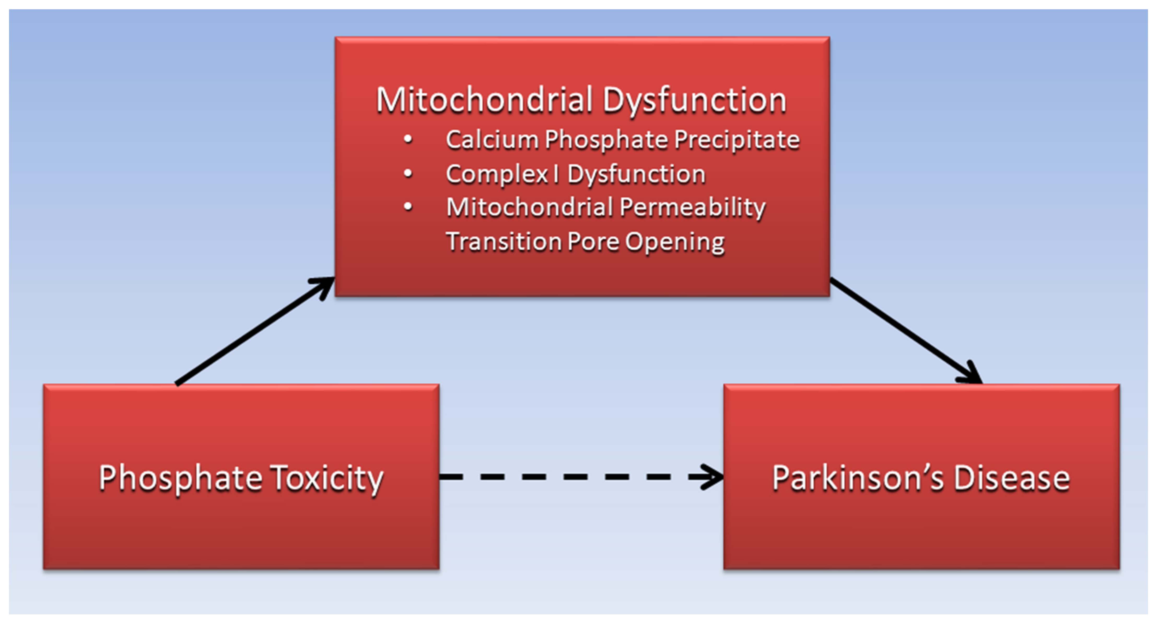
Excessive phosphate is proposed to reduce Complex I function of the mitochondrial electron transport chain in Parkinson’s disease and is linked to opening of the mitochondrial permeability transition pore, resulting in increased reactive oxygen species, inflammation, DNA damage, mitochondrial membrane depolarization, and ATP depletion causing cell death. Parkinson’s disease is associated with α-synuclein and Lewy body dementia, a secondary tauopathy related to hyperphosphorylation of tau protein, and tauopathy is among several pathophysiological pathways shared between Parkinson’s disease and diabetes. Excessive phosphate is also associated with ectopic calcification, bone mineral disorders, and low levels of serum vitamin D in patients with Parkinson’s disease. Sarcopenia and cancer in Parkinson’s disease patients are also associated with phosphate toxicity. Additionally, Parkinson’s disease benefits are related to low dietary phosphate intake.

Parkinson’s disease phosphate toxicity mitochondrial dysfunction calcium phosphate tauopathy ectopic calcification bone mineral disorders vitamin D sarcopenia cancer

1. Introduction

Parkinson’s disease (PD) is the second most prevalent neurodegenerative disorder, after Alzheimer’s disease, and hallmarks of PD include neuronal losses in the substantia nigra causing deficiencies in dopamine [1]. In addition to movement disorders (rigidity, resting tremor, and bradykinesia), conditions associated with PD include cognitive impairment, sleep disorders, and depression [2]. Globally, the burden of PD has more than doubled over the past generation and is predicted to increase substantially [3]. No curative treatments exist for PD, and “modifying disease progression and further delaying disability are the key unmet needs to be addressed by current and future research efforts” [1]. Ongoing clinical trials are testing whether dopamine production can be strengthened through central nervous system gene therapy that transfers glial-cell-line-derived neurotrophic factor (GDNF) to PD patients [4]. Although results of the gene therapy indicate clinical benefits and improved response to levodopa medication, “it remains to be seen whether the many debilitating non-motor features will also show relevant responses to treatment”.

2. Calcium Phosphate and Mitochondrial Dysfunction in PD

Experiments demonstrated that brain and neuron mitochondria are capable of forming “remarkably high levels” of calcium phosphate precipitates within the mitochondrial matrix, which are “retained in damaged mitochondria for prolonged periods” [5]. Mitochondrial precipitates are “composed primarily of tribasic calcium phosphate [Ca3(PO4)2] and/or dibasic calcium phosphate (CaHPO4)”. Of relevance, mitochondrial damage involving accumulated Pi was found in cultured pancreatic beta cells and islet cells [6][7], which is associated with reduced insulin biosynthesis and secretion, reactive oxygen species, and cell apoptosis in diabetes mellitus. This pathophysiological mechanism appears strikingly similar to reduced dopamine biosynthesis and secretion in the substantia nigra associated with PD, suggesting that increased Pi concentrations and calcium phosphate accumulation in the mitochondrial matrix may be included among shared etiological factors in PD and DM [8].
A recent systematic review and meta-analysis confirmed a significant 21% increased odds of PD associated with type II diabetes mellitus (T2DM) [9]. Experiments conducted on the mitochondrial electron transport chain in guinea pig hearts found that increased accumulations of calcium phosphate inhibited proton pumping at Complex I (NADH CoQ reductase) and reduced ATP synthesis necessary to power cellular functions [10]. Researchers suggested that calcium phosphate particles act as a physical barrier within the cristae of the mitochondrial inner membrane, which disrupts the operation of Complex I. Coincidently, a postmortem study found “a specific defect of Complex I activity in the substantia nigra of deceased patients with Parkinson’s disease” [11]. Additionally, the researchers noted that an inhibiting effect on Complex I from the neurotoxin derived from 1-methyl-4-phenyl-1,2,3,6-tetrahydropyridine (MPTP) was found to cause symptoms of PD, which “adds further support to the proposition that Parkinson’s disease may be due to an environmental toxin with action(s) similar to those of MPTP”.
Reduction in Complex I function in PD is linked to opening of the mitochondrial permeability transition pore (mPTP), allowing an influx of ions and small molecules, which is associated with increased reactive oxygen species (ROS), inflammation, DNA damage, mitochondrial membrane depolarization, and ATP depletion causing cell death [12][13][14]. “Phosphate activation of the mitochondrial permeability transition pore opening is well documented” [15]. Of relevance, reduction of polyphosphate (polyP)—a phosphate chain linked by ATP bonds in mitochondria—reduces calcium-induced mitochondrial permeability transition [16]. Furthermore, researchers using fluorescent probes estimated higher levels of endogenous polyP in models of living cells “with Parkinson’s disease related mutations” [17]. Studies are needed to further investigate the role of high Pi concentration and accumulated calcium phosphate precipitates on mitochondrial Complex I function and the opening of the mPTP in patients with PD.
The present research’s proposed association of phosphate toxicity with PD, mediated by mitochondrial dysfunction, is illustrated in Figure 1.
Figure 1. Mitochondrial dysfunction is proposed to mediate the association of phosphate toxicity with Parkinson’s disease.

3. Sarcopenia, Phosphate Toxicity, and PD

Sarcopenia, low muscle mass, and muscle function increase disability, frailty, morbidity, and mortality and lowers quality of life in the elderly population [18]. Core muscle loss was associated with reduced brain gray matter volume in patients with PD [19]. A recent systematic review and meta-analysis found that “the pooled prevalence of sarcopenia was 29% in PD, which was higher than the healthy older control group” [20]. The researchers suggested that sarcopenia and PD may share a common pathway of neuroinflammation. Coincidentally, a higher prevalence of sarcopenia was found in diabetic compared to non-diabetic individuals in a meta-analysis of an Asian population [21], yet again implying common pathways shared with PD.
Of relevance, increasing dietary Pi fed to lab animals in a model of chronic kidney disease showed a dose-dependent increase in levels of tumor necrosis factor-alpha, a biomarker of systemic inflammation, as well as decreased animal body weight, a biomarker of malnutrition, and increased vascular calcification with reduced lifespan [22]. These findings are consistent with sarcopenia as well as inflammation in PD [23], providing additional support for phosphate toxicity in PD etiology. Moreover, concentrated levels of Pi added to cultured muscle cells directly produced cell autophagy [24], and future studies should investigate muscle cell autophagy associated with phosphate-induced mitochondrial damage.

4. Cancer, Phosphate Toxicity, and PD

Tumorigenesis is associated with phosphate toxicity [25], briefly summarized here. As excessive amounts of dysregulated Pi are sequestered into precancerous cells through overexpressed sodium phosphate cotransporters, cell signaling pathways stimulate tumor growth. For example, the phosphoinositide 3 kinase (PI3K) pathway phosphorylates Akt (protein kinase B), leading to activation of mTOR (mammalian target of rapamycin), which upregulates protein synthesis in tumor growth. Phosphorus is a rate-limiting factor in biological growth, and reducing phosphorus transport into a tumor by half is predicted to reduce a tumor’s size by 75% [26].
Positive associations have been found between PD and cancers of the breast, brain, and melanoma, and cancer incidence may occur either before or after PD incidence, which is consistent with a common causative pathway in both of these “pathologically convergent diseases” [27]. Nevertheless, “the lower risks of lung, bladder, and colorectal cancer, all smoking-related cancers, in PD patients are generally undisputed”. Feasibly, lower risks of smoking-related cancers could be explained by the fact that “smokers reported reduced compliance with the DRI [dietary reference intake] for iron, phosphorus, vitamin C, riboflavin, and folate compared to nonsmokers” [28]. With a DRI of 700 mg phosphorus per day for U.S. adults, “the average daily phosphorus intake from foods is 1189 mg for women and 1596 mg for men” [29], implying that smokers’ phosphorus intake is approximately twice as low as the average intake. Moreover, lower phosphorus intake could explain “a causally protective effect of current smoking on the risk of PD” [30].

5. High Dietary Phosphate and PD

Globally, “the presence of any known causal PD mutation is rare, occurring in less than 2% of the PD population”, inferring that environmental factors such as diet are likely to play a significant role in PD etiology [31]. Relevant to the potential contribution of phosphate toxicity to PD pathophysiology, “high phosphorus intake is associated with increased mortality in a healthy US population” [32]. Importantly, “dairy products, fish, and other types of meat are a major source of phosphate in the human diet”, and “preservatives and additive salts commonly used in processed foods contain large amounts of phosphate” [33].
A prospective study of the EPIC-Greece cohort found that “incident PD exhibited strong positive association with consumption of milk, but not cheese or yoghurt”, and an “inverse association was found between polyunsaturated fat intake and incident PD” [34]. Milk consumption, but not fermented milk, was also weakly associated with increased risk of PD in a recent Swedish study [35]. Associations between various food items and PD could be mediated by an inverse relationship between the phosphorus and fat content in foods—phosphorus in food is naturally found in combination with protein [36], not with fat. Full-fat dairy products provide more calories and have lower phosphorus caloric densities (lower phosphorus levels per kcal), which helps meet caloric dietary needs with overall less phosphate, compared to low-fat or non-fat dairy products that provide fewer calories and have higher phosphorus caloric densities, which may drive up overall phosphorus intake to meet calorie requirements. Accordingly, a Harvard analysis of data from the Nurses’ Health Study and the Health Professionals’ Follow-Up Study found that three or more servings of low-fat dairy was associated with a higher risk of PD diagnosis, but no association was found with consumption of full-fat dairy [37].
Low phosphorus in a high-fat ketogenic diet (KD) could also explain neuroprotection of the KD, although “the literature does not yet support a neuroprotective effect of the KD in PD” [38]. Nevertheless, current data suggest benefits in non-motor symptoms of PD patients using a ketogenic diet [39]. Additionally, whole-food plant-based diets such as the Mediterranean diet, with reduced Pi intake from animal food products and highly processed foods, have been associated with delayed onset of PD [40]. A recent analysis of dietary patterns in the Rotterdam Study reported reduced associated risks of PD in the Netherlands general population that “corroborate previous findings of a possible protective effect of the Mediterranean diet” [41]. This type of plant-based diet often contains an abundance of whole fruit, which has low phosphorus caloric density. Table 1 shows the phosphorus caloric density of various food items based on data from the U.S. Department of Agriculture (USDA) National Nutrient Database for Standard Reference, Legacy 2018 [42]. Note that whole fruit, although high in sugar, is also high in fiber with a low glycemic index [43], and whole fruit is associated with decreased risk of diabetes [44][45]. Future studies should investigate potential PD benefits specifically associated with lower phosphate intake in whole-food plant-based diets.
Table 1. Phosphorus caloric density of selected food items.
Based on data from USDA National Nutrient Database for Standard Reference, Legacy (2018).
Restriction of dietary phosphate, 800–1000 mg/day, is currently used in managing phosphate serum levels in patients with chronic kidney disease [46], and similar strategies should be investigated to prevent and delay progression of PD. Dietary counseling from renal dietitians and other trained healthcare providers “can lead to better control of phosphorus intake” [36]. Importantly, in vivo regression of ectopic calcification is associated with the phosphoprotein osteopontin (OPN) [47], and vascular calcification reversal in rats fed low-phosphate diets was suggested to be linked to OPN [48]. Phosphorylation regulates the inhibitory effect of OPN on vascular calcification [49], providing a potential compensatory response to calcification associated with elevated phosphate levels [50]. This evidence implies that some degree of regression of mitochondrial calcification in PD may be possible by placing patients with PD on restricted-phosphate diets.

References

  1. Poewe, W.; Seppi, K.; Tanner, C.M.; Halliday, G.M.; Brundin, P.; Volkmann, J.; Schrag, A.-E.; Lang, A.E. Parkinson disease. Nat. Rev. Dis. Primers 2017, 3, 17013.
  2. Surguchov, A. Biomarkers in Parkinson’s Disease. In Neurodegenerative Diseases Biomarkers: Towards Translating Research to Clinical Practice; Peplow, P.V., Martinez, B., Gennarelli, T.A., Eds.; Springer: New York, NY, USA, 2022; pp. 155–180.
  3. Dorsey, E.R.; Elbaz, A.; Nichols, E.; Abbasi, N.; Abd-Allah, F.; Abdelalim, A.; Adsuar, J.C.; Ansha, M.G.; Brayne, C.; Choi, J.-Y.J.; et al. Global, regional, and national burden of Parkinson’s disease, 1990–2016: A systematic analysis for the Global Burden of Disease Study 2016. Lancet Neurol. 2018, 17, 939–953.
  4. Fiandaca, M.S.; Lonser, R.R.; Elder, J.B.; Ząbek, M.; Bankiewicz, K.S. Advancing gene therapies, methods, and technologies for Parkinson’s Disease and other neurological disorders. Neurol. Neurochir. Pol. 2020, 54, 220–231.
  5. Kristian, T.; Pivovarova, N.B.; Fiskum, G.; Andrews, S.B. Calcium-induced precipitate formation in brain mitochondria: Composition, calcium capacity, and retention. J. Neurochem. 2007, 102, 1346–1356.
  6. Nguyen, T.T.; Quan, X.; Hwang, K.-H.; Xu, S.; Das, R.; Choi, S.-K.; Wiederkehr, A.; Wollheim, C.B.; Cha, S.-K.; Park, K.-S. Mitochondrial oxidative stress mediates high-phosphate-induced secretory defects and apoptosis in insulin-secreting cells. Am. J. Physiol. Endocrinol. Metab. 2015, 308, E933–E941.
  7. Nguyen, T. Mitochondrial reactive oxygen species, endoplasmic reticulum stress and phosphate toxicity in impairment of pancreatic cells. TTU Rev. 2016, 1, 73–78.
  8. Hassan, A.; Sharma Kandel, R.; Mishra, R.; Gautam, J.; Alaref, A.; Jahan, N. Diabetes Mellitus and Parkinson’s Disease: Shared Pathophysiological Links and Possible Therapeutic Implications. Cureus 2020, 12, e9853.
  9. Chohan, H.; Senkevich, K.; Patel, R.K.; Bestwick, J.P.; Jacobs, B.M.; Bandres Ciga, S.; Gan-Or, Z.; Noyce, A.J. Type 2 Diabetes as a Determinant of Parkinson’s Disease Risk and Progression. Mov. Disord. 2021, 36, 1420–1429.
  10. Malyala, S.; Zhang, Y.; Strubbe, J.O.; Bazil, J.N. Calcium phosphate precipitation inhibits mitochondrial energy metabolism. PLoS Comput. Biol. 2019, 15, e1006719.
  11. Schapira, A.H.; Cooper, J.M.; Dexter, D.; Clark, J.B.; Jenner, P.; Marsden, C.D. Mitochondrial complex I deficiency in Parkinson’s disease. J. Neurochem. 1990, 54, 823–827.
  12. Kent, A.C.; El Baradie, K.B.Y.; Hamrick, M.W. Targeting the Mitochondrial Permeability Transition Pore to Prevent Age-Associated Cell Damage and Neurodegeneration. Oxid. Med. Cell Longev. 2021, 2021, 6626484.
  13. Elustondo, P.A.; Nichols, M.; Negoda, A.; Thirumaran, A.; Zakharian, E.; Robertson, G.S.; Pavlov, E.V. Mitochondrial permeability transition pore induction is linked to formation of the complex of ATPase C-subunit, polyhydroxybutyrate and inorganic polyphosphate. Cell Death Discov. 2016, 2, 16070.
  14. Phadwal, K.; Vrahnas, C.; Ganley, I.G.; MacRae, V.E. Mitochondrial Dysfunction: Cause or Consequence of Vascular Calcification? Front. Cell Dev. Biol. 2021, 9, 611922.
  15. Varanyuwatana, P.; Halestrap, A.P. The roles of phosphate and the phosphate carrier in the mitochondrial permeability transition pore. Mitochondrion 2012, 12, 120–125.
  16. Abramov, A.Y.; Fraley, C.; Diao, C.T.; Winkfein, R.; Colicos, M.A.; Duchen, M.R.; French, R.J.; Pavlov, E. Targeted polyphosphatase expression alters mitochondrial metabolism and inhibits calcium-dependent cell death. Proc. Natl. Acad. Sci. USA 2007, 104, 18091–18096.
  17. Angelova, P.R.; Agrawalla, B.K.; Elustondo, P.A.; Gordon, J.; Shiba, T.; Abramov, A.Y.; Chang, Y.T.; Pavlov, E.V. In situ investigation of mammalian inorganic polyphosphate localization using novel selective fluorescent probes JC-D7 and JC-D8. ACS Chem. Biol. 2014, 9, 2101–2110.
  18. Tsekoura, M.; Kastrinis, A.; Katsoulaki, M.; Billis, E.; Gliatis, J. Sarcopenia and Its Impact on Quality of Life. Adv. Exp. Med. Biol. 2017, 987, 213–218.
  19. Wu, Y.N.; Chen, M.H.; Chiang, P.L.; Lu, C.H.; Chen, H.L.; Yu, C.C.; Chen, Y.S.; Chang, Y.Y.; Lin, W.C. Associations between Brain Structural Damage and Core Muscle Loss in Patients with Parkinson’s Disease. J. Clin. Med. 2020, 9, 239.
  20. Cai, Y.; Feng, F.; Wei, Q.; Jiang, Z.; Ou, R.; Shang, H. Sarcopenia in Patients With Parkinson’s Disease: A Systematic Review and Meta-Analysis. Front. Neurol. 2021, 12, 598035.
  21. Chung, S.M.; Moon, J.S.; Chang, M.C. Prevalence of Sarcopenia and Its Association with Diabetes: A Meta-Analysis of Community-Dwelling Asian Population. Front. Med. 2021, 8, 681232.
  22. Yamada, S.; Tokumoto, M.; Tatsumoto, N.; Taniguchi, M.; Noguchi, H.; Nakano, T.; Masutani, K.; Ooboshi, H.; Tsuruya, K.; Kitazono, T. Phosphate overload directly induces systemic inflammation and malnutrition as well as vascular calcification in uremia. Am. J. Physiol. Ren. Physiol. 2014, 306, F1418–F1428.
  23. Adams, B.; Nunes, J.M.; Page, M.J.; Roberts, T.; Carr, J.; Nell, T.A.; Kell, D.B.; Pretorius, E. Parkinson’s Disease: A Systemic Inflammatory Disease Accompanied by Bacterial Inflammagens. Front. Aging Neurosci. 2019, 11, 210.
  24. Zhang, Y.-Y.; Yang, M.; Bao, J.-F.; Gu, L.-J.; Yu, H.-L.; Yuan, W.-J. Phosphate stimulates myotube atrophy through autophagy activation: Evidence of hyperphosphatemia contributing to skeletal muscle wasting in chronic kidney disease. BMC Nephrol. 2018, 19, 45.
  25. Brown, R.B.; Razzaque, M.S. Phosphate toxicity and tumorigenesis. Biochim. et Biophys. Acta Rev. Cancer 2018, 1869, 303–309.
  26. Kuang, Y.; Nagy, J.D.; Elser, J.J. Biological stoichiometry of tumor dynamics: Mathematical models and analysis. Discret. Contin. Dyn. Syst. Ser. B 2004, 4, 221–240.
  27. Feng, D.D.; Cai, W.; Chen, X. The associations between Parkinson’s disease and cancer: The plot thickens. Transl. Neurodegener. 2015, 4, 20.
  28. Raatz, S.K.; Jahns, L.; Johnson, L.K.; Scheett, A.; Carriquiry, A.; Lemieux, A.; Nakajima, M.; al’Absi, M. Smokers report lower intake of key nutrients than nonsmokers, yet both fall short of meeting recommended intakes. Nutr. Res. 2017, 45, 30–37.
  29. Phosphorus Fact Sheet for Health Professionals. Available online: https://ods.od.nih.gov/factsheets/Phosphorus-HealthProfessional/#:~:text=Phosphorus%20Intakes%20and%20Status,-Most%20Americans%20consume&text=In%20adults%20aged%2020%20and,mg%20for%20men%20%5B30%5D. (accessed on 3 July 2022).
  30. Mappin-Kasirer, B.; Pan, H.; Lewington, S.; Kizza, J.; Gray, R.; Clarke, R.; Peto, R. Tobacco smoking and the risk of Parkinson disease. Neurology 2020, 94, e2132–e2138.
  31. Tran, J.; Anastacio, H.; Bardy, C. Genetic predispositions of Parkinson’s disease revealed in patient-derived brain cells. NPJ Parkinson’s Dis. 2020, 6, 8.
  32. Chang, A.R.; Lazo, M.; Appel, L.J.; Gutiérrez, O.M.; Grams, M.E. High dietary phosphorus intake is associated with all-cause mortality: Results from NHANES III. Am. J. Clin. Nutr. 2013, 99, 320–327.
  33. Erem, S.; Razzaque, M.S. Dietary phosphate toxicity: An emerging global health concern. Histochem. Cell Biol. 2018, 150, 711–719.
  34. Kyrozis, A.; Ghika, A.; Stathopoulos, P.; Vassilopoulos, D.; Trichopoulos, D.; Trichopoulou, A. Dietary and lifestyle variables in relation to incidence of Parkinson’s disease in Greece. Eur. J. Epidemiol. 2013, 28, 67–77.
  35. Olsson, E.; Byberg, L.; Höijer, J.; Kilander, L.; Larsson, S.C. Milk and Fermented Milk Intake and Parkinson’s Disease: Cohort Study. Nutrients 2020, 12, 2763.
  36. D’Alessandro, C.; Piccoli, G.B.; Cupisti, A. The “phosphorus pyramid”: A visual tool for dietary phosphate management in dialysis and CKD patients. BMC Nephrol. 2015, 16, 9.
  37. Hughes, K.C.; Gao, X.; Kim, I.Y.; Wang, M.; Weisskopf, M.G.; Schwarzschild, M.A.; Ascherio, A. Intake of dairy foods and risk of Parkinson disease. Neurology 2017, 89, 46–52.
  38. Gough, S.M.; Casella, A.; Ortega, K.J.; Hackam, A.S. Neuroprotection by the Ketogenic Diet: Evidence and Controversies. Front. Nutr. 2021, 8, 782657.
  39. Choi, A.; Hallett, M.; Ehrlich, D. Nutritional Ketosis in Parkinson’s Disease—A Review of Remaining Questions and Insights. Neurotherapeutics 2021, 18, 1637–1649.
  40. Metcalfe-Roach, A.; Yu, A.C.; Golz, E.; Cirstea, M.; Sundvick, K.; Kliger, D.; Foulger, L.H.; Mackenzie, M.; Finlay, B.B.; Appel-Cresswell, S. MIND and Mediterranean Diets Associated with Later Onset of Parkinson’s Disease. Mov. Disord. 2021, 36, 977–984.
  41. Strikwerda, A.J.; Dommershuijsen, L.J.; Ikram, M.K.; Voortman, T. Diet Quality and Risk of Parkinson’s Disease: The Rotterdam Study. Nutrients 2021, 13, 3970.
  42. USDA. National Nutrient Database for Standard Reference, Legacy. 2018. Available online: https://fdc.nal.usda.gov/index.html (accessed on 16 July 2022).
  43. Jenkins, D.J.; Wolever, T.M.; Taylor, R.H.; Barker, H.; Fielden, H.; Baldwin, J.M.; Bowling, A.C.; Newman, H.C.; Jenkins, A.L.; Goff, D.V. Glycemic index of foods: A physiological basis for carbohydrate exchange. Am. J. Clin. Nutr. 1981, 34, 362–366.
  44. Du, H.; Li, L.; Bennett, D.; Guo, Y.; Turnbull, I.; Yang, L.; Bragg, F.; Bian, Z.; Chen, Y.; Chen, J.; et al. Fresh fruit consumption in relation to incident diabetes and diabetic vascular complications: A 7-y prospective study of 0.5 million Chinese adults. PLoS Med. 2017, 14, e1002279.
  45. Li, S.; Miao, S.; Huang, Y.; Liu, Z.; Tian, H.; Yin, X.; Tang, W.; Steffen, L.M.; Xi, B. Fruit intake decreases risk of incident type 2 diabetes: An updated meta-analysis. Endocrine 2015, 48, 454–460.
  46. Barreto, F.C.; Barreto, D.V.; Massy, Z.A.; Drüeke, T.B. Strategies for Phosphate Control in Patients with CKD. Kidney Int. Rep. 2019, 4, 1043–1056.
  47. Steitz, S.A.; Speer, M.Y.; McKee, M.D.; Liaw, L.; Almeida, M.; Yang, H.; Giachelli, C.M. Osteopontin inhibits mineral deposition and promotes regression of ectopic calcification. Am. J. Pathol. 2002, 161, 2035–2046.
  48. Finch, J.L.; Lee, D.H.; Liapis, H.; Ritter, C.; Zhang, S.; Suarez, E.; Ferder, L.; Slatopolsky, E. Phosphate restriction significantly reduces mortality in uremic rats with established vascular calcification. Kidney Int. 2013, 84, 1145–1153.
  49. Jono, S.; Peinado, C.; Giachelli, C.M. Phosphorylation of osteopontin is required for inhibition of vascular smooth muscle cell calcification. J. Biol. Chem. 2000, 275, 20197–20203.
  50. Hunter, G.K. Role of Osteopontin in Modulation of Hydroxyapatite Formation. Calcif. Tissue Int. 2013, 93, 348–354.
More
Information
Subjects: Pathology
Contributor MDPI registered users' name will be linked to their SciProfiles pages. To register with us, please refer to https://encyclopedia.pub/register :
View Times: 59
Revisions: 2 times (View History)
Update Date: 14 Mar 2024
1000/1000