Submitted Successfully!
To reward your contribution, here is a gift for you: A free trial for our video production service.
Thank you for your contribution! You can also upload a video entry or images related to this topic.
Version Summary Created by Modification Content Size Created at Operation
1 -- 3940 2024-03-07 09:57:59 |
2 Corrections of references Meta information modification 3940 2024-03-07 10:01:27 | |
3 Reference format revised. Meta information modification 3940 2024-03-08 02:19:35 | |
4 figure captions revised Meta information modification 3940 2024-03-08 06:16:08 |

Video Upload Options

Do you have a full video?

Confirm

Are you sure to Delete?
Cite
If you have any further questions, please contact Encyclopedia Editorial Office.
Chato-Astrain, I.; Pronot, M.; Coppola, T.; Martin, S. Mammalian Synapse by the Post-Translational Modification SUMOylation. Encyclopedia. Available online: https://encyclopedia.pub/entry/55961 (accessed on 05 May 2024).
Chato-Astrain I, Pronot M, Coppola T, Martin S. Mammalian Synapse by the Post-Translational Modification SUMOylation. Encyclopedia. Available at: https://encyclopedia.pub/entry/55961. Accessed May 05, 2024.
Chato-Astrain, Isabel, Marie Pronot, Thierry Coppola, Stéphane Martin. "Mammalian Synapse by the Post-Translational Modification SUMOylation" Encyclopedia, https://encyclopedia.pub/entry/55961 (accessed May 05, 2024).
Chato-Astrain, I., Pronot, M., Coppola, T., & Martin, S. (2024, March 07). Mammalian Synapse by the Post-Translational Modification SUMOylation. In Encyclopedia. https://encyclopedia.pub/entry/55961
Chato-Astrain, Isabel, et al. "Mammalian Synapse by the Post-Translational Modification SUMOylation." Encyclopedia. Web. 07 March, 2024.
Mammalian Synapse by the Post-Translational Modification SUMOylation
Edit

Neurotransmission occurs within highly specialized compartments forming the active synapse where the complex organization and dynamics of the interactions are tightly orchestrated both in time and space. Post-translational modifications (PTMs) are central to these spatiotemporal regulations to ensure an efficient synaptic transmission. SUMOylation is a dynamic PTM that modulates the interactions between proteins and consequently regulates the conformation, the distribution and the trafficking of the SUMO-target proteins. SUMOylation plays a crucial role in synapse formation and stabilization, as well as in the regulation of synaptic transmission and plasticity. 

post-translational modification SUMO SUMOylation LLPS synapse biomolecular condensate

1. Introduction

Neurons are polarized cells with one axon and several highly branched dendrites, which connect through synapses for rapid information transfer in the brain. Brain activity relies on the establishment of functional networks of interconnected neuronal cells. This process involves the accurate formation and maturation of synapses which are fundamental to establishing efficient neuronal wiring in the developing brain. The coordinated organization of complex protein networks on both sides of the synapse enables synaptic transmission and plasticity that supports cognitive processes. This structural organization encompasses many scaffolding and adaptor proteins and thus multiple protein–protein interactions to efficiently position the machinery for neurotransmitter release, post-synaptic neurotransmitter receptors and the associated signaling cascade components and their regulators.
Among the key regulatory mechanisms controlling the interactions and function of proteins within pre- and post-synaptic compartments are post-translational modifications (PTMs) including phosphorylation and ubiquitination. SUMOylation is another PTM playing important roles in the organization and function of the mammalian synapse. The SUMOylation process was first reported around 1996 [1][2] and for many years was exclusively associated with the nuclear compartment. Extranuclear SUMOylation is now well documented and occurs in all tissues, cells and organelles [3][4]. Although SUMOylation is also predominantly associated with the nucleus in neurons, it is also present in pre- and post-synaptic compartments [5][6][7][8][9].
SUMOylation is an enzymatic process that consists in the covalent but reversible isopeptidic binding of the Small Ubiquitin-like MOdifier (SUMO) to the lateral chain of specific lysine residues within target proteins. This modification alters the interacting properties of the SUMO-modified proteins through conformational changes, by preventing the binding of some proteins initially interacting with the non-SUMOylated form of the target protein, or by providing a binding platform for specific sets of new interacting proteins. The functional impact of SUMOylation is thus multiple and depends on the modified proteins. SUMOylation can affect the subcellular localization of the modified protein through its binding to novel protein partners or change its solubility, degradation or stability properties, but can also directly impact its intrinsic activity.

2. The SUMOylation/deSUMOylation Enzymatic Pathway at Synapses

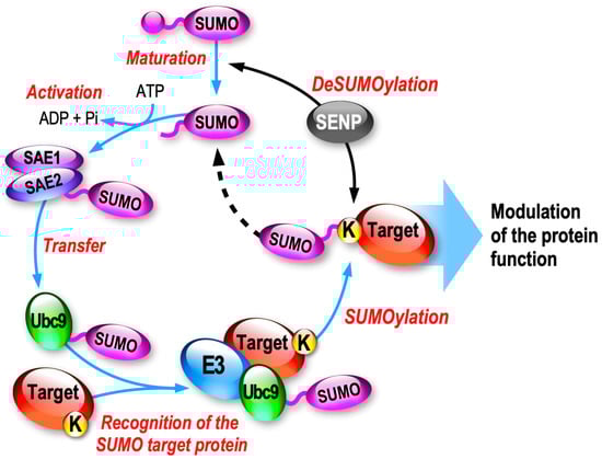
2.1. SUMO Paralogs and the Associated Enzymatic SUMOylation/deSUMOylation Cascade

Small Ubiquitin-like Modifiers (SUMOs) are small proteins of ~100 amino acids (~11 kDa) sharing 18% sequence homology with Ubiquitin. Among the five known SUMO paralogs existing in mammals, SUMO1, SUMO2 and SUMO3, referred as SUMO2/3 due to their high homology (97% identity), are the only three isoforms expressed in the brain. Despite sharing less than 50% identity, the mature forms of SUMO1 and SUMO2/3 are conjugated to substrate proteins through a specific enzymatic pathway (Figure 1).
Figure 1. Schematic representation of the enzymatic SUMOylation/deSUMOylation cycle. The different phases of the enzymatic cascade leading to the SUMOylation of the target protein are highlighted in red. SUMO, Small Ubiquitin-like MOdifier; SAE, SUMO-activating enzyme; Ubc9, SUMO-conjugating enzyme; SENP, Sentrin protease.
SUMOylation is highly dynamic and the equilibrium between SUMOylation and deSUMOylation is driven by a dedicated cascade of enzymatic reactions. First, the inactive SUMO precursors are matured by the hydrolase activity of certain SUMO proteases (SENP1,2,5) leading to the exposure of a C-terminal di-Glycine motif. Next, the mature form of SUMO (SUMO-GG) is activated by the heteromeric E1-activating complex formed by SAE1/SAE2 in an ATP-dependent manner, generating a thioester bond between the cysteine residue of SAE2 and SUMO-GG. The activated SUMO is then transferred to the active cysteine site of Ubc9, the only E2 SUMO-conjugating enzyme of the system. Ubc9 can now covalently promote the binding of the SUMO-GG to the lateral chain of the targeted lysine residue of the substrate protein, usually with the support of an E3 enzyme (Figure 1). SUMO E3s will help the SUMOylation process by enhancing the SUMO conjugation rate to the substrate protein.

2.2. Synaptic Localization of the SUMOylation/deSUMOylation Machinery

Although SUMOylation acts predominantly within the nuclear compartment or to allow the nucleocytoplasmic exchange of various target proteins, there is no doubt that SUMOylation can also occur at synapses regulating many important aspects of the pre- and post-synaptic function [6][7][8][9].
A group of recent publications reporting the partial localization of the components of the SUMOylation and deSUMOylation machinery at both pre- and post-synaptic sites in high-resolution microscopy [10][11][12] reinforced the previous findings that SUMOylation and deSUMOylation enzymes are indeed targeted to synapses [13][14][15][16][17][18][19]. Colnaghi and colleagues first used structured illumination microscopy to confirm that the immunoreactivities for SUMO1, SUMO2/3 and the SUMO-conjugating enzyme Ubc9 are preponderantly present in the nuclei of mouse hippocampal neurons as expected, but that a significant proportion of the labelling is also partially colocalized with pre- and post-synaptic markers [10]. They later reported that the deSUMOylation machinery, and in particular the deSUMOylation enzymes SENP1, SENP6 and SENP7, are also localized to some extent in the hippocampal synapses [11] in line with earlier reports. More recently, they further used super-resolution microscopy to show that the SUMO E3 ligases PIAS1 and PIAS3 are also expressed in pre- and post-synaptic compartments in mouse hippocampal neurons as for the other members of the SUMOylation machinery, but interestingly, this synaptic colocalization was much more limited in cortical neurons, suggesting distinct regulatory mechanisms driving the synaptic targeting and/or compartmentalization of the SUMO enzymes between different neuronal subtypes [12].
As the other enzyme of the SUMO system, SENP3 is mainly expressed in the nucleus but is also present in the synapses in the rat cerebrum and cerebellum [19]. SENP3 is involved in mitochondria fragmentation with a functional impact on the SUMOylation levels of the dynamin-related protein 1 Drp1 [20], but also by acting on the mitochondrial fission 1 protein (Fis1) deSUMOylation in mitochondrial autophagy [21]; both of these effects occur under stress conditions.

2.3. Synaptic Regulation of the SUMO Pathway

While a growing number of studies have demonstrated the functional consequences of specific SUMO substrates at synapses, few data are available in the literature regarding the mechanisms driving the targeting, anchoring and functional regulation of the SUMO enzymes at synapses.
Among the first evidence that the synaptic redistribution of the components of the SUMO system is regulated by changes in neuronal activity is the work achieved by the Henley group in 2009 [14]. They showed that the pharmacological stimulation of purified synaptosomal preparations by α-amino-3-hydroxy-5-methyl-4-isoxazolepropionic acid (AMPA), a specific agonist of the ionotropic glutamate AMPA receptors (AMPAR) prior to a subsynaptic fractionation, leads to a significant increase in the association of the SUMO-conjugating enzyme Ubc9 in the presynaptic active zone. This redistribution of Ubc9 to the presynaptic region following AMPA receptor stimulation was accompanied by an increase in the total level of SUMOylated proteins in the presynaptic fraction [14].
The same group later showed that the mRNA levels for both SUMO1 and Ubc9 are increased in dendrites following a chemical protocol to trigger long-term potentiation (LTP). This pharmacological treatment also led to an increased immunoreactivity for both SUMO1 and Ubc9 at post-synaptic sites, suggesting an activity-dependent local translation mechanism for the SUMO pathway [22].

An additional level of activity-dependent regulation of the SUMO/deSUMOylation pathway occurs directly at the synapse and relies on the activation of type 1 metabotropic glutamate mGlu1 and mGluR5 receptors [1[9][23][24]. A short activation of mGlu5 receptors triggers a Protein Kinase C (PKC) phosphorylation-dependent trapping of the SUMO-conjugating enzyme Ubc9 at post-synaptic sites, leading to an increased SUMOylation and to the modulation of neuronal excitability [1[9]. The sustained activation of mGlu5R then leads to a decrease in the exit rate of the deSUMOylation enzyme SENP1 from dendritic spines resulting in the post-synaptic accumulation of SENP1 to bring SUMOylation back to initial levels [23]. Blocking the activation of mGlu1R enhances the mGlu5R-induced post-synaptic accumulation of SENP1 indicating that the SUMOylation/deSUMOylation balance is bidirectionally regulated by type I mGluRs to control the levels of synaptic SUMOylation [24]. Altogether, these findings revealed that type I mGlu1/5 receptors are central to dynamically maintain the homeostasis of SUMOylation at the mammalian synapse but also indicate that the relative amount of mGlu1 and mGlu5 receptors at individual synapses will drive the diversity of the post-synaptic SUMOylation responses.

3. SUMOylation in Neurite Growth, Synapse Formation, Elimination and Maturation

3.1. SUMOylation of Transcription Factors in Neurite Growth and Branching

3.1.1. MEF2 SUMOylation

The transcription factor family of Myocyte-specific enhancer factor 2 (MEF2) is central to brain development and particularly abundant in the cerebellar cortex [25]. MEF2s play critical roles in cell differentiation, dendritic morphogenesis, synapse formation, pruning and synaptic plasticity. Mutations of these genes are associated with various pathological conditions, including epilepsies and autism spectrum disorders, among others [26]. The function of MEF2 is regulated by several PTMs, including acetylation, phosphorylation and SUMOylation.
SUMO1-ylation of MEF2A at the K403 residue inhibits its transcription activity and thus promotes the differentiation of dendritic protrusions [27]. The engineering of SENP2 knockout embryos and the use of in vitro SUMO assays in SHSY5Y cells showed that the deSUMOylase SENP2, but not SENP1, regulates MEF2A deSUMOylation in response to activity-dependent stimuli [28]. Co-expression of SENP2 and MEF2A leads to the deSUMOylation of MEF2A and to an increase in its transcriptional activity [28]. These data highlight that the SUMOylation/deSUMOylation equilibrium is important for regulating the transcriptional activity of MEF2A.

3.1.2. FOXP2 SUMOylation

The transcription factor Forkhead box P2 (FOXP2) is SUMOylated at K674 (K673 in mouse) [29][30][31]. Mutations within the FOXP2 gene lead to brain developmental abnormalities with reduced gray matter associated with speech and language deficits. In an elegant study, Usui and collaborators characterized the SUMOylation of FOXP2 in the developing mouse cerebellum and showed that it modifies transcriptional regulation by FOXP2 [31]. In utero electroporation of specific shRNAs to FOXP2 in Purkinje cells led to a significant reduction in dendritic outgrowth and arborization. This reduction of dendritic outgrowth and arborization in Purkinje cells that was rescued by the WT expression of FOXP2 but not by its SUMO-deficient mutant KR revealed the functional impact of FOXP2 SUMOylation in neuronal differentiation through neurite/dendritic outgrowth and arborization.

3.1.3. MeCP2 SUMOylation

Methyl-CpG-binding protein 2 (MeCP2) acts as a transcriptional repressor, and mutations within the human MECP2 gene on the X chromosome cause a severe neurodevelopmental disease in females called Rett syndrome. SUMOylation of the MeCP2 protein at the K223 was initially reported [32], and this modification is essential for its transcriptional repression activity in mouse primary cortical neurons. Using in utero electroporation of specific MeCP2 RNAi in the developing rat hippocampus reduced the density of excitatory synapses. This neurodevelopmental defect was fully rescued upon the co-expression of the WT form of MeCP2 in the rat hippocampus, but not when the non-SUMOylatable K223R form of MeCP2 was expressed [32].

3.1.4. ZBTB20 SUMOylation

ZBTB20 is a zinc finger protein and a transcriptional repressor [33]. ZBTB20 is involved in neurite growth and branching in the developing brain. Defects in the function of ZBTB20 is linked to a wide range of neurodevelopmental disorders, including intellectual disability and autism [34][35].
Ripamonti and colleagues reported the SUMOylation of the ZBTB20 protein at the K330 and K371 residues [36]. They showed that the lack of ZBTB20 SUMOylation has no effect on its nuclear localization or dimerization. Its SUMOylation instead modifies the interacting pool of transcriptional co-regulators, leading to a change in ZBTB20-target gene expression. Deletion of Zbtb20 in hippocampal neurons results in a decrease in the complexity of the neurite branching in immature neurons but also in more mature cells, revealing a functional role for this translational repressor in the control of neurite growth and ramification. Overexpression of the WT form of ZBTB20 in Zbtb20-lacking neurons is able to rescue neurite branching defects, while re-expression of its non-SUMOylatable mutant cannot, further suggesting a functional role for the SUMOylation of the ZBTB20 protein in neurite growth and branching [36].

3.2. Extranuclear SUMOylation in Synapse Formation and Maturation

3.2.1. CASK SUMOylation

CASK (Ca2+/Calmodulin-dependent serine protein kinase) is a member of the membrane-associated guanylate kinase (MAGUK) family and plays an important role in the formation of dendritic spines through its interaction with the actin cytoskeleton via the adhesion protein 4.1 [37]. Knockdown of the endogenous CASK expression in hippocampal neurons results in a reduction in the density, length and width of dendritic spines via its interaction with the protein 4.1 [38]. They also showed that SUMOylation of CASK at K679, a site very close to the protein 4.1 binding site, impairs its interaction with the protein 4.1, which in turn reduces the association of CASK with the actin cytoskeleton.

3.2.2. Local Protein Synthesis and Dendritic SUMOylation

CPEB3 SUMOylation

The RNA-binding CPEB3 belongs to a family of four proteins in vertebrates and is involved in the regulation of local translation in the brain, and consequently in synaptic plasticity and memory formation. The Kandel lab first reported the SUMO2-ylated CPEB3 in hippocampal neurons both in vitro and in vivo [39]. They showed that the SUMOylation of CPEB3 regulates its oligomerization. In a basal state, CPEB3 is SUMOylated and acts as a local translational repressor. CPEB3 is rapidly deSUMOylated upon neuronal activation, leading to its aggregation and the translation of target mRNAs, including the sumo2 mRNA [39].

FMRP SUMOylation

The RNA-binding protein FMRP participates in the formation and organization of membraneless RNA-containing structures in dendrites and axons to transport and locally regulate the translation of several mRNAs essential for synaptic formation and plasticity in a mGlu5 receptor-dependent manner [40][41][42][43]. Loss of FMRP expression or function leads to the most common inherited cause of intellectual disability and autism called Fragile X Syndrome (FXS; [42][43]). At the cellular level, the absence of FMRP leads to a pathological hyperabundance of immature spines in FXS patients and mouse models of the disease [44][45]. The consequences of these synaptic defects are synaptic transmission and plasticity alterations leading to socio-cognitive impairments.

3.3. SUMOylation and Microtubules (MTs)

Microtubules (MTs) are abundant in neurons occupying axons and dendrites to provide a structure allowing them to acquire or maintain their specialized morphologies [46]. MTs are also present in spines, and knockdown of the MT-plus-end-binding protein EB3 significantly reduces spine formation, confirming the role of MTs in the regulation of dendritic spine development [47]. Molecularly, MTs are polymers of tubulin heterodimers consisting of α-tubulin and β-tubulin subunits interacting with high affinity. The polymerization/depolymerization of MTs are governed by a mechanism called dynamic instability [48], which involves several post-translational modifications of tubulin as well as interactions with various MT-associated proteins [46].
The Katanin p60 ATPase-containing subunit A1 (KATNA1) is a microtubule-cleaving enzyme which has been reported to be SUMO2-ylated at the K330 residue [49]. At the molecular level, KATNA1 hydrolyses ATP to release energy to sever MTs [50]. Overexpression of KATNA1 in hippocampal neurons leads to an increased neurite length and promotes branching [51], while knockdown of KATNA1 inhibits axonal growth [52]. Overexpression of the SUMO-deficient K330R KATNA1 mutant in cultured hippocampal neurons decreases the total neurite length as well as the axonal and dendritic length [49]. This indicates that the SUMO-modification at K330 in KATNA1 enhances the severing activity of the enzyme, promoting neurite outgrowth by cleaving acetylated MT.
Spastin is another MT-severing protein shown to be SUMO-modified at K427 [53]. The non-SUMOylatable K427R impacts the Spastin conformation and in turn abolishes the ability of Spastin to cleave MTs, leading to their stability. Interestingly, overexpression of the non-SUMOylated K427R Spastin mutant in cultured hippocampal neurons led to an increased number of mushroom-like spines, suggesting that the balance between the SUMOylation and deSUMOylation of Spastin plays a role in the maturation of dendritic spines [53].
KATNA1 and Spastin exert differential actions in axonal growth [54]. While SUMOylation of KATNA1 promotes axonal growth, nothing has been reported regarding the role of Spastin SUMOylation in axonal formation and/or branching. It would thus be of interest to evaluate the effect of Spastin SUMOylation on axonal growth to gain further insights into the role of SUMOylation in MT-driven axonal formation.

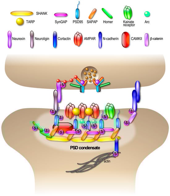
4. SUMOylation, Biomolecular Condensates and Compartmentalization of the Synapse

4.1. En Masse SUMOylation at Synapses?

The physiology and physiopathology associated with the equilibrium between the SUMOylation and deSUMOylation of nuclear proteins has been extensively reviewed [55][56], but several proteins are also SUMO-modified in extranuclear compartments in neurons [6][7][8][9]. However, given the low proportion of SUMOylation outside of the nucleus and the high SUMO protease activities [57], identification of SUMOylated proteins in other subcellular compartments and especially at synapses remains particularly challenging.
Scholars recently published a unique mass spectrometry dataset highlighting the composition of the rat synapse in SUMO2/3-ylated proteins [58]. This represents about 18% of the synaptic proteome and includes adhesion molecules, vesicle trafficking and cytoskeleton-associated proteins, scaffolding proteins and neurotransmitter receptors and transporters. These potential SUMO2/3-ylated targeted proteins have also been functionally associated with synaptic processes like synapse formation, synaptic vesicle cycling and neurotransmission and synaptic plasticity.
The interesting concept of ‘en masse SUMOylation’, also referred to as ‘Protein-Group SUMOylation’ emerged from quantitative mass spectrometry (MS) experiments primarily aiming to investigate DNA repair [59]. These data revealed that SUMOylation most frequently targets entire groups of proteins that are physically interacting or in an immediate proximity, rather than individual proteins.

4.2. SUMOylation and LLPS

A growing number of studies now report potential roles for protein SUMOylation in liquid–liquid phase separation (LLPS) and in the formation of biomolecular condensates in response to various cellular stresses.
LLPS, phase transitions and formation of biomolecular condensates are interconnected events with essential roles in the organization of specific subcellular domains and are thus detrimental processes to cell function. Despite extensive investigation in recent years, little is known about how the formation and dissociation of these biomolecular condensates are regulated, especially within the synapse. Interestingly, there is increasing evidence for the pivotal role of SUMOylation in LLPS-based formation/dissociation of these biomolecular condensates [60][61]. The best-described role for SUMOylation in LLPS is linked to the nucleus, where SUMOylation regulates the formation of promyelocytic leukemia protein (PML) bodies [62]. Formation of these biomolecular SUMO-containing condensates is partly based on the SUMO from a substrate protein and its non-covalent interaction via the SIM of another protein, bridging many proteins and their bound molecules together into membraneless condensates. SIM domains are usually composed of three exposed hydrophobic residues close to negatively-charged residues allowing the non-covalent binding of SUMO and are thus present in many different proteins [63][64]. SIM thus represents a prevalent and well-characterized motif involved in the recognition of SUMO and mediating non-covalent protein–protein contacts in the context of SUMOylation by increasing the allosteric interactions between SUMO-modified and non-SUMOylated proteins [65].

4.3. SUMOylation and Compartmentalization of Pre- and Post-Synaptic Sites

Presynaptic compartmentalization is essential to ensure the activity-dependent and calcium-sensitive control of neurotransmitter release. The mechanism of exocytosis has been studied for many years, which was detrimental to defining the precise role of the proteins involved and to understanding the sequence of molecular events occurring at each step of the process [66]. The release of neurotransmitters in the synaptic cleft is the direct consequence of axonal plasma membrane depolarization, which leads to the opening of presynaptic calcium channels. The increase in presynaptic calcium concentration triggers neurotransmitter exocytosis via complex and rapid molecular reorganization events. The precise control of ion input and output is therefore essential to this process. In the past few years, numerous studies have shown that several types of ion channels are SUMOylated [67].
The first example in the literature is the SUMOylation of synapsin-1a [68]. Synapsins ensure that SVs are maintained in a reserved accessible pool which is essential for delivering SVs to the active zone of the synapse. The authors demonstrated that the SUMOylation of synapsin-1a at K687 is required to maintain the size of the reserve pool of SVs and that the SUMO-modified form of synapsin-1a interacts more efficiently with exocytosis vesicles [68]. Synapsin-rich condensates recruit additional SVs or soluble proteins including Synaptophysin, VGLUT1 and α-synuclein [69][70], which are known as SUMOylated or potential SUMO targets [58][71]
At the center of the exocytosis machinery are VGCCs, the site of calcium entry, and synaptotagmin-1, which is the calcium sensor triggering SV exocytosis. The exocytosis machinery on the presynaptic side is highly regulated by post-translational modifications. Thus, the activity of several kinases, including the well-described PKA, PKC and CaMK, is crucial for modulating neurotransmitter release [72].
RIM1α is a Rab3a effector protein, capable of multiple interactions with key synaptic proteins, including VGCC, syntaxin-1a, SNAP25 and synaptotagmin-1, to shape the active zone [73]. These interactions are regulated by calcium ions, inositol phosphates or phosphorylation. Interestingly, the SUMOylation of RIM1α at K502 concentrates ion channels in the active zone [74], whereas its non-SUMOylated form is instead involved in vesicle priming.
Synaptotagmin-1 is a calcium sensor essential for fast SV exocytosis reported to be SUMO1-ylated [75]. To date, the functional consequences of this SUMOylation remain elusive but may be linked to the clustering of molecules around the neurotransmitter releasing site.
Many synaptic proteins including SUMOylated proteins also possess SIM domains that non-covalently bind SUMO moieties, and thus may facilitate complex assemblies by trapping other SUMO-modified proteins in their vicinity, consequently leading to clustering of specific SIM/SUMO-proteins in sub-synaptic domains. SUMOylation might therefore have additional functions, by organizing subdomains of the synapse, in shaping the protein content of presynaptic condensates to achieve a proper SV cycle and neurotransmitter release.
Another key protein of the SNARE protein complex essential for SV exocytosis and endocytosis is syntaxin-1. The syntaxin-1A is SUMOylated within its C-terminal region at K252, 253 and K256, close to the transmembrane domain [76]. The interaction of the SNARE proteins SNAP-25 and VAMP-2, but not Munc-18, with syntaxin-1A, is significantly reduced by its SUMOylation. Using real-time presynaptic imaging experiments, the authors also showed that the SUMOylation of syntaxin-1A instead regulates the rate of SV endocytosis without impacting the exocytosis process [76].
The case of the type III metabotropic glutamate receptor mGluR7 is particularly interesting [77]. The amount of glutamate released at the synapse is modulated by a feedback control loop involving mGluR7. The phosphorylation of mGluR7 at S862 facilitates its SUMOylation at K889 [77].
The process of protein cluster/phase separation is not restricted to the compartmentalization of the presynaptic site and can be extended to the post-synaptic compartment. Indeed, recent exciting studies have reported that the assembly and activity-dependent modulation of the post-synaptic density (PSD) composition involves LPPS [78][79][80]. The PSD is composed of hundreds of different proteins, including neurotransmitter receptors, scaffold proteins and many signaling molecules, with these dynamically interconnected assemblies creating a defined synaptic subdomain. It has been shown that four major post-synaptic proteins, PSD-95, SAPAP, Shank and Homer, form the PSD condensate [78]. Formation of condensed molecular assemblies has also been suggested for AMPAR synaptic clustering and transmission [79]. The binding of PSD95 to transmembrane AMPAR regulatory proteins (TARPs), which are essential AMPAR auxiliary subunits, helps to regulate the number and density of surface-expressed AMPARs at the PSD and consequently strengthen the synapse [81]. In addition, several enzymes such as SynGAP interact with PSD95, increasing their concentration within the constrained space of the PSD [78]). Upon synaptic stimulation, SynGAP is dispersed from the PSD condensates, highlighting the dynamic nature of PSD assemblies [82]. The mechanisms by which the assembly and disassembly of PSD condensates are regulated to allow changes in post-synaptic surface-expressed AMPARs are still unclear. However, 7% of the identified SUMO target proteins at synapses (55 out of 803) are directly linked to the PSD [58], including the major PSD components PSD95, Homer, SAPAP and SynGAP1 (Figure 2).
Figure 2. Landscape of SUMOylated proteins in the compartmentalization of post-synaptic sites. Consistent with the emerging post-synaptic functions of SUMOylation, several post-synaptic proteins are known SUMO substrates, including Kainate receptors [13][83][84] and Arc [85][86]. In addition, SynGAP, SAPAP3, PSD95, Homer1, CaMKII and Adhesion molecules like Neuroligin, N-Cadherin and β-catenin have been identified as potential SUMO targets in [58].

References

  1. Matunis, M.J.; Coutavas, E.; Blobel, G. A novel ubiquitin-like modification modulates the partitioning of the Ran-GTPase-activating protein RanGAP1 between the cytosol and the nuclear pore complex. J. Cell Biol. 1996, 135, 1457–1470.
  2. Mahajan, R.; Delphin, C.; Guan, T.; Gerace, L.; Melchior, F. A small ubiquitin-related polypeptide involved in targeting RanGAP1 to nuclear pore complex protein RanBP2. Cell 1997, 88, 97–107.
  3. Flotho, A.; Melchior, F. Sumoylation: A regulatory protein modification in health and disease. Annu. Rev. Biochem. 2013, 82, 357–385.
  4. Celen, A.B.; Sahin, U. Sumoylation on its 25th anniversary: Mechanisms, pathology, and emerging concepts. FEBS J. 2020, 287, 3110–3140.
  5. Gwizdek, C.; Casse, F.; Martin, S. Protein sumoylation in brain development, neuronal morphology and spinogenesis. Neuromol. Med. 2013, 15, 677–691.
  6. Schorova, L.; Martin, S. Sumoylation in Synaptic Function and Dysfunction. Front. Synaptic Neurosci. 2016, 8, 9.
  7. Henley, J.M.; Carmichael, R.E.; Wilkinson, K.A. Extranuclear SUMOylation in Neurons. Trends Neurosci. 2018, 41, 198–210.
  8. Folci, A.; Mirabella, F.; Fossati, M. Ubiquitin and Ubiquitin-Like Proteins in the Critical Equilibrium between Synapse Physiology and Intellectual Disability. eNeuro 2020, 7, 1–25.
  9. Henley, J.M.; Seager, R.; Nakamura, Y.; Talandyte, K.; Nair, J.; Wilkinson, K.A. SUMOylation of synaptic and synapse-associated proteins: An update. J. Neurochem. 2021, 156, 145–161.
  10. Colnaghi, L.; Russo, L.; Natale, C.; Restelli, E.; Cagnotto, A.; Salmona, M.; Chiesa, R.; Fioriti, L. Super Resolution Microscopy of SUMO Proteins in Neurons. Front. Cell. Neurosci. 2019, 13, 486.
  11. Colnaghi, L.; Rondelli, D.; Muzi-Falconi, M.; Sertic, S. Tau and DNA Damage in Neurodegeneration. Brain Sci. 2020, 10, 946.
  12. Conz, A.; Musi, C.A.; Russo, L.; Borsello, T.; Colnaghi, L. Super-resolution study of PIAS SUMO E3-ligases in hippocampal and cortical neurons. Eur. J. Histochem. 2021, 65, 3241.
  13. Martin, S.; Nishimune, A.; Mellor, J.R.; Henley, J.M. SUMOylation regulates kainate-receptor-mediated synaptic transmission. Nature 2007, 447, 321–325.
  14. Feligioni, M.; Nishimune, A.; Henley, J.M. Protein SUMOylation modulates calcium influx and glutamate release from presynaptic terminals. Eur. J. Neurosci. 2009, 29, 1348–1356.
  15. Loriol, C.; Parisot, J.; Poupon, G.; Gwizdek, C.; Martin, S. Developmental regulation and spatiotemporal redistribution of the sumoylation machinery in the rat central nervous system. PLoS ONE 2012, 7, e33757.
  16. Loriol, C.; Khayachi, A.; Poupon, G.; Gwizdek, C.; Martin, S. Activity-dependent regulation of the sumoylation machinery in rat hippocampal neurons. Biol. Cell 2013, 105, 30–45.
  17. Loriol, C.; Casse, F.; Khayachi, A.; Poupon, G.; Chafai, M.; Deval, E.; Gwizdek, C.; Martin, S. mGlu5 receptors regulate synaptic sumoylation via a transient PKC-dependent diffusional trapping of Ubc9 into spines. Nat. Commun. 2014, 5, 5113.
  18. Hasegawa, Y.; Yoshida, D.; Nakamura, Y.; Sakakibara, S. Spatiotemporal distribution of SUMOylation components during mouse brain development. J. Comp. Neurol. 2014, 522, 3020–3036.
  19. Josa-Prado, F.; Luo, J.; Rubin, P.; Henley, J.M.; Wilkinson, K.A. Developmental profiles of SUMOylation pathway proteins in rat cerebrum and cerebellum. PLoS ONE 2019, 14, e0212857.
  20. Guo, C.; Hildick, K.L.; Luo, J.; Dearden, L.; Wilkinson, K.A.; Henley, J.M. SENP3-mediated deSUMOylation of dynamin-related protein 1 promotes cell death following ischaemia. EMBO J. 2013, 32, 1514–1528.
  21. Waters, E.; Wilkinson, K.A.; Harding, A.L.; Carmichael, R.E.; Robinson, D.; Colley, H.E.; Guo, C. The SUMO protease SENP3 regulates mitochondrial autophagy mediated by Fis1. EMBO Rep. 2022, 23, e48754.
  22. Jaafari, N.; Konopacki, F.A.; Owen, T.F.; Kantamneni, S.; Rubin, P.; Craig, T.J.; Wilkinson, K.A.; Henley, J.M. SUMOylation is required for glycine-induced increases in AMPA receptor surface expression (ChemLTP) in hippocampal neurons. PLoS ONE 2013, 8, e52345.
  23. Lyons, G.E.; Micales, B.K.; Schwarz, J.; Martin, J.F.; Olson, E.N. Expression of mef2 genes in the mouse central nervous system suggests a role in neuronal maturation. J. Neurosci. 1995, 15, 5727–5738.
  24. Flavell, S.W.; Greenberg, M.E. Signaling mechanisms linking neuronal activity to gene expression and plasticity of the nervous system. Annu. Rev. Neurosci. 2008, 31, 563–590.
  25. Shalizi, A.; Gaudilliere, B.; Yuan, Z.; Stegmuller, J.; Shirogane, T.; Ge, Q.; Tan, Y.; Schulman, B.; Harper, J.W.; Bonni, A. A calcium-regulated MEF2 sumoylation switch controls postsynaptic differentiation. Science 2006, 311, 1012–1017.
  26. Lu, H.; Liu, B.; You, S.; Chen, L.; Dongmei, Q.; Gu, M.; Lu, Y.; Chen, Y.; Zhang, F.; Yu, B. SENP2 regulates MEF2A de-SUMOylation in an activity dependent manner. Mol. Biol. Rep. 2013, 40, 2485–2490.
  27. Meredith, L.J.; Wang, C.M.; Nascimento, L.; Liu, R.; Wang, L.; Yang, W.H. The Key Regulator for Language and Speech Development, FOXP2, is a Novel Substrate for SUMOylation. J. Cell. Biochem. 2016, 117, 426–438.
  28. Estruch, S.B.; Graham, S.A.; Deriziotis, P.; Fisher, S.E. The language-related transcription factor FOXP2 is post-translationally modified with small ubiquitin-like modifiers. Sci. Rep. 2016, 6, 20911.
  29. Usui, N.; Co, M.; Harper, M.; Rieger, M.A.; Dougherty, J.D.; Konopka, G. Sumoylation of FOXP2 Regulates Motor Function and Vocal Communication Through Purkinje Cell Development. Biol. Psychiatry 2017, 81, 220–230.
  30. Cheng, J.; Huang, M.; Zhu, Y.; Xin, Y.J.; Zhao, Y.K.; Huang, J.; Yu, J.X.; Zhou, W.H.; Qiu, Z. SUMOylation of MeCP2 is essential for transcriptional repression and hippocampal synapse development. J. Neurochem. 2014, 128, 798–806.
  31. Zhang, W.; Mi, J.; Li, N.; Sui, L.; Wan, T.; Zhang, J.; Chen, T.; Cao, X. Identification and characterization of DPZF, a novel human BTB/POZ zinc finger protein sharing homology to BCL-6. Biochem. Biophys. Res. Commun. 2001, 282, 1067–1073.
  32. Gilman, S.R.; Iossifov, I.; Levy, D.; Ronemus, M.; Wigler, M.; Vitkup, D. Rare de novo variants associated with autism implicate a large functional network of genes involved in formation and function of synapses. Neuron 2011, 70, 898–907.
  33. Jones, K.A.; Luo, Y.; Dukes-Rimsky, L.; Srivastava, D.P.; Koul-Tewari, R.; Russell, T.A.; Shapiro, L.P.; Srivastava, A.K.; Penzes, P. Neurodevelopmental disorder-associated ZBTB20 gene variants affect dendritic and synaptic structure. PLoS ONE 2018, 13, e0203760.
  34. Ripamonti, S.; Shomroni, O.; Rhee, J.S.; Chowdhury, K.; Jahn, O.; Hellmann, K.P.; Bonn, S.; Brose, N.; Tirard, M. SUMOylation controls the neurodevelopmental function of the transcription factor Zbtb20. J. Neurochem. 2020, 154, 647–661.
  35. Biederer, T.; Sudhof, T.C. CASK and protein 4.1 support F-actin nucleation on neurexins. J. Biol. Chem. 2001, 276, 47869–47876.
  36. Chao, H.W.; Hong, C.J.; Huang, T.N.; Lin, Y.L.; Hsueh, Y.P. SUMOylation of the MAGUK protein CASK regulates dendritic spinogenesis. J. Cell Biol. 2008, 182, 141–155.
  37. Drisaldi, B.; Colnaghi, L.; Fioriti, L.; Rao, N.; Myers, C.; Snyder, A.M.; Metzger, D.J.; Tarasoff, J.; Konstantinov, E.; Fraser, P.E.; et al. SUMOylation Is an Inhibitory Constraint that Regulates the Prion-like Aggregation and Activity of CPEB3. Cell Rep. 2015, 11, 1694–1702.
  38. Huber, K.M.; Roder, J.C.; Bear, M.F. Chemical induction of mGluR5- and protein synthesis--dependent long-term depression in hippocampal area CA1. J. Neurophysiol. 2001, 86, 321–325.
  39. Bear, M.F.; Huber, K.M.; Warren, S.T. The mGluR theory of fragile X mental retardation. Trends Neurosci. 2004, 27, 370–377.
  40. Bassell, G.J. Fragile balance: RNA editing tunes the synapse. Nat. Neurosci. 2011, 14, 1492–1494.
  41. Darnell, J.C.; Van Driesche, S.J.; Zhang, C.; Hung, K.Y.; Mele, A.; Fraser, C.E.; Stone, E.F.; Chen, C.; Fak, J.J.; Chi, S.W.; et al. FMRP stalls ribosomal translocation on mRNAs linked to synaptic function and autism. Cell 2011, 146, 247–261.
  42. The Dutch-Belgian Fragile X Consortium; Bakker, C.E.; Verheij, C.; Willemsen, R.; van der Helm, R.; Oerlemans, F.; Vermey, M.; Bygrave, A.; Hoogeveen, A.; Oostra, B.A.; et al. Fmr1 knockout mice: A model to study fragile X mental retardation. The Dutch-Belgian Fragile X Consortium. Cell 1994, 78, 23–33.
  43. Mientjes, E.J.; Nieuwenhuizen, I.; Kirkpatrick, L.; Zu, T.; Hoogeveen-Westerveld, M.; Severijnen, L.; Rife, M.; Willemsen, R.; Nelson, D.L.; Oostra, B.A. The generation of a conditional Fmr1 knock out mouse model to study Fmrp function in vivo. Neurobiol. Dis. 2006, 21, 549–555.
  44. Baas, P.W.; Rao, A.N.; Matamoros, A.J.; Leo, L. Stability properties of neuronal microtubules. Cytoskeleton 2016, 73, 442–460.
  45. Gu, J.; Firestein, B.L.; Zheng, J.Q. Microtubules in dendritic spine development. J. Neurosci. 2008, 28, 12120–12124.
  46. Mitchison, T.; Kirschner, M. Dynamic instability of microtubule growth. Nature 1984, 312, 237–242.
  47. Li, S.; Liang, Y.; Zou, J.; Cai, Z.; Yang, H.; Yang, J.; Zhang, Y.; Lin, H.; Zhang, G.; Tan, M. SUMOylation of microtubule-cleaving enzyme KATNA1 promotes microtubule severing and neurite outgrowth. J. Biol. Chem. 2022, 298, 102292.
  48. McNally, F.J.; Vale, R.D. Identification of katanin, an ATPase that severs and disassembles stable microtubules. Cell 1993, 75, 419–429.
  49. Chen, K.; Ye, Y.; Ji, Z.; Tan, M.; Li, S.; Zhang, J.; Guo, G.; Lin, H. Katanin p60 promotes neurite growth and collateral formation in the hippocampus. Int. J. Clin. Exp. Med. 2014, 7, 2463–2470.
  50. Shin, S.C.; Im, S.K.; Jang, E.H.; Jin, K.S.; Hur, E.M.; Kim, E.E. Structural and Molecular Basis for Katanin-Mediated Severing of Glutamylated Microtubules. Cell Rep. 2019, 26, 1357–1367.e5.
  51. Ji, Z.S.; Liu, Q.L.; Zhang, J.F.; Yang, Y.H.; Li, J.; Zhang, G.W.; Tan, M.H.; Lin, H.S.; Guo, G.Q. SUMOylation of spastin promotes the internalization of GluA1 and regulates dendritic spine morphology by targeting microtubule dynamics. Neurobiol. Dis. 2020, 146, 105133.
  52. Yu, W.; Qiang, L.; Solowska, J.M.; Karabay, A.; Korulu, S.; Baas, P.W. The microtubule-severing proteins spastin and katanin participate differently in the formation of axonal branches. Mol. Biol. Cell 2008, 19, 1485–1498.
  53. Vertegaal, A.C.O. Signalling mechanisms and cellular functions of SUMO. Nat. Rev. Mol. Cell Biol. 2022, 23, 715–731.
  54. Gasser, S.M.; Stutz, F. SUMO in the regulation of DNA repair and transcription at nuclear pores. FEBS Lett. 2023, 597, 2833–2850.
  55. Esteras, M.; Liu, I.C.; Snijders, A.P.; Jarmuz, A.; Aragon, L. Identification of SUMO conjugation sites in the budding yeast proteome. Microb. Cell 2017, 4, 331–341.
  56. Pronot, M.; Kieffer, F.; Gay, A.S.; Debayle, D.; Forquet, R.; Poupon, G.; Schorova, L.; Martin, S.; Gwizdek, C. Proteomic Identification of an Endogenous Synaptic SUMOylome in the Developing Rat Brain. Front. Mol. Neurosci. 2021, 14, 780535.
  57. Psakhye, I.; Jentsch, S. Protein group modification and synergy in the SUMO pathway as exemplified in DNA repair. Cell 2012, 151, 807–820.
  58. Cheng, X.; Yang, W.; Lin, W.; Mei, F. Paradoxes of Cellular SUMOylation Regulation: A Role of Biomolecular Condensates? Pharmacol. Rev. 2023, 75, 979–1006.
  59. Keiten-Schmitz, J.; Roder, L.; Hornstein, E.; Muller-McNicoll, M.; Muller, S. SUMO: Glue or Solvent for Phase-Separated Ribonucleoprotein Complexes and Molecular Condensates? Front. Mol. Biosci. 2021, 8, 673038.
  60. Bratek-Skicki, A.; Pancsa, R.; Meszaros, B.; Van Lindt, J.; Tompa, P. A guide to regulation of the formation of biomolecular condensates. FEBS J. 2020, 287, 1924–1935.
  61. Yau, T.Y.; Sander, W.; Eidson, C.; Courey, A.J. SUMO Interacting Motifs: Structure and Function. Cells 2021, 10, 2825.
  62. Lascorz, J.; Codina-Fabra, J.; Reverter, D.; Torres-Rosell, J. SUMO-SIM interactions: From structure to biological functions. Semin. Cell Dev. Biol. 2022, 132, 193–202.
  63. Rodriguez, M.S.; Dargemont, C.; Hay, R.T. SUMO-1 conjugation in vivo requires both a consensus modification motif and nuclear targeting. J. Biol. Chem. 2001, 276, 12654–12659.
  64. Rizo, J. Mechanism of neurotransmitter release coming into focus. Protein Sci. 2018, 27, 1364–1391.
  65. Benson, M.; Iniguez-Lluhi, J.A.; Martens, J. Sumo Modification of Ion Channels. Adv. Exp. Med. Biol. 2017, 963, 127–141.
  66. Tang, L.T.; Craig, T.J.; Henley, J.M. SUMOylation of synapsin Ia maintains synaptic vesicle availability and is reduced in an autism mutation. Nat. Commun. 2015, 6, 7728.
  67. Sansevrino, R.; Hoffmann, C.; Milovanovic, D. Condensate biology of synaptic vesicle clusters. Trends Neurosci. 2023, 46, 293–306.
  68. Rousseaux, M.W.C.; Vazquez-Velez, G.E.; Al-Ramahi, I.; Jeong, H.H.; Bajic, A.; Revelli, J.P.; Ye, H.; Phan, E.T.; Deger, J.M.; Perez, A.M.; et al. A Druggable Genome Screen Identifies Modifiers of α-Synuclein Levels via a Tiered Cross-Species Validation Approach. J. Neurosci. 2018, 38, 9286–9301.
  69. Krumova, P.; Meulmeester, E.; Garrido, M.; Tirard, M.; Hsiao, H.H.; Bossis, G.; Urlaub, H.; Zweckstetter, M.; Kugler, S.; Melchior, F.; et al. Sumoylation inhibits α-synuclein aggregation and toxicity. J. Cell Biol. 2011, 194, 49–60.
  70. Leenders, A.G.; Sheng, Z.H. Modulation of neurotransmitter release by the second messenger-activated protein kinases: Implications for presynaptic plasticity. Pharmacol. Ther. 2005, 105, 69–84.
  71. Coppola, T.; Magnin-Luthi, S.; Perret-Menoud, V.; Gattesco, S.; Schiavo, G.; Regazzi, R. Direct interaction of the Rab3 effector RIM with Ca2+ channels, SNAP-25, and synaptotagmin. J. Biol. Chem. 2001, 276, 32756–32762.
  72. Girach, F.; Craig, T.J.; Rocca, D.L.; Henley, J.M. RIM1α SUMOylation is required for fast synaptic vesicle exocytosis. Cell Rep. 2013, 5, 1294–1301.
  73. Matsuzaki, S.; Lee, L.; Knock, E.; Srikumar, T.; Sakurai, M.; Hazrati, L.N.; Katayama, T.; Staniszewski, A.; Raught, B.; Arancio, O.; et al. SUMO1 Affects Synaptic Function, Spine Density and Memory. Sci. Rep. 2015, 5, 10730.
  74. Craig, T.J.; Anderson, D.; Evans, A.J.; Girach, F.; Henley, J.M. SUMOylation of Syntaxin1A regulates presynaptic endocytosis. Sci. Rep. 2015, 5, 17669.
  75. Choi, J.H.; Park, J.Y.; Park, S.P.; Lee, H.; Han, S.; Park, K.H.; Suh, Y.H. Regulation of mGluR7 trafficking by SUMOylation in neurons. Neuropharmacology 2016, 102, 229–235.
  76. Zeng, M.; Shang, Y.; Araki, Y.; Guo, T.; Huganir, R.L.; Zhang, M. Phase Transition in Postsynaptic Densities Underlies Formation of Synaptic Complexes and Synaptic Plasticity. Cell 2016, 166, 1163–1175.e12.
  77. Zeng, M.; Diaz-Alonso, J.; Ye, F.; Chen, X.; Xu, J.; Ji, Z.; Nicoll, R.A.; Zhang, M. Phase Separation-Mediated TARP/MAGUK Complex Condensation and AMPA Receptor Synaptic Transmission. Neuron 2019, 104, 529–543.e6.
  78. Wu, X.; Hu, S.; Kang, X.; Wang, C. Synaptotagmins: Beyond Presynaptic Neurotransmitter Release. Neuroscientist 2020, 26, 9–15.
  79. Nicoll, R.A.; Tomita, S.; Bredt, D.S. Auxiliary subunits assist AMPA-type glutamate receptors. Science 2006, 311, 1253–1256.
  80. Araki, Y.; Zeng, M.; Zhang, M.; Huganir, R.L. Rapid dispersion of SynGAP from synaptic spines triggers AMPA receptor insertion and spine enlargement during LTP. Neuron 2015, 85, 173–189.
  81. Chamberlain, S.E.; Gonzalez-Gonzalez, I.M.; Wilkinson, K.A.; Konopacki, F.A.; Kantamneni, S.; Henley, J.M.; Mellor, J.R. SUMOylation and phosphorylation of GluK2 regulate kainate receptor trafficking and synaptic plasticity. Nat. Neurosci. 2012, 15, 845–852.
  82. Konopacki, F.A.; Jaafari, N.; Rocca, D.L.; Wilkinson, K.A.; Chamberlain, S.; Rubin, P.; Kantamneni, S.; Mellor, J.R.; Henley, J.M. Agonist-induced PKC phosphorylation regulates GluK2 SUMOylation and kainate receptor endocytosis. Proc. Natl. Acad. Sci. USA 2011, 108, 19772–19777.
  83. Craig, T.J.; Henley, J.M. SUMOylation, Arc and the regulation homeostatic synaptic scaling: Implications in health and disease. Commun. Integr. Biol. 2012, 5, 634–636.
  84. Nair, R.R.; Patil, S.; Tiron, A.; Kanhema, T.; Panja, D.; Schiro, L.; Parobczak, K.; Wilczynski, G.; Bramham, C.R. Dynamic Arc SUMOylation and Selective Interaction with F-Actin-Binding Protein Drebrin A in LTP Consolidation In Vivo. Front. Synaptic Neurosci. 2017, 9, 8.
More
Information
Subjects: Cell Biology
Contributors MDPI registered users' name will be linked to their SciProfiles pages. To register with us, please refer to https://encyclopedia.pub/register : , , ,
View Times: 116
Revisions: 4 times (View History)
Update Date: 08 Mar 2024
1000/1000