Your browser does not fully support modern features. Please upgrade for a smoother experience.
Submitted Successfully!
 +1 credit
Thank you for your contribution! You can also upload a video entry or images related to this topic.
For video creation, please contact our Academic Video Service.
| Version | Summary | Created by | Modification | Content Size | Created at | Operation |
|---|---|---|---|---|---|---|
| 1 | Mugur Cristian Grasu | -- | 5603 | 2024-03-05 00:05:40 | | | |
| 2 | Catherine Yang | + 1 word(s) | 5604 | 2024-03-05 02:26:29 | | |
Video Upload Options
We provide professional Academic Video Service to translate complex research into visually appealing presentations. Would you like to try it?
Cite
If you have any further questions, please contact Encyclopedia Editorial Office.
Anghel, C.; Grasu, M.C.; Anghel, D.A.; Rusu-Munteanu, G.; Dumitru, R.L.; Lupescu, I.G. AI Applications in Pancreatic Ductal Adenocarcinoma. Encyclopedia. Available online: https://encyclopedia.pub/entry/55846 (accessed on 03 July 2026).
Anghel C, Grasu MC, Anghel DA, Rusu-Munteanu G, Dumitru RL, Lupescu IG. AI Applications in Pancreatic Ductal Adenocarcinoma. Encyclopedia. Available at: https://encyclopedia.pub/entry/55846. Accessed July 03, 2026.
Anghel, Cristian, Mugur Cristian Grasu, Denisa Andreea Anghel, Gina-Ionela Rusu-Munteanu, Radu Lucian Dumitru, Ioana Gabriela Lupescu. "AI Applications in Pancreatic Ductal Adenocarcinoma" Encyclopedia, https://encyclopedia.pub/entry/55846 (accessed July 03, 2026).
Anghel, C., Grasu, M.C., Anghel, D.A., Rusu-Munteanu, G., Dumitru, R.L., & Lupescu, I.G. (2024, March 05). AI Applications in Pancreatic Ductal Adenocarcinoma. In Encyclopedia. https://encyclopedia.pub/entry/55846
Anghel, Cristian, et al. "AI Applications in Pancreatic Ductal Adenocarcinoma." Encyclopedia. Web. 05 March, 2024.
Copy Citation
Pancreatic ductal adenocarcinoma (PDAC) stands out as the predominant malignant neoplasm affecting the pancreas, characterized by a poor prognosis, in most cases patients being diagnosed in a nonresectable stage. Image-based artificial intelligence (AI) models implemented in tumor detection, segmentation, and classification could improve diagnosis with better treatment options and increased survival.
pancreatic adenocarcinoma
artificial intelligence
pancreas imaging
1. Detection
Pancreatic ductal adenocarcinoma (PDAC) carries a poor prognosis because of the delayed diagnosis and the lack of screening modalities and protocols in the general population. Studies indicate that the detection of PDAC in high-risk individuals leads to a median overall survival of 9.8 years (compared to 1.5 years for those diagnosed outside of surveillance) [1]. This can be correlated to the fact that early detected tumors are smaller in size and do not infiltrate the surrounding vessels. Identifying small and iso-attenuating tumors can be sometimes difficult, especially in asymptomatic patients, and can lead to delayed recognition [2][3]. There is a great interest in developing an AI model that could identify PDAC on prediagnostic CTs before the clinical diagnosis. AI algorithms show promise in detecting subtle changes in pancreas and small pancreatic tumors, that can be missed by human observers.
Because most of the tumors are large at diagnosis, the AI models are trained on data sets that contain a small proportion of small tumors, making it hard for the model to achieve good results in detecting small and isoattenuating PDAC. Large datasets that contain an increased number of small tumors should be made publicly available in order for researchers to be able to train their models on and have better results in detecting small (even <1 cm) tumors in high-risk patients. Selected papers that focused on detection can be seen in Table 1.
Cao et al. present a novel deep learning framework named Pancreatic Cancer Detection with Artificial Intelligence (PANDA) aimed at detecting and categorizing pancreatic lesions utilizing non-contrast CT images [4]. This model underwent training using a dataset comprising non-contrast CT scans from 3208 patients sourced from a single center. In a multicenter validation encompassing 6239 patients from 10 distinct centers, the model demonstrated exceptional performance with an area under the receiver operating curve (AUC) ranging between 0.986 and 0.996 for lesion detection. Their model could represent a screening method in the high-risk population, given the lower dose compared to CECT and the absence of risk given by the administration of intravenous contrast.
Korfiatis et al. conducted a case–control study to develop an automated 3-dimensional (3D) Convolutional Neural Network (CNN) for detecting PDAC on diagnostic computer tomography scans [5]. One of their primary aims was to assess the model’s capability to identify visually imperceptible preinvasive cancer on prediagnostic CT scans. The 3D-CNN was trained using a dataset consisting of 696 portal phase diagnostic CT scans featuring PDAC cases and 1080 control images displaying non-neoplastic pancreatic conditions. Despite being exclusively trained on CT images depicting larger tumors, the model successfully detected occult PDAC on prediagnostic CT scans (incidentally acquired 3–36 months before the clinical diagnosis of PDAC) with AUC curve 0.91, sensitivity 0.75, and specificity 0.90 at a median of 475 days before clinical diagnosis. Their model shows great promise in future screening of high-risk individuals and could alert readers for small lesions and secondary signs of PDAC (atrophy and ductal dilatation), but further training on CTs images with smaller tumors will increase the sensitivity and the specificity.
Chu et al. engineered a deep-learning algorithm designed to differentiate CT cases from patients afflicted with PDAC versus those from control subjects [6]. Initial findings from an analysis comprising 156 PDAC cases and 300 normal cases revealed a sensitivity of 94.1% and a specificity of 98.5% for the detection of PDAC. The deep network performed well on larger tumors and often missed small tumors, but this problem could be overcome by adding more small tumors in the training data set and modifying the model structure to focus on finding smaller lesions [6]. Their model shows good results, but needs to be validated on larger data sets, including images from other institutions.
Another study determined the utility of radiomics features in differentiating CT cases of PDAC from normal pancreas [7]. In this study, 190 patients with PDAC and 190 healthy potential renal donors were included, with 40 radiomic features selected for analysis. The dataset was partitioned into 255 training cases, comprising 125 normal control cases and 130 PDAC cases, and 125 validation cases, including 65 normal control cases and 60 PDAC cases. The random forest binary classification achieved an accuracy of 99.2% and an AUC of 99.9%. However, it is noteworthy that the mean tumor size within this dataset was 4.1 cm, a dimension typically within the range of detection capability for the average radiologist. The results need to be validated on datasets containing much smaller tumors (even as small as 1–2 cm) in order to have a clinical impact and assist the radiologist in detecting tumors while still at a resectable stage.
Chu et al. have also published a paper comparing the diagnostic performance of commercially available vs. in-house radiomics software in the classification of CT images from patients with PDAC vs. healthy controls [8]. When employing 40 features in the random forest classification, the in-house software demonstrated superior sensitivity at 100% and accuracy at 99.2%, surpassing the performance of the commercially available research prototype, which exhibited a sensitivity of 95% and accuracy of 96.8%. However, when the number of features was reduced to five, the diagnostic performance of the in-house software decreases to sensitivity (95%) and accuracy (93.6%), while the diagnostic performance of the commercially available prototype was unchanged. The study population was selected based on a previously published study [7]. Future studies are needed to determine if other commercially available radiomics software will achieve similar results.
Ma et al. constructed a CNN model using a dataset of 3494 CT images from 222 patients confirmed with PDAC and 3751 images from 190 patients with normal pancreas with the aim to distinguish pancreatic cancer from benign tissue [9]. The plain phase had the same sensitivity (91.58%) as the arterial and venous phases, with an accuracy of 95.47% and specificity of 98.27%. Given the lower radiation dose and easier access to non-contrast CT, their model is suitable for PDAC screening, especially in patients with contraindications for the intravenous administration of contrast media.
Alves et al. developed a deep learning model used for PDAC detection, focusing on small lesions [10]. The study included CECT images from 119 pathology proven PDAC patients and 123 patients without PDAC that were used to train a nnUnet for automatic lesions detection and segmentation. The proposed model achieved good results with a maximum AUC of 0.914 in the whole external test and 0.876 in the subgroup of tumors < 2 cm. However, their study included only tumors that were located in the pancreatic head and further studies are needed on data sets including lesions located in the body and tail of the pancreas.
Mukherjee et al. developed a radiomics-based machine-learning (ML) model to detect PDAC at the prediagnostic stage [11]. They included prediagnostic CTs (median interval between CT and PDAC diagnosis: 398 days) of 155 patients and an age-matched cohort of 265 patients with normal pancreas, with 34 radiomics features selected. An internal set of 176 patients and 80 publicly available control cases were used for validation. A machine learning classifier, support vector machine (SVM), had a sensitivity of 95.5% and AUC of 0.98. Their study shows good results in detecting PDAC at a prediagnostic stage, which could greatly benefit the patients by identifying the tumors in a resectable stage.
Wang et al. addressed the issues of consistency and reproducibility of feature extraction (radiomics) and subsequent modeling by exploring the resilience of radiomics features against various sources of uncertainty including bin width resampling, image transformation, image noise, and segmentation growth/shrinkage. [12]. They collected venous-phase CECT images from 181 control subjects and 85 PDAC case subjects and extracted 924 radiomics features using PyRadiomics. The training set was formed from 189 cases (60 cancer and 129 control) and the remaining 77 cases (25 cancer and 52 control) formed the testing set. They trained a Random Forest model using eight nonredundant radiomics features to distinguish the cancer patients from healthy individuals, which achieved a mean AUC of 0.99 ± 0.01 on the training dataset by cross-validation and an AUC of 0.910 on the independent data set. However, the sample size was limited compared to other published studies [7][13]. Their model needs validation on bigger data sets.
Viviers et al. proposed a model based on a U-Net-Like Deep CNN that incorporates the external secondary features such as the pancreatic duct, common bile duct, and the pancreas, in conjunction with a processed CECT scan [14]. The study included a dataset of 99 cases of PDAC located in the pancreatic head and 97 control cases without a pancreatic tumor. The model achieved a performance score for classification and localization of 99% sensitivity and 99% specificity. However, the CNN model cannot discover the causal relationship between these secondary features and the presence of the tumor, with future work needed to enable CNN to fully integrate all the information. The study focuses on secondary features of the PDAC, which is of great interest especially in isoattenuating and small tumors. Their model also included only patients with PDAC located in the pancreatic head, with further research needed to see if the model obtains the same results for tumors located in the pancreatic body or tail.
Table 1. AI detection models.
| Author | Year | Modality | Approach | Sensitivity |
|---|---|---|---|---|
| Cao et al. [4] | 2023 | Non-contrast CT | Deep learning | 92.9% |
| Korifatis et al. [5] | 2023 | CECT | 3D-CNN | 75% |
| Alves et al. [10] | 2022 | CECT | CNN | N.A. |
| Wang et al. [12] | 2022 | CECT | Radiomics | 84% |
| Mukherjee et al. [11] | 2022 | CECT | Radiomics-ML | 95.5% |
| Viviers et al. [14] | 2022 | CECT | U-Net-Like Deep CNN | 99% |
| Ma et al. [9] | 2020 | Non-contrast CT | CNN | 91.58% |
| Chu et al. [6] | 2019 | CECT | Deep learning | 94.1% |
| Chu et al. [7] | 2019 | CECT | Radiomics | 100% |
2. Segmentation
Most of the AI radiology models are supervised and need large amounts of images for training [15]. Image preparation including lesion and organ segmentation is performed in most instances manually or in a semi-automated manner by the radiologist. This process needs a huge amount of work, and given the fact that for AI models to be validated large datasets are needed for training and testing, a great interest in the automatic segmentation of the pancreas and pancreatic lesions is seen in the last years. An example of semi-automated tumor segmentation using 3D Slicer software ver. 5.6.1 [16] is shown in Figure 2.
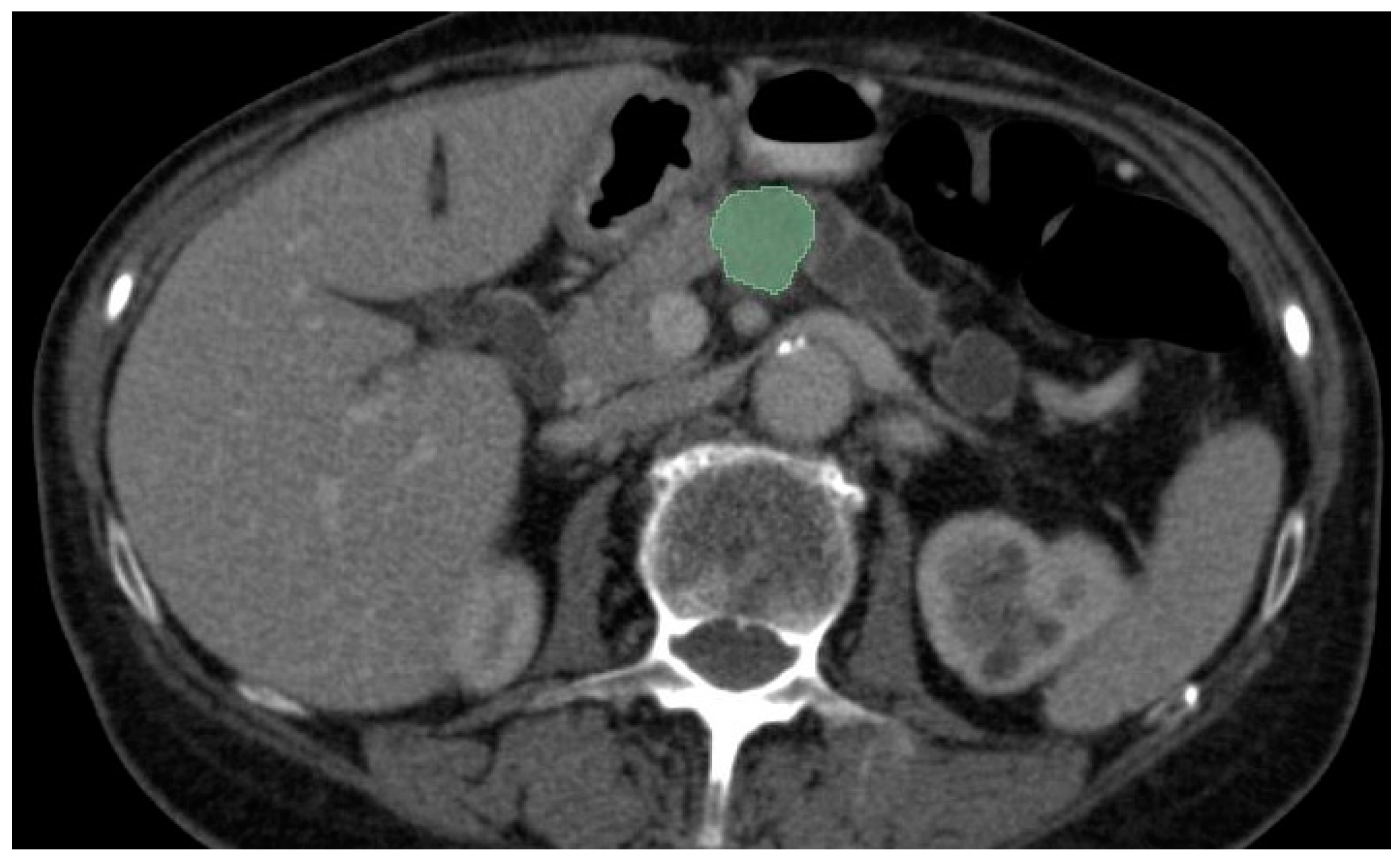
Figure 2. CECT portal venous-phase showing semi-automated corporeal pancreatic tumor segmentation (depicted in green) using “growing-seeds” method.
The automated segmentation of PDAC in CT scans represents a challenge given (1) the variability in size, shape, and location of the tumor in the pancreas, (2) the small size of the pancreas and the tumor by comparison with the entire abdomen and (3) the poor contrast of the pancreas with the surrounding structures [17][18][19]. AI segmentation algorithms include multiorgan atlas-based, landmark-based, shape model-based, and neural network-based [20]. The Dice similarity coefficient (DSC) is used to evaluate the overlap between a predicted segmentation mask and the ground truth segmentation (performed manually by the radiologist), with a score that ranges from 0 (no spatial overlap between two sets of binary segmentation results) to 1 (complete overlap) [20]. Selected papers that focused on segmentation can be seen in Table 2.
Ni et al. investigated the reliability of radiomic features extracted from CECT by AX-Unet, a pancreas segmentation model that integrates the strengths of deepLabV series [21], Unet, and Xception networks [22], to analyze the recurrence of PDAC after radical surgery [23]. The model was trained on a set of 205 PDAC patients and evaluated on an independent testing set of 64 patients with clear prognoses. The Dice score for their dataset reached 85.9%, showing great effectiveness for PDAC image segmentation. The study shows good results, with great potential in accurately identifying and segmenting the tumor and maximizing the tumor surgical removal.
Turečková et al. proposed the extension of CNN by including deep supervision and attention gates [24]. The model examined a dataset comprising 421 portal venous-phase CT images and corresponding segmentation masks delineating the pancreas and tumor, collected from the Memorial Sloan Kettering Cancer Center. Among these, 282 images were included in the training set, which was publicly released for the 2018 MICCAI Medical Decathlon Challenge [25]. During training, they conducted five-fold cross-validation, employing 26 images for validation and 105 images for training. The Dice scores achieved by their VNet-AG-DSV model were 81.22% for pancreas labels and 52.99% for tumor labels, respectively. In comparison, Isensee et al. achieved in the MICCAI Medical Decathlon Challenge a Dice score for the pancreas and the pancreas tumor segmentation of 79.30% and 52.12% using a nnU-Net model [26].
The algorithms studied for segmenting PDAC are optimized for only one type of input (one phase of CT acquisition); therefore; the models cannot be implemented in a multi-phase instance. To address this problem, Zhou et al. proposed a multi-phase segmentation model, Hyper-Pairing Network (HPN) for better performance for detecting pancreatic tumors [18]. They constructed a dual-path network for handling multi-phase data and applied skip connections across different paths of the network (referred to as hyper-connections) to enable information exchange between different phases. They also applied an additional pairing loss that encourages the commonality between the two sets of high-level semantic representations to reduce view divergence. The model was trained on arterial and venous phase CT images from 239 patients with pathologically proven PDAC. Compared to single-phase algorithms, multi-phase algorithms had a superior DSC (57.10% ± 24.76% for 3D-UNet-multi-phase-HPN and 63.94% ± 22.74% for 3D-ResDSN-multi-HPN). This study opens the pathway for further research on multi-phase segmentation, that is much needed in PDAC diagnosis because some tumors have a heterogeneous appearance and could benefit from both arterial and venous phase analysis.
Mahmoudi et al. used a 3D-CNN architecture localized in the pancreas region from the whole CT volume using 3D Local Binary Pattern (LBP) map of the original image [27]. The pancreatic tumor was subsequently segmented using 2D attenuation U-Net and Texture Attention U-Net (TAU-Net) that was introduced by the fusion of dense Scale-Invariant Feature Transform (SIFT) and LBP descriptors intro the attention U-Net. An ensemble model was used to summarize the advantages of both networks using a 3D-CNN. The Dice global score for the proposed hybrid model auto-localization was 60.6%. The model showed a good performance in segmentation on small sample size; however, further research is needed on larger data sets for this algorithm to be validated.
Most of the proposed algorithms for PDAC segmentations have been trained on large tumors, but there is little research focused on the early signs of PDAC (such as dilated pancreatic duct). Shen et al. addressed this problem and trained a fully convolutional networks (FCN) to generate an ROI covering the pancreas and used a 3D U-Net-like FCN for coarse pancreas segmentation [28]. The average Dice score and sensitivity was 49.9% and 51.9%, respectively. Their work shows the potential of using AI models for dilated pancreatic duct segmentation, which could also be beneficial in the detection of small tumors.
Table 2. AI segmentation models for PDAC.
3. Classification
3.1. Differential Diagnosis
One IA research area in PDAC represents the use of algorithms in differential diagnosis with other benign or maligning conditions. This could be of great use because in some instances the treatment is different and could improve patient management by avoiding unnecessary surgery. Selected papers that focused on differential diagnosis can be seen in Table 3.
Focal-type autoimmune pancreatitis (fAIP) represents a segmental involvement of the pancreatic parenchyma and can mimic PDAC, making the differential diagnosis very difficult. Most patients with fAIP will undergo surgical resection for a clear histopathological diagnosis. However, with the development of new AI models, there is hope that a clear discrimination between these entities will be achieved, eliminating the need for patients to undergo future surgical resection. Lu et al. developed a CT-based radiomics nomogram combining subjective CT findings and radiomic features for the preoperative differentiation of fAIP from PDAC [29]. The study included CT images from 96 patients (32 with AIP and 64 with PDAC) with 158 features extracted from each image. Seven features were selected by the LASSO algorithm and a multiparametric radiomics signature was established. They constructed a combined model of radiomics nomogram using multivariate logistic analysis of five characteristics (capsule-like rim, pancreatic atrophy, biliary wall thickening, vascular invasion, and Rad-Score). The nomogram model exhibited robust sensitivity and specificity, achieving AUC values of 0.87 and 0.83 in the training and test cohorts, respectively. The study had some limitations, one of them being the low number of fAIP cases acquired over a period of 11 years (2011 to 2021), which are not enough to validate the proposed radiomics model. However, further studies on larger data sets from multiple centers could improve the differential diagnosis between PDAC and fAIP.
Another study was conducted by Park et al. on differentiating autoimmune pancreatitis (AIP) from PDAC with radiomics features that included CECT images from 89 patients with AIP and 93 patients with PDAC [30]. Using the CT radiomics features in the test set, AIP could be distinguished from PDAC with a sensitivity of 89.7% (95% CI: 78.6–100%), a specificity of 100% (95% CI: 93–100%), an accuracy of 95.2% (95% CI: 89.8–100%), and an AUC of 0.975 (95% CI: 0.936–1.0). Limitations of the study were the small sample size that included both focal and diffuse forms of AIP and the period of time in which the images were acquired for the patients with AIP (2004 to 2018).
Anai et al., constructed a support vector machine (SVM) classifier using CT textured analysis for differentiating fAIP and PDAC and to assess the radiologist’s (4) diagnostic performance with or without SVM [31]. The study included CECT images from 20 patients with fAIP and 30 patients with PDAC from which 62 texture-based features were extracted. The SVM classifier had strong performance in differentiating fAIP and PDAC, with an AUC of 0.920. The SVM classifier increased the AUC for all four readers, but the reader with less experience benefitted the most from the SVM outputs. However, given the small number of patients included in the study, there is need for a larger study population to confirm the results.
Mass-forming chronic pancreatitis (MFCP) is another benign condition that can easily mimic PDAC lesions. A study addressed the problem of discriminating between PDAC and mass-forming chronic pancreatitis lesions (MFCP) [32]. This retrospective study included 119 patients from two independent institutions that had performed preoperative MRI. They extracted four feature sets from T1-weighted imaging (T1WI), T2-weighted imaging (T2WI), arterial phase, and portal phase of dynamic contrast-enhanced MRI, and radiomics models were constructed. The study shows good results with AUCs for the T1WI, T2WI, arterial and portal phases of 0.893, 0.911, 0.958, and 0.997 in the primary cohort and 0.882, 0.902, 0.920, and 0.962 in the validation cohort.
Ren et al. proposed a CT-based radiomics signature for the differential diagnosis between pancreatic adenosquamous carcinoma (PASC) and PDAC [33]. They used CECT images (both late arterial phase and portal venous phase) from 81 patients with PDAC and 31 patients with PASC to extract radiomics features. Seven radiomics features from late arterial phase images and three from portal venous phase images were selected and a radiomics signature was constructed. Using a 10-times leave group our-cross-validation method (LGOCV), the radiomics signature was proven to be robust and reliable with an average AUC of 0.82. However, the limitations of the study were the small number of PASC patients, different scanners used for image acquisition, and a selection bias due to retrospective nature of the study.
Shi et al. evaluated the performance of the histogram array and CNN based on diffusion-weighted imaging (DWI) with multiple b-values to distinguish PDAC from solid pseudopapillary neoplasm (SPN) and pancreatic neuroendocrine neoplasm (PNEN) [34]. They included 231 patients (132 PDACs, 45 PNENs, and 54 SPNs), which were further divided into a training group (n = 92), validation group (n = 46), and testing group (n = 93). The DWI images from 10 b values were converted into a histogram array that was entered into the CNN model. The AUCs of CNN for distinguishing PDACs from non-PDACS were 0.896, 0.846, and 0.839 in the training, validation, and testing groups. One of the limitations of the study was the low number of PNENs and SPNs used for training the CNN models, given the fact that these pancreatic tumors are rare compared to PDAC [35]. The study also did not include other types of pancreatic tumors such as metastases, mucinous tumors, or lymphoma.
Zhang et al. evaluated the diagnostic efficacy of radiomics in conjunction with multiple machine learning algorithms for distinguishing between PDAC and pancreatic neuroendocrine tumors (pNET) [36]. This was achieved by constructing 45 discriminative models using five selection algorithms and nine classification algorithms. [36]. The study included CECT images from 238 patients (156 diagnosed with PDAC and 82 diagnosed with pNET). By using the combination of Gradient Boosting Decision Tree (GBDT) as the selection algorithm and Random Forest (RF) as the classification algorithm, the model reached the highest AUC: 0.971 in the training set and 0.930 in the validation set. The study reinforces the idea that multi-algorithm modeling should be considered when predicting subtypes of cancer with machine learning.
Current research that analyzed the differential diagnosis of PDAC from other types of pancreatic lesions has been conducted in a binary fashion (distinguishing PDAC from other types of pancreatic lesions, for example, neuroendocrine tumors); however, for a model to be clinically relevant it should be able to distinguish between a more complex number of pathologies (for example, autoimmune pancreatitis, chronic pancreatitis, cystic lesions, and other malignancies).
Table 3. AI models studied in differential diagnosis of PDAC.
| Author | Year | Modality | Scope | Approach | AUC |
|---|---|---|---|---|---|
| Lu et al. [29] | 2023 | CECT | fAPI vs. PDAC | Radiomics | 0.83 |
| Shi et al. [34] | 2023 | MRI | PDAC vs. PNEN and SPN | CNN | 0.839 |
| Zhang et al. [36] | 2022 | CECT | PDAC vs. pNET | Radiomics | 0.930 |
| Anai et al. [31] | 2022 | CECT | fAPI vs. PDAC | Radiomics | 0.920 |
| Deng et al. [32] | 2021 | MRI | MFCP vs. PDAC | Radiomics | 0.962 |
| Ren et al. [33] | 2020 | CECT | PASC vs. PDAC | Radiomics | 0.82 |
| Park et al. [30] | 2020 | CECT | AIP vs. PDAC | Radiomics | 0.975 |
3.2. Histopathological Subtype and Genomic Features
The histopathological subtype is a crucial factor in PDAC prognosis, with poorly differentiated tumors showing higher aggression and shorter survival than well-differentiated PDAC [37][38]. Poor differentiation represents an independent prognostic factor that affects overall survival in patients with PDAC [39]. Knowing the histopathological grade before the surgical intervention might change the treatment options for patients with poorly differentiated tumors; however, the histopathological grade is often determined after surgery. It still remains a challenge to determine the histopathological grade of the tumor due to marked morphological tumor heterogeneity and the limited amount of tumor tissue samples in cases of tumor biopsy [40][41][42][43]. Developing a noninvasive technique to differentiate between the subtypes of PDAC before treatment could help to maximize patient survival and avoid the risks associated with surgical resection. Selected papers that focused on histopathological subtype and genomic features can be seen in Table 4.
Qiu et al. constructed a score using a support vector machine (SVM) to predict the histopathological grade (low/high-grade) of PDAC based on preoperative CECT images from 56 patients [44]. They developed four models: all texture features, histogram features, run-length features, and co-occurrence features. With all texture features, their model predicted low-grade/high-grade PDAC with 86% accuracy, 78% sensitivity, and 95% specificity. The study enrolled a small number of patients and needs further validation in a prospective multicenter study.
In another retrospective investigation, preoperative clinical radiomics nomograms were examined, employing features extracted from CECT images to differentiate between high-grade and low-grade PDAC and to forecast overall survival (OS) [45]. A model was developed to anticipate histological grade based on radiomics scores extracted from CECT scans (HGrad), yielding an area under the curve (AUC) of 0.75 (95% CI: 0.64, 0.85) in the test cohort and 0.76 (95% CI: 0.60, 0.91) in the validation cohort. However, their model was based on information obtained in part from biopsy, which can be potentially inaccurate given the fact that only a small sample of the tumor is analyzed.
The most frequent genes that are altered in PDAC are KRAS, CDKN2A, TP53, and SMAD4 [46]. Unfortunately, these markers can only be obtained from biopsy specimens or resected lesions. Developing AI models to facilitate the prediction of these markers in PDAC could provide a better therapy option for the patient, without the need for tumor resection.
Hinzpeter et al. investigated the correlation between CECT-derived radiomics and driver gene mutations in patients with PDAC in a retrospective study that included 47 patients treated surgically for PDAC [47]. They constructed two statistical models to evaluate the predictive ability of CT-derived radiomics features for driver gene mutations. They obtained an acceptable predictive ability for KRAS and TP52 with a Youden Index of 0.56 and 0.67, and mild-to-acceptable predictive ability for SMAD4 and CDKN2A with a Youden Index of 0.5. One limitation of the study was that they did not discriminate between different molecular subtypes of PDAC, which may have important prognostic implications.
In a study conducted by Gao et al., the preoperative prediction of TP53 status was assessed based on MRI radiomics extracted from seven different sequences: dynamic contrast-enhanced (DCE) T1-weighted imaging (pre-contrast, late arterial phase (ap), portal venous phase, delayed phase), T2-weighted imaging (T2WI), diffusion-weighted imaging (DWI), and apparent diffusion coefficient (ADC) [48]. They used the PyRadiomics package to generate 558 two-dimensional (2D) and 994 three-dimensional (3D) images’ features and further constructed models using SVM. The best performance was achieved by the 3D ADC-ap-DWI-T2WI model with 11 selected features, with an accuracy of 91% and an AUC of 0.96. Some limitations of the study were the small number of patients (57) and the fact that the images were acquired on 1.5 T MRI scanners or 3.0 T MRI scanners, and there are limited data on whether there is a clear effect of field strength on radiomics characteristics.
The high expression of Mucin 4 (MUC4), a highly glycosylated membrane-bound protein, promotes tumor cell metastasis and tumor cell resistance to chemotherapy [49][50]. The overexpression of MUC4 is associated with an unfavorable prognosis in patients diagnosed with PDAC [51]. Deng et al. investigated the potential of radiomics derived from magnetic resonance imaging (MRI) to preoperatively predict the status of MUC4 expression in PDAC within a retrospective study involving 52 patients [52]. They extracted two sets of features from the arterial and portal phases, and after univariate analysis followed by minimum redundancy maximum relevance and principal component analysis, features with a cumulative variance of 90% were selected to construct a radiomics model. Additionally, they developed a clinical model. The AUC values for the arterial model, portal model, and combined model were 0.732, 0.709, and 0.861, respectively. The study indicates that integrating the arterial phase model, portal phase model, and clinical model enhances the predictive capacity of MUC4 expression status. Their results reinforce the need to develop models that can analyze multi-phase images. However, the model needs further external validation on larger samples.
Iwatate et al. investigated the relationship between p53 mutations, PD-L1 abnormal expression, and clinicopathological factors in order to construct prediction models [53]. The retrospective study included 107 patients diagnosed with PDAC with CECT images (early and late-phase images) that were used for feature extraction. The AUC for p53 and PD-L1 predictive models were 0.795 and 0.683. The p53-positive and PD-L1-positive groups were associated with poor prognosis (p = 0.008, 0.013). Larger prospective studies need to be conducted to support the results of this study.
The prediction of fibroblast activation protein (FAP) expression in patients with PDAC was studied by Meng et al. on a population of 129 patients with pathology-confirmed PDAC [54]. They constructed a multiplayer perceptron (MLP) network classifier based on radiomics features from noncontrast MRI (breath-hold single-shot fast-spin echo T2-weighted sequence and unenhanced and non-contrast T1-weighted fat-suppressed sequences). Their predictive model had an AUC of 0.84 for the training set and 0.77 for the validation set. Their study shows good results using noncontrast MRI images, which could open the door for this modality to become a screening tool in the future. One limitation of the study was that the images were obtained from a single MRI machine, so further validation on images from other machines needs to be conducted.
Knowing the histopathological grade and the expression of different genes before the surgical intervention might change the treatment options for selected patients; however, the histopathological analysis is often determined after surgery.
Table 4. AI models studied in histopathological and genomic features.
| Author | Year | Modality | Scope | Approach | Performance |
|---|---|---|---|---|---|
| Cen et al. [45] | 2023 | CECT | Histopathological grade | Radiomics | AUC 0.76 |
| Deng et al. [52] | 2022 | MRI | MUC4 expression | Radiomics | AUC 0.861 |
| Hinzpeter et al. [47] | 2022 | CECT | Correlation with driver gene mutations | Radiomics | Youden Index 0.56 (KRAS), 0.67 (TP53), 0.5 (SMAD4 and CDKN2A) |
| Gao et al. [48] | 2021 | MRI | TP53 mutation | Radiomics | AUC 0.96 |
| Meng et al. [54] | 2021 | MRI | FAP expression | Radiomics | AUC 0.77 |
| Iwatate et al. [53] | 2020 | CECT | P53 and PD-L1 expression | Radiomics | AUC 0.795 and 0.683 |
| Qiu et al. [44] | 2019 | CECT | Histopathological grade | Radiomics | Accuracy 86% |
3.3. Prognosis
In recent years, there has been a notable surge in interest regarding the utilization of radiomic features for predicting the prognosis of PDAC. Selected papers that focused on prognosis can be seen in Table 5. Vezakis et al. evaluated a fully automated pipeline for survival prediction [55]. They trained a nnU-Net (3D-CNN) on an external dataset and generated automated pancreas and tumor segmentations. The radiomics features were extracted from CECT images from a population of 40 PDAC patients. These features were combined with the TNM system staging parameters and the patient’s age. A random forest model was trained to perform an overall prediction over time and random forest classifier for the binary classification of two-year survival. Their results show promise, achieving a mean C-index of 0.731 for survival modeling and a mean accuracy of 0.76 in two-year survival prediction. One limitation of the study was that given the fully automated manner of the model, segmentation inaccuracies may lead to suboptimal radiomics features extraction.
Xu et al. developed an MRI–radiomics nomogram for the prognosis of PDAC that achieved a C-index of 0.780 [56]. The limitations of the study were the small population (78 PDAC patients) and the absence of external validation. The study used images from the arterial phase of T1 sequences with contrast, but further research should also include other sequences.
Qiu et al. developed a prognostic model utilizing a preoperative MRI-based radiomics nomogram for patients diagnosed with PDAC [57]. To mitigate bias stemming from confounding factors, they opted for T2-weighted imaging sequences for the radiomics analysis. They derived a radiomics signature (Rad-score) based on radiomics features that was significantly associated with overall survival (OS) and progression-free survival (PFS). By integrating the Rad-score with clinical parameters, they constructed a clinical–radiomics nomogram. In the development cohort, this nomogram yielded a C-index of 0.814 for OS and 0.767 for PFS. They manually delineated the region of interest (ROI) for radiomics feature extraction, which is time consuming and hard to implement in a clinical setting.
In another study conducted by Li et al. a radiomics–clinical nomogram integrating intra- and peritumoral CT radiomics signature and clinical factors was built in order to assess the recurrence risk of PDAC after radical resection [58]. The radiomics nomogram showed an AUC of 0.764 (95% CI: 0.644–0.859) in the validation test for predicting 1-year recurrence and an AUC of 0.773 (95% CI: 0.654–0.866) in the validation test for predicting 2-year recurrence.
Xie et al. constructed a radiomics nomogram integrating the Rad-score (from CT images) and clinical data that showed better performance of the survival prediction than the clinical model and TNM staging system with a C-index of 0.742 (95% CI: 0.697–0.787) for the training cohort and 0.726 (95% CI: 0.646–0.806) for the validation cohort [59]. The Rad-score was calculated in a manual fashion, which is time consuming and not feasible in a clinical setting.
In an alternative investigation assessing preoperative CT radiomics features for predicting postoperative survival in patients with PDAC, the C-index for survival prediction based solely on clinical parameters was 0.6785. However, the incorporation of CT radiomics features enhanced the C-index to 0.7414, underscoring an improvement in predictive accuracy [60]. This enforces the need to include radiomics features in addition to clinical parameters for postoperative survival prediction in the upcoming studies.
Ni et al. investigated the use of radiomic features extracted from CECT by a pancreas segmentation model to analyze the recurrence of PDAC after radical surgery [23]. The AUC for the nomogram predicting whether the patients will have recurrence after surgery was 0.92 (95% CI: 0.78–0.99), and the C-index was 0.62 (95% CI: 0.48–0.78).
Another study analyzed the use of radiomics features extracted from portal venous CT for predicting surgical portal-superior mesenteric vein (PV-SMV) invasion in patients with PDAC [61]. The radiomics signature had an AUC of 0.848 (95% CI: 0.724–0.971) in the validation cohort. One limitation of the study was the exclusion of patients that received neoadjuvant therapy before surgery. Their model shows promising results and should be tested on larger data sets, including patients who received neoadjuvant therapy, because conventional cross-sectional imaging often cannot precisely identify the extent of the remaining viable tumor [62][63].
The prediction of lymph node (LN) status before surgical resection could have an important impact in the treatment of PDAC. Three papers focused on predicting lymph node involvement [64][65][66]. Bian et al. constructed a prediction nomogram that incorporated the radiomics signature (features extracted from arterial CT scans) and CT-reported LN status to LN metastasis, which had an AUC of 0.75 (95% CI: 0.68–0.82) in the training cohort and 0.81 (95% CI: 0.69–0.94) in the validation cohort [65]. Shi et al. made a radiomics nomogram constructed by radiomics score for T2WI combined with portal venous phase T1TW and MRI-reported LN status that showed an AUC of 0.845 for the training cohort and 0.816 for the validation cohort [64]. Chang et al. constructed a 3D-CNN model that predicted lymph node status with an accuracy of 90% for per-patient analysis and 75% for per-scan analysis [66].
Table 5. AI models studied in prognosis.
| Author | Year | Modality | Scope | Approach | Performance |
|---|---|---|---|---|---|
| Vezakis et al. [55] | 2023 | CECT | Survival prediction | Radiomics | C-index of 0.731 |
| Xu et al. [56] | 2023 | MRI | Survival prediction | Radiomics | C-index of 0.780 |
| Qiu et al. [57] | 2022 | MRI | Survival prediction | Radiomics | C-index of 0.814 |
| Li et al. [58] | 2022 | CECT | Risk of recurrence | Radiomics | AUC 0.764 for 1-year recurrence and AUC 0.773 for 2 year recurrence |
| Xie et al. [59] | 2020 | CECT | Survival prediction | Radiomics | C-index of 0.742 |
| Park et al. [60] | 2021 | CECT | Post-operative survival | Radiomics | C-index 0.7414 |
| Ni et al. [23] | 2023 | CECT | Recurrenceafter surgery | Radiomics | C-index 0.62 |
| Chen et al. [61] | 2020 | CECT | Surgical portal-superior mesenteric vein invasion | Radiomics | AUC 0.848 |
| Shi et al. [64] | 2022 | MRI | Lymph node metastasis | Radiomics | AUC 0.845 |
| Bian et al. [65] | 2022 | CECT | Lymph node metastasis | Radiomics | AUC 0.81 |
| Chang et al. [66] | 2022 | CECT | Lymph node metastasis and | 3D-CNN | Accuracy 90% for per-patient analysis and 75% for per-scan analysis |
In terms of survival predictions, the majority of the papers the researchers reviewed focused on patients with resectable disease. Nonetheless, it is crucial to emphasize that a large number of PDAC patients are diagnosed at an unresectable stage. We advocate for further research to explore multiple treatment options, employing models that predict responses to chemo/radiotherapy.
References
- Dbouk, M.; Katona, B.W.; Brand, R.E.; Chak, A.; Syngal, S.; Farrell, J.J.; Kastrinos, F.; Stoffel, E.M.; Blackford, A.L.; Rustgi, A.K.; et al. The Multicenter Cancer of Pancreas Screening Study: Impact on Stage and Survival. J. Clin. Oncol. 2022, 40, 3257–3266.
- Ramaekers, M.; Viviers, C.G.A.; Janssen, B.V.; Hellström, T.A.E.; Ewals, L.; van der Wulp, K.; Nederend, J.; Jacobs, I.; Pluyter, J.R.; Mavroeidis, D.; et al. Computer-Aided Detection for Pancreatic Cancer Diagnosis: Radiological Challenges and Future Directions. J. Clin. Med. 2023, 12, 4209.
- Jang, K.M.; Kim, S.H.; Kim, Y.K.; Song, K.D.; Lee, S.J.; Choi, D. Missed Pancreatic Ductal Adenocarcinoma: Assessment of Early Imaging Findings on Prediagnostic Magnetic Resonance Imaging. Eur. J. Radiol. 2015, 84, 1473–1479.
- Cao, K.; Xia, Y.; Yao, J.; Han, X.; Lambert, L.; Zhang, T.; Tang, W.; Jin, G.; Jiang, H.; Fang, X.; et al. Large-Scale Pancreatic Cancer Detection via Non-Contrast CT and Deep Learning. Nat. Med. 2023, 29, 3033–3043.
- Korfiatis, P.; Suman, G.; Patnam, N.G.; Trivedi, K.H.; Karbhari, A.; Mukherjee, S.; Cook, C.; Klug, J.R.; Patra, A.; Khasawneh, H.; et al. Automated Artificial Intelligence Model Trained on a Large Data Set Can Detect Pancreas Cancer on Diagnostic Computed Tomography Scans as Well as Visually Occult Preinvasive Cancer on Prediagnostic Computed Tomography Scans. Gastroenterology 2023, 165, 1533–1546.e4.
- Chu, L.C.; Park, S.; Kawamoto, S.; Wang, Y.; Zhou, Y.; Shen, W.; Zhu, Z.; Xia, Y.; Xie, L.; Liu, F.; et al. Application of Deep Learning to Pancreatic Cancer Detection: Lessons Learned From Our Initial Experience. J. Am. Coll. Radiol. 2019, 16, 1338–1342.
- Chu, L.C.; Park, S.; Kawamoto, S.; Fouladi, D.F.; Shayesteh, S.; Zinreich, E.S.; Graves, J.S.; Horton, K.M.; Hruban, R.H.; Yuille, A.L.; et al. Utility of CT Radiomics Features in Differentiation of Pancreatic Ductal Adenocarcinoma From Normal Pancreatic Tissue. Am. J. Roentgenol. 2019, 213, 349–357.
- Chu, L.C.; Solmaz, B.; Park, S.; Kawamoto, S.; Yuille, A.L.; Hruban, R.H.; Fishman, E.K. Diagnostic Performance of Commercially Available vs. in-House Radiomics Software in Classification of CT Images from Patients with Pancreatic Ductal Adenocarcinoma vs. Healthy Controls. Abdom. Radiol. 2020, 45, 2469–2475.
- Ma, H.; Liu, Z.-X.; Zhang, J.-J.; Wu, F.-T.; Xu, C.-F.; Shen, Z.; Yu, C.-H.; Li, Y.-M. Construction of a Convolutional Neural Network Classifier Developed by Computed Tomography Images for Pancreatic Cancer Diagnosis. World J. Gastroenterol. 2020, 26, 5156–5168.
- Alves, N.; Schuurmans, M.; Litjens, G.; Bosma, J.S.; Hermans, J.; Huisman, H. Fully Automatic Deep Learning Framework for Pancreatic Ductal Adenocarcinoma Detection on Computed Tomography. Cancers 2022, 14, 376.
- Mukherjee, S.; Patra, A.; Khasawneh, H.; Korfiatis, P.; Rajamohan, N.; Suman, G.; Majumder, S.; Panda, A.; Johnson, M.P.; Larson, N.B.; et al. Radiomics-Based Machine-Learning Models Can Detect Pancreatic Cancer on Prediagnostic Computed Tomography Scans at a Substantial Lead Time before Clinical Diagnosis. Gastroenterology 2022, 163, 1435–1446.e3.
- Wang, S.; Lin, C.; Kolomaya, A.; Ostdiek-Wille, G.P.; Wong, J.; Cheng, X.; Lei, Y.; Liu, C. Compute Tomography Radiomics Analysis on Whole Pancreas Between Healthy Individual and Pancreatic Ductal Adenocarcinoma Patients: Uncertainty Analysis and Predictive Modeling. Technol. Cancer Res. Treat. 2022, 21, 15330338221126869.
- Chen, P.-T.; Chang, D.; Yen, H.; Liu, K.-L.; Huang, S.-Y.; Roth, H.; Wu, M.-S.; Liao, W.-C.; Wang, W. Radiomic Features at CT Can Distinguish Pancreatic Cancer from Noncancerous Pancreas. Radiol. Imaging Cancer 2021, 3, e210010.
- Viviers, C.G.A.; Ramaekers, M.; de With, P.H.N.; Mavroeidis, D.; Nederend, J.; Luyer, M.; van der Sommen, F. Improved Pancreatic Tumor Detection by Utilizing Clinically-Relevant Secondary Features. In MICCAI Workshop on Cancer Prevention through Early Detection; Springer: Cham, Switzerland, 2022; pp. 139–148.
- Weisberg, E.M.; Chu, L.C.; Park, S.; Yuille, A.L.; Kinzler, K.W.; Vogelstein, B.; Fishman, E.K. Deep Lessons Learned: Radiology, Oncology, Pathology, and Computer Science Experts Unite around Artificial Intelligence to Strive for Earlier Pancreatic Cancer Diagnosis. Diagn. Interv. Imaging 2020, 101, 111–115.
- Available online: https://www.slicer.org/ (accessed on 10 February 2024).
- Farag, A.; Lu, L.; Roth, H.R.; Liu, J.; Turkbey, E.; Summers, R.M. A Bottom-Up Approach for Pancreas Segmentation Using Cascaded Superpixels and (Deep) Image Patch Labeling. IEEE Trans. Image Process. 2017, 26, 386–399.
- Zhou, Y.; Li, Y.; Zhang, Z.; Wang, Y.; Wang, A.; Fishman, E.K.; Yuille, A.L.; Park, S. Hyper-Pairing Network for Multi-Phase Pancreatic Ductal Adenocarcinoma Segmentation. In Proceedings of the Medical Image Computing and Computer Assisted Intervention–MICCAI 2019: 22nd International Conference, Shenzhen, China, 13–17 October 2019; Shen, D., Ed.; Springer: Cham, Switzerland, 2019; pp. 155–163.
- Zhu, Z.; Xia, Y.; Xie, L.; Fishman, E.K.; Yuille, A.L. Multi-Scale Coarse-to-Fine Segmentation for Screening Pancreatic Ductal Adenocarcinoma. In Proceedings of the Medical Image Computing and Computer Assisted Intervention–MICCAI 2019: 22nd International Conference, Shenzhen, China, 13–17 October 2018; Shen, D., Ed.; Springer: Cham, Switzerland, 2019.
- Barat, M.; Chassagnon, G.; Dohan, A.; Gaujoux, S.; Coriat, R.; Hoeffel, C.; Cassinotto, C.; Soyer, P. Artificial Intelligence: A Critical Review of Current Applications in Pancreatic Imaging. Jpn. J. Radiol. 2021, 39, 514–523.
- Chen, L.-C.; Papandreou, G.; Kokkinos, I.; Murphy, K.; Yuille, A.L. DeepLab: Semantic Image Segmentation with Deep Convolutional Nets, Atrous Convolution, and Fully Connected CRFs. IEEE Trans. Pattern Anal. Mach. Intell. 2018, 40, 834–848.
- Chollet, F. Xception: Deep Learning with Depthwise Separable Convolutions. In Proceedings of the 2017 IEEE Conference on Computer Vision and Pattern Recognition (CVPR), Honolulu, HI, USA, 21–26 July 2017; IEEE: Piscataway, NJ, USA, 2017; pp. 1800–1807.
- Ni, H.; Zhou, G.; Chen, X.; Ren, J.; Yang, M.; Zhang, Y.; Zhang, Q.; Zhang, L.; Mao, C.; Li, X. Predicting Recurrence in Pancreatic Ductal Adenocarcinoma after Radical Surgery Using an AX-Unet Pancreas Segmentation Model and Dynamic Nomogram. Bioengineering 2023, 10, 828.
- Turečková, A.; Tureček, T.; Komínková Oplatková, Z.; Rodríguez-Sánchez, A. Improving CT Image Tumor Segmentation Through Deep Supervision and Attentional Gates. Front. Robot. AI 2020, 7, 106.
- Simpson, A.L.; Antonelli, M.; Bakas, S.; Bilello, M.; Farahani, K.; van Ginneken, B.; Kopp-Schneider, A.; Landman, B.A.; Litjens, G.; Menze, B.; et al. A Large Annotated Medical Image Dataset for the Development and Evaluation of Segmentation Algorithms. arXiv 2019, arXiv:1902.09063.
- Isensee, F.; Petersen, J.; Klein, A.; Zimmerer, D.; Jaeger, P.F.; Kohl, S.; Wasserthal, J.; Koehler, G.; Norajitra, T.; Wirkert, S.; et al. NnU-Net: Self-Adapting Framework for U-Net-Based Medical Image Segmentation. arXiv 2018, arXiv:1809.10486.
- Mahmoudi, T.; Kouzahkanan, Z.M.; Radmard, A.R.; Kafieh, R.; Salehnia, A.; Davarpanah, A.H.; Arabalibeik, H.; Ahmadian, A. Segmentation of Pancreatic Ductal Adenocarcinoma (PDAC) and Surrounding Vessels in CT Images Using Deep Convolutional Neural Networks and Texture Descriptors. Sci. Rep. 2022, 12, 3092.
- Shen, C.; Roth, H.R.; Hayashi, Y.; Oda, M.; Miyamoto, T.; Sato, G.; Mori, K. A Cascaded Fully Convolutional Network Framework for Dilated Pancreatic Duct Segmentation. Int. J. Comput. Assist. Radiol. Surg. 2022, 17, 343–354.
- Lu, J.; Jiang, N.; Zhang, Y.; Li, D. A CT Based Radiomics Nomogram for Differentiation between Focal-Type Autoimmune Pancreatitis and Pancreatic Ductal Adenocarcinoma. Front. Oncol. 2023, 13, 979437.
- Park, S.; Chu, L.C.; Hruban, R.H.; Vogelstein, B.; Kinzler, K.W.; Yuille, A.L.; Fouladi, D.F.; Shayesteh, S.; Ghandili, S.; Wolfgang, C.L.; et al. Differentiating Autoimmune Pancreatitis from Pancreatic Ductal Adenocarcinoma with CT Radiomics Features. Diagn. Interv. Imaging 2020, 101, 555–564.
- Anai, K.; Hayashida, Y.; Ueda, I.; Hozuki, E.; Yoshimatsu, Y.; Tsukamoto, J.; Hamamura, T.; Onari, N.; Aoki, T.; Korogi, Y. The Effect of CT Texture-Based Analysis Using Machine Learning Approaches on Radiologists’ Performance in Differentiating Focal-Type Autoimmune Pancreatitis and Pancreatic Duct Carcinoma. Jpn. J. Radiol. 2022, 40, 1156–1165.
- Deng, Y.; Ming, B.; Zhou, T.; Wu, J.L.; Chen, Y.; Liu, P.; Zhang, J.; Zhang, S.Y.; Chen, T.W.; Zhang, X.M. Radiomics Model Based on MR Images to Discriminate Pancreatic Ductal Adenocarcinoma and Mass-Forming Chronic Pancreatitis Lesions. Front Oncol 2021, 11, 620981.
- Ren, S.; Zhao, R.; Cui, W.; Qiu, W.; Guo, K.; Cao, Y.; Duan, S.; Wang, Z.; Chen, R. Computed Tomography-Based Radiomics Signature for the Preoperative Differentiation of Pancreatic Adenosquamous Carcinoma From Pancreatic Ductal Adenocarcinoma. Front. Oncol. 2020, 10, 1618.
- Shi, Y.J.; Zhu, H.T.; Li, X.T.; Zhang, X.Y.; Liu, Y.L.; Wei, Y.Y.; Sun, Y.S. Histogram Array and Convolutional Neural Network of DWI for Differentiating Pancreatic Ductal Adenocarcinomas from Solid Pseudopapillary Neoplasms and Neuroendocrine Neoplasms. Clin. Imaging 2023, 96, 15–22.
- Younan, G. Pancreas Solid Tumors. Surg. Clin. N. Am. 2020, 100, 565–580.
- Zhang, T.; Xiang, Y.; Wang, H.; Yun, H.; Liu, Y.; Wang, X.; Zhang, H. Radiomics Combined with Multiple Machine Learning Algorithms in Differentiating Pancreatic Ductal Adenocarcinoma from Pancreatic Neuroendocrine Tumor: More Hands Produce a Stronger Flame. J. Clin. Med. 2022, 11, 6789.
- Macías, N.; Sayagués, J.M.; Esteban, C.; Iglesias, M.; González, L.M.; Quiñones-Sampedro, J.; Gutiérrez, M.L.; Corchete, L.A.; Abad, M.M.; Bengoechea, O.; et al. Histologic Tumor Grade and Preoperative Bilary Drainage Are the Unique Independent Prognostic Factors of Survival in Pancreatic Ductal Adenocarcinoma Patients After Pancreaticoduodenectomy. J. Clin. Gastroenterol. 2018, 52, e11–e17.
- Kuhlmann, K.F.D.; de Castro, S.M.M.; Wesseling, J.G.; ten Kate, F.J.W.; Offerhaus, G.J.A.; Busch, O.R.C.; van Gulik, T.M.; Obertop, H.; Gouma, D.J. Surgical Treatment of Pancreatic Adenocarcinoma. Eur. J. Cancer 2004, 40, 549–558.
- Han, S.H.; Heo, J.S.; Choi, S.H.; Choi, D.W.; Han, I.W.; Han, S.; You, Y.H. Actual Long-Term Outcome of T1 and T2 Pancreatic Ductal Adenocarcinoma after Surgical Resection. Int. J. Surg. 2017, 40, 68–72.
- Schuurmans, M.; Alves, N.; Vendittelli, P.; Huisman, H.; Hermans, J. Setting the Research Agenda for Clinical Artificial Intelligence in Pancreatic Adenocarcinoma Imaging. Cancers 2022, 14, 3498.
- Fu, H.; Mi, W.; Pan, B.; Guo, Y.; Li, J.; Xu, R.; Zheng, J.; Zou, C.; Zhang, T.; Liang, Z.; et al. Automatic Pancreatic Ductal Adenocarcinoma Detection in Whole Slide Images Using Deep Convolutional Neural Networks. Front. Oncol. 2021, 11, 665929.
- Sántha, P.; Lenggenhager, D.; Finstadsveen, A.; Dorg, L.; Tøndel, K.; Amrutkar, M.; Gladhaug, I.P.; Verbeke, C. Morphological Heterogeneity in Pancreatic Cancer Reflects Structural and Functional Divergence. Cancers 2021, 13, 895.
- Baxi, V.; Edwards, R.; Montalto, M.; Saha, S. Digital Pathology and Artificial Intelligence in Translational Medicine and Clinical Practice. Mod. Pathol. 2022, 35, 23–32.
- Qiu, W.; Duan, N.; Chen, X.; Ren, S.; Zhang, Y.; Wang, Z.; Chen, R. Pancreatic Ductal Adenocarcinoma: Machine Learning–Based Quantitative Computed Tomography Texture Analysis for Prediction of Histopathological Grade. Cancer Manag. Res. 2019, 11, 9253–9264.
- Cen, C.; Wang, C.; Wang, S.; Wen, K.; Liu, L.; Li, X.; Wu, L.; Huang, M.; Ma, L.; Liu, H.; et al. Clinical-Radiomics Nomogram Using Contrast-Enhanced CT to Predict Histological Grade and Survival in Pancreatic Ductal Adenocarcinoma. Front. Oncol. 2023, 13, 1218128.
- Makohon-Moore, A.; Iacobuzio-Donahue, C.A. Pancreatic Cancer Biology and Genetics from an Evolutionary Perspective. Nat. Rev. Cancer 2016, 16, 553–565.
- Hinzpeter, R.; Kulanthaivelu, R.; Kohan, A.; Avery, L.; Pham, N.A.; Ortega, C.; Metser, U.; Haider, M.; Veit-Haibach, P. CT Radiomics and Whole Genome Sequencing in Patients with Pancreatic Ductal Adenocarcinoma: Predictive Radiogenomics Modeling. Cancers 2022, 14, 6224.
- Gao, J.; Chen, X.; Li, X.; Miao, F.; Fang, W.; Li, B.; Qian, X.; Lin, X. Differentiating TP53 Mutation Status in Pancreatic Ductal Adenocarcinoma Using Multiparametric MRI-Derived Radiomics. Front. Oncol. 2021, 11, 632130.
- Chaturvedi, P.; Singh, A.P.; Moniaux, N.; Senapati, S.; Chakraborty, S.; Meza, J.L.; Batra, S.K. MUC4 Mucin Potentiates Pancreatic Tumor Cell Proliferation, Survival, and Invasive Properties and Interferes with Its Interaction to Extracellular Matrix Proteins. Mol. Cancer Res. 2007, 5, 309–320.
- Mimeault, M.; Johansson, S.L.; Senapati, S.; Momi, N.; Chakraborty, S.; Batra, S.K. MUC4 Down-Regulation Reverses Chemoresistance of Pancreatic Cancer Stem/Progenitor Cells and Their Progenies. Cancer Lett. 2010, 295, 69–84.
- Saitou, M. MUC4 Expression Is a Novel Prognostic Factor in Patients with Invasive Ductal Carcinoma of the Pancreas. J. Clin. Pathol. 2005, 58, 845–852.
- Deng, Y.; Li, Y.; Wu, J.L.; Zhou, T.; Tang, M.Y.; Chen, Y.; Zuo, H.D.; Tang, W.; Chen, T.W.; Zhang, X.M. Radiomics Models Based on Multi-Sequence MRI for Preoperative Evaluation of MUC4 Status in Pancreatic Ductal Adenocarcinoma: A Preliminary Study. Quant. Imaging Med. Surg. 2022, 12, 5129–5139.
- Iwatate, Y.; Hoshino, I.; Yokota, H.; Ishige, F.; Itami, M.; Mori, Y.; Chiba, S.; Arimitsu, H.; Yanagibashi, H.; Nagase, H.; et al. Radiogenomics for Predicting P53 Status, PD-L1 Expression, and Prognosis with Machine Learning in Pancreatic Cancer. Br. J. Cancer 2020, 123, 1253–1261.
- Meng, Y.; Zhang, H.; Li, Q.; Xing, P.; Liu, F.; Cao, K.; Fang, X.; Li, J.; Yu, J.; Feng, X.; et al. Noncontrast Magnetic Resonance Radiomics and Multilayer Perceptron Network Classifier: An Approach for Predicting Fibroblast Activation Protein Expression in Patients with Pancreatic Ductal Adenocarcinoma. J. Magn. Reson. Imaging 2021, 54, 1432–1443.
- Vezakis, I.; Vezakis, A.; Gourtsoyianni, S.; Koutoulidis, V.; Polydorou, A.A.; Matsopoulos, G.K.; Koutsouris, D.D. An Automated Prognostic Model for Pancreatic Ductal Adenocarcinoma. Genes 2023, 14, 1742.
- Xu, X.; Qu, J.; Zhang, Y.; Qian, X.; Chen, T.; Liu, Y. Development and Validation of an MRI-Radiomics Nomogram for the Prognosis of Pancreatic Ductal Adenocarcinoma. Front. Oncol. 2023, 13, 1074445.
- Qiu, H.; Xu, M.; Wang, Y.; Wen, X.; Chen, X.; Liu, W.; Zhang, N.; Ding, X.; Zhang, L. Original Article A Novel Preoperative MRI-Based Radiomics Nomogram Outperforms Traditional Models for Prognostic Prediction in Pancreatic Ductal Adenocarcinoma. Am. J. Cancer Res. 2022, 12, 2032.
- Li, X.; Wan, Y.; Lou, J.; Xu, L.; Shi, A.; Yang, L.; Fan, Y.; Yang, J.; Huang, J.; Wu, Y.; et al. Preoperative Recurrence Prediction in Pancreatic Ductal Adenocarcinoma after Radical Resection Using Radiomics of Diagnostic Computed Tomography. EClinicalMedicine 2022, 43, 101215.
- Xie, T.; Wang, X.; Li, M.; Tong, T.; Yu, X.; Zhou, Z. Pancreatic Ductal Adenocarcinoma: A Radiomics Nomogram Outperforms Clinical Model and TNM Staging for Survival Estimation after Curative Resection. Eur. Radiol. 2020, 30, 2513–2524.
- Park, S.; Sham, J.G.; Kawamoto, S.; Blair, A.B.; Rozich, N.; Fouladi, D.F.; Shayesteh, S.; Hruban, R.H.; He, J.; Wolfgang, C.L.; et al. CT Radiomics–Based Preoperative Survival Prediction in Patients With Pancreatic Ductal Adenocarcinoma. Am. J. Roentgenol. 2021, 217, 1104–1112.
- Chen, F.; Zhou, Y.; Qi, X.; Zhang, R.; Gao, X.; Xia, W.; Zhang, L. Radiomics-Assisted Presurgical Prediction for Surgical Portal Vein-Superior Mesenteric Vein Invasion in Pancreatic Ductal Adenocarcinoma. Front. Oncol. 2020, 10, 523543.
- Jang, J.K.; Byun, J.H.; Kang, J.H.; Son, J.H.; Kim, J.H.; Lee, S.S.; Kim, H.J.; Yoo, C.; Kim, K.; Hong, S.-M.; et al. CT-Determined Resectability of Borderline Resectable and Unresectable Pancreatic Adenocarcinoma Following FOLFIRINOX Therapy. Eur. Radiol. 2021, 31, 813–823.
- Zins, M.; Matos, C.; Cassinotto, C. Pancreatic Adenocarcinoma Staging in the Era of Preoperative Chemotherapy and Radiation Therapy. Radiology 2018, 287, 374–390.
- Shi, L.; Wang, L.; Wu, C.; Wei, Y.; Zhang, Y.; Chen, J. Preoperative Prediction of Lymph Node Metastasis of Pancreatic Ductal Adenocarcinoma Based on a Radiomics Nomogram of Dual-Parametric MRI Imaging. Front. Oncol. 2022, 12, 927077.
- Bian, Y.; Guo, S.; Jiang, H.; Gao, S.; Shao, C.; Cao, K.; Fang, X.; Li, J.; Wang, L.; Ma, C.; et al. Radiomics Nomogram for the Preoperative Prediction of Lymph Node Metastasis in Pancreatic Ductal Adenocarcinoma. Cancer Imaging 2022, 22, 4.
- Chang, J.; Liu, Y.; Saey, S.A.; Chang, K.C.; Shrader, H.R.; Steckly, K.L.; Rajput, M.; Sonka, M.; Chan, C.H.F. Machine-Learning Based Investigation of Prognostic Indicators for Oncological Outcome of Pancreatic Ductal Adenocarcinoma. Front. Oncol. 2022, 12, 6660.
More
Information
Subjects:
Oncology
Contributors
MDPI registered users' name will be linked to their SciProfiles pages. To register with us, please refer to https://encyclopedia.pub/register
:
View Times:
792
Revisions:
2 times
(View History)
Update Date:
05 Mar 2024
Table of Contents


Notice
You are not a member of the advisory board for this topic. If you want to update advisory board member profile, please contact office@encyclopedia.pub.
OK
Confirm
Only members of the Encyclopedia advisory board for this topic are allowed to note entries. Would you like to become an advisory board member of the Encyclopedia?
Yes
No
${ textCharacter }/${ maxCharacter }
Submit
Cancel
Back
Comments
${ item }
|
${ item.createdUser.fullName }
${ item.createdAt }
${ item.vote }
${ item.reply }
Delete
${ reply.createdUser.fullName }
${ reply.createdAt }
${ reply.vote }
Delete
There is no reply to this comment~
${ item.replyTextCharacter }/${ item.replyMaxCharacter }
Submit
Cancel
More
No more~
There is no comment~
${ textCharacter }/${ maxCharacter }
Submit
Cancel
${ selectedItem.replyTextCharacter }/${ selectedItem.replyMaxCharacter }
Submit
Cancel
Confirm
Are you sure to Delete?
Yes
No