Submitted Successfully!
To reward your contribution, here is a gift for you: A free trial for our video production service.
Thank you for your contribution! You can also upload a video entry or images related to this topic.
Version Summary Created by Modification Content Size Created at Operation
1 -- 2338 2024-03-02 23:38:08 |
2 format correct + 1 word(s) 2339 2024-03-04 02:01:32 |

Video Upload Options

Do you have a full video?

Confirm

Are you sure to Delete?
Cite
If you have any further questions, please contact Encyclopedia Editorial Office.
Covington, B.A.; Chen, W. Type 2 Diabetes: Beta Cell Compensation and Death. Encyclopedia. Available online: https://encyclopedia.pub/entry/55785 (accessed on 19 May 2024).
Covington BA, Chen W. Type 2 Diabetes: Beta Cell Compensation and Death. Encyclopedia. Available at: https://encyclopedia.pub/entry/55785. Accessed May 19, 2024.
Covington, Brittney A., Wenbiao Chen. "Type 2 Diabetes: Beta Cell Compensation and Death" Encyclopedia, https://encyclopedia.pub/entry/55785 (accessed May 19, 2024).
Covington, B.A., & Chen, W. (2024, March 02). Type 2 Diabetes: Beta Cell Compensation and Death. In Encyclopedia. https://encyclopedia.pub/entry/55785
Covington, Brittney A. and Wenbiao Chen. "Type 2 Diabetes: Beta Cell Compensation and Death." Encyclopedia. Web. 02 March, 2024.
Type 2 Diabetes: Beta Cell Compensation and Death
Edit

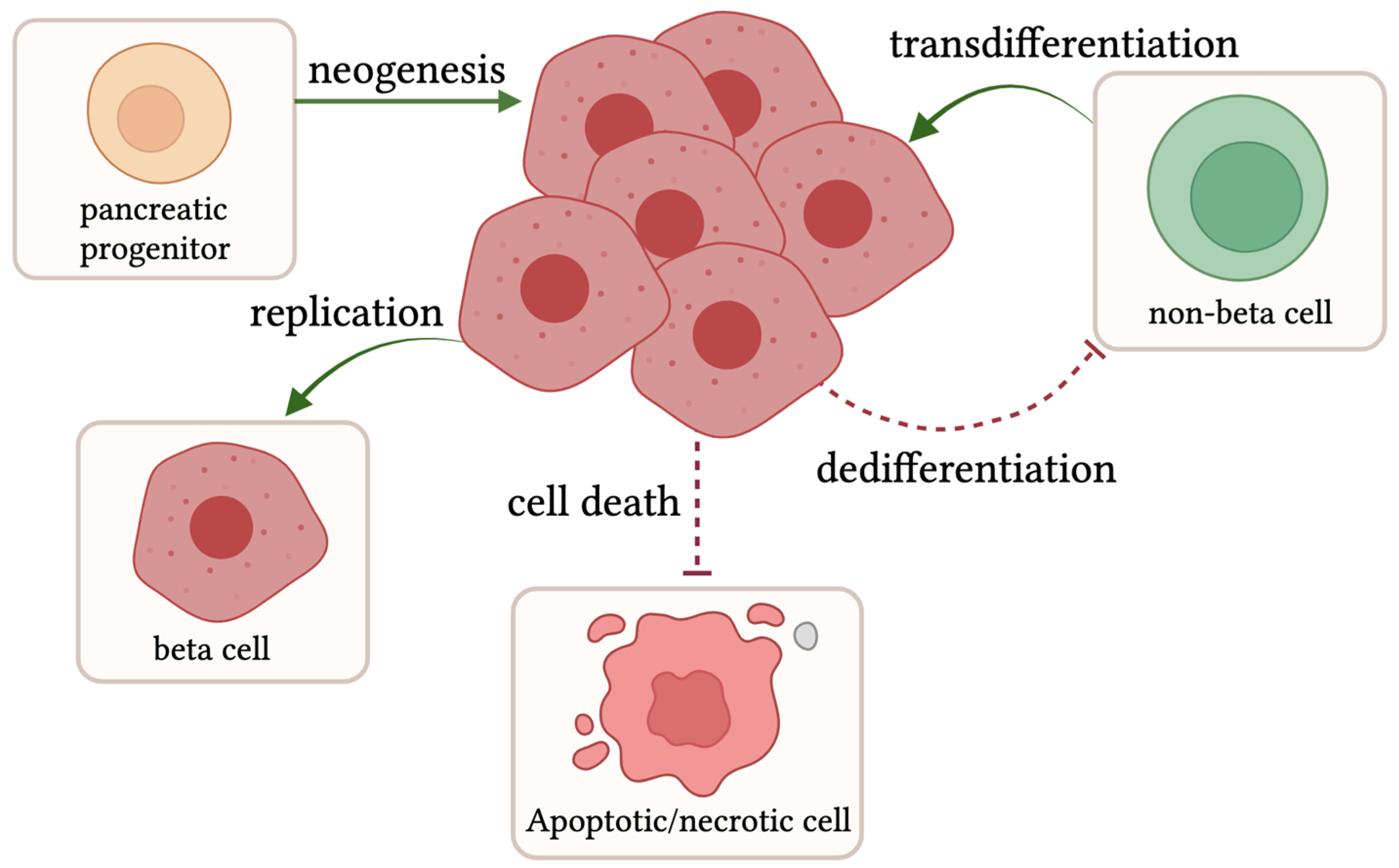
Type 2 diabetes (T2D) has become a worldwide epidemic, primarily driven by obesity from overnutrition and sedentariness. Physiologically, T2D manifests as an inability of the pancreatic beta cells to produce and secrete a sufficient bolus of insulin to elicit a response in target cells to transport glucose from the blood and properly regulate glucose levels. Insulin is synthesized in the endoplasmic reticulum (ER) of pancreatic beta cells where it undergoes a series of post-translational modifications to form mature insulin. Insulin resistance requires more insulin to be produced by beta cells to compensate for these desensitized cells. Consequently, this compensation causes additional strain on beta cells. This stress primarily originates from the ER and can also trigger oxidative stress. These cellular stresses can lead to beta cell decompensation, manifested by dysfunction and eventually a loss of beta cell mass.

type 2 diabetes beta cell death islet inflammation

1. Compensation: Beta Cell Proliferation, Transdifferentiation, and Neogenesis

The expansion of beta cell mass is infrequent in adult humans, with an estimated rate of 0.1–0.5% proliferating cells versus a 4% peak in fetal development [1][2][3]. However, under conditions of stress and heightened insulin demand due to factors including excess calorie intake, insulin resistance, and pregnancy, not only beta cell function but also beta cell mass increases to compensate for an amplified insulin need [4][5]. Beta cell mass expansion can occur through a variety of mechanisms, including neogenesis, proliferation, and transdifferentiation (Figure 1). Neogenesis arises when endocrine progenitor cells become beta cells. Proliferation refers to the replication of existing beta cells. Transdifferentiation is the shift of a non-beta cell in the islet into a beta cell. These new beta cells are functional and increase insulin content to help combat hyperglycemia and T2D [6][7]. In combination with additive factors, reductive processes, including cell death and dedifferentiation, or a loss of beta cell identity can be inhibited to promote beta cell mass expansion (Figure 1).
Figure 1. Routes promoting beta cell expansion: Beta cell numbers can expand through different pathways, including neogenesis from pancreatic progenitor cells, intrinsic cell replication, or transdifferentiation of non-beta cells into beta cells, as shown by the green arrows. Other factors promoting beta cell mass expansion are the inhibition of both beta cell death and the dedifferentiation of beta cells. Created with Biorender.com (2 February 2024).
Beta cell compensation has been reported in various animal models, including zebrafish, rodents, non-human primates, and humans [4][5][8]. Butler et al. showed evidence of beta cell compensation in humans by their work with human cadavers. The study revealed that individuals who were classified as obese had a larger beta cell volume than individuals with (a) both obesity and T2D and (b) normal-weight individuals [8]. The results imply that obesity can induce a rise in beta cell mass, which is decreased in diabetic obese patients.
Though these results suggest a change in beta cell mass at different stages of T2D development, there are natural variations in beta cell mass in healthy individuals [9]. Furthermore, obesity state and beta cell mass do not track perfectly in all ethnicities. In a study by Inaishi and colleagues, Japanese individuals had no significant difference in beta cell mass between lean and obese subjects regardless of T2D [10]. Therefore, although there is evidence that supports beta cell mass compensation in humans until beta cell mass can be tracked from birth to disease state, there is not a definitive answer for the extent to which beta cell mass compensation occurs in humans.

1.1. Beta Cell Expansion in Rodent Models

Expansions in beta cell mass can occur through multiple biological pathways, including hypertrophy, transdifferentiation, neogenesis, and beta cell replication. In the diabetes field, there is a long history of debate regarding the primary pathway responsible for beta cell mass expansion in adulthood. Early studies by Bonner-Weir and colleagues found when rats were exposed to short bouts of hyperglycemia through glucose infusions, beta cell mass increased by 50% and the mitotic index, a measurement attained from the accumulation of mitotic frequency, increased by 5-fold [11]. The increase in mitotic frequency suggests that the major pathway of beta cell mass expansion was replication in this study [11].
In other rodent models, including mice, beta cell replication also appeared to be the primary means of mass expansion instead of neogenesis or endocrine transdifferentiation [12][13]. Dor et al. found via genetic lineage tracing that pre-existing beta cells rather than pluripotent stem cells were the major avenue of mass expansion in mice [13]. Indeed, Dalboge et al. and colleagues found that the total beta cell mass more than doubled in db/db mice from 5 weeks to 12 weeks of age [14]. The major route of cell expansion in these studies was concluded to stem from beta cell proliferation driven via increases in islet size and not islet number [14].
Pick et al. and colleagues found while investigating ZDF rats that at 5–7 weeks old, beta cell mass was significantly increased in the ZDF rat compared with Zucker lean control (ZLC) rats [15]. Furthermore, the increase in mass was noted as coming from proliferation, as an immunochemistry method, 6-h 5-bromo-2′-deoxyuridine (BrdU) incorporation, indicated that cell proliferation was the major source of mass increase [15].
Sand rats, when fed a high-energy diet, show dramatic increases in beta cell mass even after short 2- and 5-day overfeeding diets [16]. Interestingly, in these short-term feeding models, increases in beta cell mass mostly stemmed from increased rates of beta cell proliferation, as shown by PCNA staining. However, in sand rats fed a high-energy diet for 22 days, beta cell neogenesis increased by sixfold [16]. These studies by Kaiser et al. illustrated that the means of increased mass in sand rats may change depending on the timeline of the disease.
Although there is a bolus of studies supporting replication as a major compensatory pathway in murine animal models, other studies contest these early findings showing both neogenesis and replication in mice under different settings, including pancreatic regeneration, partial pancreatectomy, and partial duct ligation along with various drug treatments [17][18][19]. Furthermore, using control and diabetic mouse models, a new study with improved lineage tracing by Gribben and colleagues illustrated that progenitor ductal cells expressing Ngn3 contribute to adult beta cell mass in adulthood [20]. Therefore, there is still a debate on the origin of new beta cells generated during compensation. However, both neogenesis and replication probably contribute to the expansion of beta cell mass, and which is the major pathway likely depends on the individual and the specific context of the disease.

1.2. Human Beta Cell Expansion

Research in beta cell compensation in humans is a much harder feat than in animal models, as the major methods for assessing beta cell mass are from autopsies and organ donors. However, there are still a few studies that were able to evaluate the question of beta cell mass in humans. In a study by Butler and colleagues examining autopsies of pregnant women, relative beta cell volume was increased by 40% in pregnant versus non-pregnant women [21]. However, only a small increase in proliferation was observed in the pancreas assessed by the Ki67 marker for replication [21]. Instead of enlarged islets, which would be more indicative of replication, there was an increased number of small islets distributed around the pancreas, and insulin-positive cells were found within the ducts; therefore, neogenesis appeared to be the major pathway of expansion rather than replication [21]. Indeed, the presence of insulin-positive duct cells has been found in a few autopsied adult studies. Furthermore, in an obese model, when human islets were transplanted in mice, although there was a robust amount of native beta cell proliferation in response to a high-fat diet, there was little to no proliferation in the human islets [22]. These results suggest that beta cell expansion may follow neogenesis instead of the replication of existing beta cells. However, more work needs to be performed to definitively determine the major pathway of beta cell expansion in humans during pregnancy and in obesogenic settings.

1.3. Zebrafish Beta Cell Expansion

The major form of compensatory beta cell expansion in zebrafish at the larval stage may be neogenesis. The researcher's group found that when zebrafish were treated with glucose for 8 hours, beta cell mass increased by 30% in larval fish, and the increase in mass was due to the neogenesis of beta cells arising from endocrine precursor cells expressing mnx1 or nkx2.2 [23]. This expansion of beta cell mass is attributed to persistent insulin secretion, as prolonged pharmacologic activation of beta cell insulin secretion is sufficient to induce a compensatory response in zebrafish without feeding [24]. Interestingly, a similar mechanism also regulates beta cell proliferation in mice [25]. In contrast, drugs that block insulin secretion inhibit overnutrition-induced beta cell mass expansion [23].
Unlike humans, zebrafish have a tremendous regenerative capacity. Several groups have found that when beta cells are abolished using chemical or genetic ablation, beta cells regenerate, and the regenerated cells are functionally competent to regulate glucose levels within 1 month of insult in both larval and adult fish [26][27][28]. The origin of the regenerated cells has been widely studied and is reviewed by Yang et al. [4]. Early studies implicate alpha cells and notch-responsive ductal cells as a resource of new beta cells [29][30]. Recently, using single-cell transcriptomics and lineage tracing, the Ninov and Manfroid groups demonstrated that a major source of new beta cells after ablation is sst1-expressing cells in the islet [31][32]. These cells become Sst1+ Ins+ bihormonal cells and eventually Ins+ monohormonal cells [31][32][33]. Another source of new beta cells is ghrelin-expressing epsilon cells [34]. To determine the relative contribution of these sources to beta cell regeneration, Mi et al. performed a series of lineage tracing studies [33]. They found the major source of regenerated beta cells was not from alpha, delta (Sst2+), or gip cells, but from Sst1+ cells. Furthermore, Mi et al. demonstrated that Sst1+ cells were derived from Krt4+ ductal cells, distinct from notch-responsive ductal cells [33]. A series of bioinformatic analyses of single-cell sequencing data revealed the trajectory of Krt4+ to Sst1+ differentiation [33]. These studies vary from mouse models that illustrate a 70–80% loss of beta cells results in regeneration via the proliferation of surviving cells, while a near complete ablation of beta cells in mice results in alpha–beta cell conversion [12]. Humans do not have this incredible capacity for regeneration upon beta cell injury and, therefore, require insulin therapy upon diabetic disease states and major pancreatic damage. A greater understanding of the pathways for regeneration in zebrafish beta cells and other animal models may lead to novel pathways capable of eliciting beta cell-specific proliferation in humans for improved treatment strategies and better glycemic control.

2. Decompensation: Beta Cell Death and Loss of Identity

When beta cells are pushed to a certain point of strain, many groups have found that there is a loss of beta cell mass. Loss of beta cell mass can happen through a variety of different mechanisms. The major avenues of beta cell loss are through increased beta cell death and loss of beta cell identity. Loss of beta cell identity occurs when beta cells stop expressing beta cell markers. The beta cell markers include insulin and several transcription factors, including NKX6.1, MAFA, and PDX1 in mice [35]. In T2D human cadavers, a reduction in these transcription factors, NKX6.1, MAFA, and PDX1, was also observed [36]. These studies show that under some conditions of T2D pathophysiology, beta cell loss of identity may occur. However, there is also evidence for loss of beta cell mass by beta cell death. As cell death occurs relatively quickly and cell corpses are rapidly cleared, it is more difficult to detect cell death. Because T2D is such a heterogeneous disease, it is very likely that either cell death, loss of identity, or a combination of both could occur depending upon the pathological setting in humans.
Beta cell necrosis has been reported in Psammomys obesus, desert sand rats, a non-insulin dependent T2D model. This model experiences reductions in beta cell mass after three weeks on a high-energy diet [37]. Reductions in beta cell volume were attributed to necrosis as cell membrane rupture, and swollen mitochondria with dilated cisternae of the Golgi complex and the rough ER in the cytoplasm of beta cells were observed, while apoptotic bodies were not found [38][39]. Therefore, necrotic cell death may be a physiologically relevant avenue of beta cell death in a subset of T2D patients. Understanding the pathway of beta cell death in these various animal models may introduce novel drug targets and alternative pathological pathways for disease progression in T2D.
The db/db mouse model also experiences a loss of beta cell mass. In a study by Dalbøge and colleagues, beta cell mass declines in db/db mice from 12 weeks of age, with a peak mean value of 4.84 mg average mass, to 34 weeks with 3.3 mg average mass [14]. The number of islets was found to be similar throughout ages 5–24 weeks, with variations being constrained to islet size and not number. Beta cell proliferation was reduced in 24-week-old mice compared to 10-week-old mice via Ki-67 analysis [14]. This study did not find any significant differences in apoptosis, as measured by the use of caspase 3 immunoreactive assays. An alternative study by Puff and colleagues found apoptosis to be increased in db/db mice. However, their studies were performed on mice at earlier time points of 5–12 weeks of age, and they were unable to ensure that these cells were truly beta cells via staining [40]. ZDF rats also experience a robust decompensation of beta cell mass, losing more than 50% in some cases [41]. Beta cell loss was thought to be an outcome of increased apoptosis, as increased DNA fragmentation was found in several studies [15][41].
In the diabetes prone zebrafish model (zMIR) beta cell loss occurs after 3 days of overnutrition feeding in larval zebrafish. The decrease in beta cell mass is not because of dedifferentiation, as all beta cells are marked with a stable fluorescent protein and, therefore, cells will continue to possess the marker even if they stop expressing insulin. Indeed, when the fish are immunostained for insulin, all marked cells still express insulin. The cell death in zMIR fish appears non-apoptotic in nature as various apoptosis inhibitors were not able to prevent cell death and there were no apoptotic bodies detected. 

References

  1. Meier, J.J. Beta cell mass in diabetes: A realistic therapeutic target? Diabetologia 2008, 51, 703–713.
  2. Spears, E.; Serafimidis, I.; Powers, A.C.; Gavalas, A. Debates in Pancreatic Beta Cell Biology: Proliferation versus Progenitor Differentiation and Transdifferentiation in Restoring β Cell Mass. Front. Endocrinol. 2021, 12, 722250.
  3. Wang, P.; Alvarez-Perez, J.C.; Felsenfeld, D.P.; Liu, H.; Sivendran, S.; Bender, A.; Kumar, A.; Sanchez, R.; Scott, D.K.; Garcia-Ocaña, A.; et al. A high-throughput chemical screen reveals that harmine-mediated inhibition of DYRK1A increases human pancreatic beta cell replication. Nat. Med. 2015, 21, 383–388.
  4. Yang, B.; Covington, B.A.; Chen, W. In vivo generation and regeneration of β cells in zebrafish. Cell Regen 2020, 9, 9.
  5. Shcheglova, E.; Blaszczyk, K.; Borowiak, M. Mitogen Synergy: An Emerging Route to Boosting Human Beta Cell Proliferation. Front. Cell Dev. Biol. 2021, 9, 734597.
  6. Cerf, M.E. Beta cell dysfunction and insulin resistance. Front. Endocrinol. 2013, 4, 37.
  7. Linnemann, A.K.; Baan, M.; Davis, D.B. Pancreatic β-cell proliferation in obesity. Adv. Nutr. 2014, 5, 278–288.
  8. Butler, A.E.; Janson, J.; Bonner-Weir, S.; Ritzel, R.; Rizza, R.A.; Butler, P.C. Beta-cell deficit and increased beta-cell apoptosis in humans with type 2 diabetes. Diabetes 2003, 52, 102–110.
  9. Olehnik, S.K.; Fowler, J.L.; Avramovich, G.; Hara, M. Quantitative analysis of intra- and inter-individual variability of human beta-cell mass. Sci. Rep. 2017, 7, 16398.
  10. Inaishi, J.; Saisho, Y. Ethnic Similarities and Differences in the Relationship between Beta Cell Mass and Diabetes. J. Clin. Med. 2017, 6, 113.
  11. Bonner-Weir, S.; Deery, D.; Leahy, J.L.; Weir, G.C. Compensatory Growth of Pancreatic β-Cells in Adult Rats After Short-Term Glucose Infusion. Diabetes 1989, 38, 49–53.
  12. Nir, T.; Melton, D.A.; Dor, Y. Recovery from diabetes in mice by β cell regeneration. J. Clin. Investig. 2007, 117, 2553–2561.
  13. Dor, Y.; Brown, J.; Martinez, O.I.; Melton, D.A. Adult pancreatic β-cells are formed by self-duplication rather than stem-cell differentiation. Nature 2004, 429, 41–46.
  14. Dalbøge, L.S.; Almholt, D.L.C.; Neerup, T.S.R.; Vassiliadis, E.; Vrang, N.; Pedersen, L.; Fosgerau, K.; Jelsing, J. Characterisation of Age-Dependent Beta Cell Dynamics in the Male db/db Mice. PLoS ONE 2013, 8, e82813.
  15. Pick, A.; Clark, J.; Kubstrup, C.; Levisetti, M.; Pugh, W.; Bonner-Weir, S.; Polonsky, K.S. Role of apoptosis in failure of beta-cell mass compensation for insulin resistance and beta-cell defects in the male Zucker diabetic fatty rat. Diabetes 1998, 47, 358–364.
  16. Kaiser, N.; Yuli, M.; Uçkaya, G.k.; Oprescu, A.I.; Berthault, M.-F.; Kargar, C.; Donath, M.Y.; Cerasi, E.; Ktorza, A. Dynamic Changes in β-Cell Mass and Pancreatic Insulin During the Evolution of Nutrition-Dependent Diabetes in Psammomys obesus: Impact of Glycemic Control. Diabetes 2005, 54, 138–145.
  17. Bonner-Weir, S.; Li, W.C.; Ouziel-Yahalom, L.; Guo, L.; Weir, G.C.; Sharma, A. Beta-cell growth and regeneration: Replication is only part of the story. Diabetes 2010, 59, 2340–2348.
  18. Wang, R.N.; Klöppel, G.; Bouwens, L. Duct- to islet-cell differentiation and islet growth in the pancreas of duct-ligated adult rats. Diabetologia 1995, 38, 1405–1411.
  19. Bonner-Weir, S. New evidence for adult beta cell neogenesis. Cell Stem Cell 2021, 28, 1889–1890.
  20. Gribben, C.; Lambert, C.; Messal, H.A.; Hubber, E.L.; Rackham, C.; Evans, I.; Heimberg, H.; Jones, P.; Sancho, R.; Behrens, A. Ductal Ngn3-expressing progenitors contribute to adult β cell neogenesis in the pancreas. Cell Stem Cell 2021, 28, 2000–2008.e4.
  21. Butler, A.E.; Cao-Minh, L.; Galasso, R.; Rizza, R.A.; Corradin, A.; Cobelli, C.; Butler, P.C. Adaptive changes in pancreatic beta cell fractional area and beta cell turnover in human pregnancy. Diabetologia 2010, 53, 2167–2176.
  22. Dai, C.; Kayton, N.S.; Shostak, A.; Poffenberger, G.; Cyphert, H.A.; Aramandla, R.; Thompson, C.; Papagiannis, I.G.; Emfinger, C.; Shiota, M.; et al. Stress-impaired transcription factor expression and insulin secretion in transplanted human islets. J. Clin. Investig. 2016, 126, 1857–1870.
  23. Li, M.; Maddison, L.A.; Page-McCaw, P.; Chen, W. Overnutrition induces β-cell differentiation through prolonged activation of β-cells in zebrafish larvae. Am. J. Physiol. Endocrinol. Metab. 2014, 306, E799–E807.
  24. Li, M.; Maddison, L.A.; Crees, Z.; Chen, W. Targeted Overexpression of CKI-Insensitive Cyclin-Dependent Kinase 4 Increases Functional β-Cell Number Through Enhanced Self-Replication in Zebrafish. Zebrafish 2013, 10, 170–176.
  25. Porat, S.; Weinberg-Corem, N.; Tornovsky-Babaey, S.; Schyr-Ben-Haroush, R.; Hija, A.; Stolovich-Rain, M.; Dadon, D.; Granot, Z.; Ben-Hur, V.; White, P.; et al. Control of pancreatic β cell regeneration by glucose metabolism. Cell Metab. 2011, 13, 440–449.
  26. Moss, J.B.; Koustubhan, P.; Greenman, M.; Parsons, M.J.; Walter, I.; Moss, L.G. Regeneration of the pancreas in adult zebrafish. Diabetes 2009, 58, 1844–1851.
  27. Curado, S.; Stainier, D.Y.; Anderson, R.M. Nitroreductase-mediated cell/tissue ablation in zebrafish: A spatially and temporally controlled ablation method with applications in developmental and regeneration studies. Nat. Protoc. 2008, 3, 948–954.
  28. Pisharath, H.; Rhee, J.M.; Swanson, M.A.; Leach, S.D.; Parsons, M.J. Targeted ablation of beta cells in the embryonic zebrafish pancreas using E. coli nitroreductase. Mech. Dev. 2007, 124, 218–229.
  29. Ye, L.; Robertson, M.A.; Hesselson, D.; Stainier, D.Y.; Anderson, R.M. Glucagon is essential for alpha cell transdifferentiation and beta cell neogenesis. Development 2015, 142, 1407–1417.
  30. Delaspre, F.; Beer, R.L.; Rovira, M.; Huang, W.; Wang, G.; Gee, S.; Vitery Mdel, C.; Wheelan, S.J.; Parsons, M.J. Centroacinar Cells Are Progenitors That Contribute to Endocrine Pancreas Regeneration. Diabetes 2015, 64, 3499–3509.
  31. Carril Pardo, C.A.; Massoz, L.; Dupont, M.A.; Bergemann, D.; Bourdouxhe, J.; Lavergne, A.; Tarifeño-Saldivia, E.; Helker, C.S.; Stainier, D.Y.; Peers, B.; et al. A δ-cell subpopulation with a pro-β-cell identity contributes to efficient age-independent recovery in a zebrafish model of diabetes. eLife 2022, 11, e67576.
  32. Singh, S.P.; Chawla, P.; Hnatiuk, A.; Kamel, M.; Silva, L.D.; Spanjaard, B.; Eski, S.E.; Janjuha, S.; Olivares-Chauvet, P.; Kayisoglu, O.; et al. A single-cell atlas of de novo β-cell regeneration reveals the contribution of hybrid β/δ-cells to diabetes recovery in zebrafish. Development 2022, 149, dev199853.
  33. Mi, J.; Liu, K.-C.; Andersson, O. Decoding pancreatic endocrine cell differentiation and β cell regeneration in zebrafish. Sci. Adv. 2023, 9, eadf5142.
  34. Yu, J.; Ma, J.; Li, Y.; Zhou, Y.; Luo, L.; Yang, Y. Pax4-Ghrelin mediates the conversion of pancreatic ε-cells to β-cells after extreme β-cell loss in zebrafish. Development 2023, 150, dev201306.
  35. Talchai, C.; Xuan, S.; Lin, H.V.; Sussel, L.; Accili, D. Pancreatic β cell dedifferentiation as a mechanism of diabetic β cell failure. Cell 2012, 150, 1223–1234.
  36. Cinti, F.; Bouchi, R.; Kim-Muller, J.Y.; Ohmura, Y.; Sandoval, P.R.; Masini, M.; Marselli, L.; Suleiman, M.; Ratner, L.E.; Marchetti, P.; et al. Evidence of β-Cell Dedifferentiation in Human Type 2 Diabetes. J. Clin. Endocrinol. Metab. 2016, 101, 1044–1054.
  37. Jörns, A.; Tiedge, M.; Ziv, E.; Shafrir, E.; Lenzen, S. Gradual loss of pancreatic beta-cell insulin, glucokinase and GLUT2 glucose transporter immunoreactivities during the time course of nutritionally induced type-2 diabetes in Psammomys obesus (sand rat). Virchows Arch. 2002, 440, 63–69.
  38. Kahn, S.E. The relative contributions of insulin resistance and beta-cell dysfunction to the pathophysiology of Type 2 diabetes. Diabetologia 2003, 46, 3–19.
  39. Kayagaki, N.; Kornfeld, O.S.; Lee, B.L.; Stowe, I.B.; O’Rourke, K.; Li, Q.; Sandoval, W.; Yan, D.; Kang, J.; Xu, M.; et al. NINJ1 mediates plasma membrane rupture during lytic cell death. Nature 2021, 591, 131–136.
  40. Puff, R.; Dames, P.; Weise, M.; Göke, B.; Seissler, J.; Parhofer, K.G.; Lechner, A. Reduced proliferation and a high apoptotic frequency of pancreatic beta cells contribute to genetically-determined diabetes susceptibility of db/db BKS mice. Horm. Metab. Res. 2011, 43, 306–311.
  41. Shimabukuro, M.; Zhou, Y.T.; Levi, M.; Unger, R.H. Fatty acid-induced beta cell apoptosis: A link between obesity and diabetes. Proc. Natl. Acad. Sci. USA 1998, 95, 2498–2502.
More
Information
Contributors MDPI registered users' name will be linked to their SciProfiles pages. To register with us, please refer to https://encyclopedia.pub/register : ,
View Times: 119
Revisions: 2 times (View History)
Update Date: 04 Mar 2024
1000/1000