Submitted Successfully!
To reward your contribution, here is a gift for you: A free trial for our video production service.
Thank you for your contribution! You can also upload a video entry or images related to this topic.
Version Summary Created by Modification Content Size Created at Operation
1 -- 2488 2024-02-29 08:29:54 |
2 Reference format revised. Meta information modification 2488 2024-02-29 09:02:18 |

Video Upload Options

Do you have a full video?

Confirm

Are you sure to Delete?
Cite
If you have any further questions, please contact Encyclopedia Editorial Office.
Kofotolios, I.; Bonios, M.J.; Adamopoulos, M.; Mourouzis, I.; Filippatos, G.; Boletis, J.N.; Marinaki, S.; Mavroidis, M. Han:SPRD Rat as Preclinical Model of Polycystic Kidney. Encyclopedia. Available online: https://encyclopedia.pub/entry/55718 (accessed on 29 April 2024).
Kofotolios I, Bonios MJ, Adamopoulos M, Mourouzis I, Filippatos G, Boletis JN, et al. Han:SPRD Rat as Preclinical Model of Polycystic Kidney. Encyclopedia. Available at: https://encyclopedia.pub/entry/55718. Accessed April 29, 2024.
Kofotolios, Ioannis, Michael J. Bonios, Markos Adamopoulos, Iordanis Mourouzis, Gerasimos Filippatos, John N. Boletis, Smaragdi Marinaki, Manolis Mavroidis. "Han:SPRD Rat as Preclinical Model of Polycystic Kidney" Encyclopedia, https://encyclopedia.pub/entry/55718 (accessed April 29, 2024).
Kofotolios, I., Bonios, M.J., Adamopoulos, M., Mourouzis, I., Filippatos, G., Boletis, J.N., Marinaki, S., & Mavroidis, M. (2024, February 29). Han:SPRD Rat as Preclinical Model of Polycystic Kidney. In Encyclopedia. https://encyclopedia.pub/entry/55718
Kofotolios, Ioannis, et al. "Han:SPRD Rat as Preclinical Model of Polycystic Kidney." Encyclopedia. Web. 29 February, 2024.
Han:SPRD Rat as Preclinical Model of Polycystic Kidney
Edit

Autosomal Dominant Polycystic Kidney Disease (ADPKD) stands as the most prevalent hereditary renal disorder in humans, ultimately culminating in end-stage kidney disease. Animal models carrying mutations associated with polycystic kidney disease have played an important role in the advancement of ADPKD research. The Han:SPRD rat model, carrying an R823W mutation in the Anks6 gene is a well-documented animal model of inherited PKD with an autosomal dominant pattern of inheritance, closely mirroring several features of human ADPKD, including renal hyperplasia, azotemia, and extrarenal manifestations. The mutated protein, named Samcystin, is localized in cilia of tubular epithelial cells and seems to be involved in cystogenesis. The homozygous Anks6 mutation leads to end-stage renal disease and death, making it a critical factor in kidney development and function. 

polycystic kidney disease animal models ADPKD

1. Introduction

Cystic kidney diseases are multisystemic disorders that manifest in both children and adults and arise from both genetic and non-genetic causes, which often lead to end-stage renal disease (ESRD) [1][2]. Genetic disorders include autosomal dominant and recessive polycystic kidney disease (ADPKD, ARPKD), nephronopthisis, von Hippel–Lindau, and tuberous sclerosis [3]. These inherited cystic kidney diseases are caused by disorders of the cilia, often referred to as ciliopathies.
ADPKD is a highly prevalent heterogeneous disease, affecting 1 in 500 to 1000 people worldwide [4][5]. Approximately 10% of all patients receiving renal replacement therapy in Europe suffer from ADPKD [6][7]. Mutations in the PKD1 (MIM# 601313) and PKD2 (MIM# 173910) genes, encoding polycystins 1 or 2, lead to the dysfunction of the polycystin complex, contributing to the vast majority of all cases of ADPKD [8]. These mutations result in a decrease in intracellular calcium concentration, thereby increasing the activity of adenylyl cyclase as well as cyclic adenosine monophosphate (cAMP) [9]. The sustained high levels of intracellular cAMP in the proximal, distal, and collecting ducts leads to abnormal proliferation of tubular epithelial cells. Chloride-controlled fluid secretion and cAMP production are two of the main components of cyst formation and growth [10][11][12]. Uncontrolled cyst growth can result in the displacement of adjacent nephrons, the destruction of the renal parenchyma, and, eventually, the enlargement of the kidneys and progressive renal failure [13][14].
Nephronophthisis (NPHP), an autosomal recessive cystic kidney disease, stands as the most prevalent genetic cause of ESRD during the first three decades of life [15][16]. The estimated incidence of new cases ranges from 1 in 50,000 to 1 in 1,000,000 live births. NPHP in three clinical forms—infantile, juvenile, and adolescent—is distinguished based on the onset of ESRD at ages 1, 13, and 19, respectively [17][18][19]. Renal histology reveals a distinctive triad of tubular basement membrane disruption, tubulointerstitial nephropathy, and corticomedullary cysts [20]. Renal size is within normal range or slightly reduced, except in infantile NPHP type 2, where moderate renal enlargement may occur. This contrasts with ADPKD, where cysts are evenly distributed throughout the organ and result in severe renal enlargement. NPHP is associated with various clinical manifestations such as tapeto-retinal degeneration (Senior–Løken syndrome), cerebellar vermis aplasia (Joubert syndrome), Cogan-type oculomotor apraxia, mental retardation, liver fibrosis, or cone-shaped epiphyses of the phalanges. Infantile NPHP type 2 may be associated with situs inversus, retinitis pigmentosa, or cardiac ventricular septal defect [21][22].

2. The Han:SPRD Rat Model

The intricacy of genetic studies of ADPKD in humans has led to the use of rodent models of spontaneous polycystic kidney disease (PKD) to characterize mechanisms involved in renal cystogenesis [23]. The Han:SPRD rat model stands as the sole well-documented animal model of inherited PKD with an autosomal dominant pattern of inheritance, closely mirroring several features of human ADPKD, including renal hyperplasia, azotemia, and extrarenal manifestations [24]. This model was first described by Dr. Deerberg as a spontaneous derived model (Central Institute for Laboratory Animal Breeding, Hannover, Germany) in 1989 [25]. Subsequently, the colony of inbred Han:SPRD rats was established in the laboratory in Mannheim, under the control of Dr. Gretz after 18 additional generations of inbreeding, and named PKD/Mhm (University of Heidelberg, Mannheim, Germany) rats [26][27][28]. A striking contrast in the severity of cystic disease is evident when comparing homozygous and heterozygous affected animals. Homozygous rats developed massive renal enlargement and marked azotemia, and died at approximately 3 weeks of age. Clinically, affected homozygous rats show a markedly distended abdomen and, in post-mortem analysis, reveal enlarged kidneys, often comprising more than 20% of total body weight [29]. At 9 weeks of age, all heterozygous animals exhibit kidney enlargement and abnormal ultrasound features. Male rats, at this age, commonly display hyperechoic renal cortex with cyst formation and loss of corticomedullary junction. On the contrary, female rats have no visible cysts at this age, and enlarged hyperechoic renal cortex is the most prevalent abnormality.

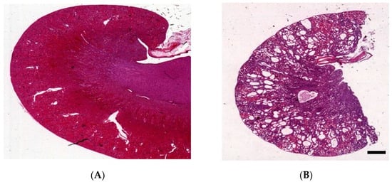
3. Kidney Histology of Han:SPRD Rats

The histological differences between homozygous (Cy/Cy) and heterozygous (Cy/+) Han:SPRD rats are apparent [30]. In homozygotes, cystic dilatation affects all segments of the nephron except the glomeruli [31]. However, the histology results in heterozygotes differ according to the age and sex of the rats. Early on, Cy/+ rats have minimal renal cyst development, mainly on the proximal tubules of juxtamedullary nephrons [32]. At 5 to 6 weeks, cysts become more prominent but still mainly affect the proximal tubules. By 8 to 10 weeks cysts fill the entire organ, with male kidneys more often affected than females (Figure 1) [33].
Figure 1. Characteristics of the kidney in wild-type (A) and Han:SPRD Cy/+ rats (B), hematoxylin and eosin (HE)-stained kidneys of 12-week-old rats (unpublished results, scale bar: 1.5 mm).

4. Genetics

The phenotypic ratios observed from mattings of rats affected by PKD, as well as from mattings of healthy and diseased rats, align consistently with those of an autosomal dominant trait transmitted by a single gene [30]. In an experimental backcross population comprising affected Han:SPRD rats and unaffected Wistar Ottawa Karlsburg rats, a comprehensive genome scan employing 117 microsatellite markers successfully identified and allowed the genetic dissection of PKD on rat chromosome 5. In a detailed linkage mapping of rat chromosome 5, the PKD locus is located approximately 25 cM from the proenkephalin gene [26]. This region serves as a quantitative trait locus that controls PKD, kidney mass, and plasma urea concentration. The gene that induces PKD in the Han:SPRD rat was neither PKD1, which is located on human chromosome 16, nor PKD2, which is located on human chromosome 4. Therefore, Bihoreau et al. denoted a new locus as PKDR1. Searching the EST database for similar sequences to rat Pkdr1, Kaisaki et al. pinpointed human ANKS6, located on human chromosome 9, as the human PKDR1 [34]. The ANKS6 gene contains 15 coding exons and spans 64.5 kb [30]. The deduced protein, which is called samcystin, was found to be expressed specifically in the proximal renal tubules, and it contains 11 tandem ankyrin repeats in the NH2 terminus and a sterile alpha motif (SAM) in the COOH terminus. Positional cloning and mutational analysis revealed a cytosine-to-thymine transition, resulting in an arginine-to-tryptophan substitution at amino acid 823 in the protein sequence. The arginine residue mutated in the rat sequence of the SAM domain, suggesting an important functional role. The removal of arginine and insertion of tryptophan disrupt the overall tertiary structure of the protein and prevent the mid-loop surface of the ANKS6–SAM domain from adopting a complementary fold for binding to RNA and other proteins, including the ANKS3 and other signaling molecules, that are critical to its function [24][31][35].

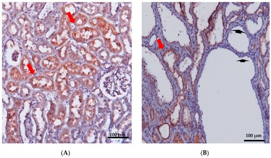
5. Localization and Function of Samcystin in Kidney

Early postnatal kidney samcystin expression was observed in wild-type rats during the final stage of renal development, with levels decreasing from 7 to 45 days. In the kidneys of heterozygous and homozygous rat kidney (Han:SPRD-Cy/+; or Han:SPRD-Cy/Cy), samcystin expression was downregulated at 3 and 7 days, and then was markedly increased compared to age-matched normal kidneys [24]. Immunohistochemical analysis revealed that samcystin was distributed on the brush border of proximal tubules in normal rat kidneys. Conversely, in Cy/+ kidneys, robust samcystin staining was observed in cyst-lining epithelial cells, accompanied by loss of apical localization and increased numbers of proliferating cell nuclear antigen-positive cells in cyst-lining epithelia (Figure 2) [24]. Anks6 is exclusively expressed in the proximal tubules of the adult kidney, thus indicating that cysts primarily originate from these tubules in mutants [32]. While anks6 (p.R823W) may be critical for the maintenance of proximal tubular function, its mild expression in other tubular segments/tubules may lead to cyst formation, albeit to a lesser extent and in older age. By inhibiting or impairing the ability of the cell to proliferate, Anks6 disrupts the delicate balance between the opposing processes of cell proliferation and apoptosis. Furthermore, in heterozygous neonatal rats Han:SPRD (Cy/+), distinct mRNA expression of anks6 (p. R823W) was observed in the tubular epithelium and podocytes. Therefore, the proteinuria of some patients could be attributed to ANKS6 mutation, which may indicate a critical function of ANKS6 not only in tubular epithelial cells but in podocytes as well [36]. Finally, molecular analysis indicates that anks6 localizes in the primary cilium of tubular epithelial cells, particularly in the inversin compartment (IC), where interacts with colocalized cystoproteins INVS, NHPH3, and NEK8 (Figure 3), [36][37][38].
Figure 2. Immunohistochemical staining of renal cortex was performed in 12-week-old wild-type (A) and PKD/Mhm (Cy/+) (B) rats for Anks6. Anks6 localized in the brush border of proximal tubules (red arrow). Cysts came mainly from proximal tubules, but there were also cysts without anks6 expression (arrowhead) (unpublished results, the Anti-Anks6 sc-515124 Ab Santa Cruz Biotechnology, was used in a 1:150 dilution; scale bar 100 μm).
Figure 3. Anks6 localizes in the inversin compartment of renal primary cilium and interacts with other nephrocystins in transition zone. The polycystin complex, which acts as a calcium channel, is localized in the primary cilia as well, however a clear interaction with anks6 has not been yet revealed. “Created with BioRender.com, accessed on 26 January 2024”.

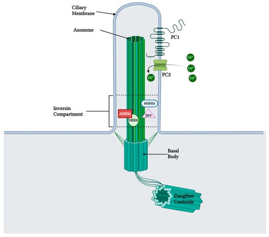
6. Role of Samcystin in Cystogenesis

Samcystin expression is consistent with the observed development of cysts in heterozygous rats, as well as the widespread cystic growth and renal enlargement in homozygous rats. The main sites of its expression were the proximal tubules, with particular emphasis on the brush border as the central point of expression [33]. Overexpression of samcystin in mutants resulted in a loss of specificity for its localization to the brush border in cyst-lined epithelial cells [39], indicating a link between the Cy mutation in the SAM domain and mislocalization of the protein. Overall, a point mutation of anks6, which results in the substitution of the amino acid “R823W” in the samcystin domain, results in the misexpression and mislocalisation of samcystin in the cystic epithelial cell [24]. The Cy mutation may interfere with samcystin’s interactions with other signaling molecules, while also increasing samcystin levels in response to the loss of a functional protein. This could lead to the accumulation of inadequate samcystin proteins and a dominant negative reaction, resulting in cellular differentiation and cyst formation [37]. Though the precise role of ANKS6 in renal cystogenesis is not fully understood, a recent study provides a compelling argument for the potential role of ANKS6 and NEK8 in regulating polycystic kidney function. The authors suggest a mechanistic connection between the IC and the phenotypic results of both ANKS6 and NEK8 polycystic kidney mutants. ANKS6 transports NEK8 from the cytoplasm to the cilia, where it is phosphorylated in the presence of INV and NPHP3, suggesting its role as both a substrate and a functional activator of NEK8 kinase activity [40]. NEK8 has already been identified as an important regulator in kidney and liver cystogenesis and seems to be involved in the same signaling cascade as PKD1 and PKD2 [41][42].

7. Extrarenal Manifestations of ADPKD

Cerebrovascular aneurysms, heart valve abnormalities, and colonic diversions are the most frequent extrarenal manifestations of ADPKD. Notably, Han:SPRD (Cy/+) rats lack cerebral aneurysms, in contrast to humans. In the heart, the splice-site and truncating mutations of ANKS6 were linked to hypertrophic obstructive cardiomyopathy, aortic stenosis, pulmonary stenosis, patent ductus arteriosus, and situs inversus [37]. Furthermore, male rats exhibit fibrosis, ranging from isolated interstitial connective tissue collections to dense collagen in regions devoted to normal cardiac fibers [25]. The involvement of ANKS6 in cardiac development and function is further supported by the observation that a patient who had a truncation in ANKS6 at the N-terminal end of the SAM domain, who also presented aortic stenosis, resulted in obstructive cardiomyopathy [43]. Regarding renal osteodystrophy, impaired kidney function in male rats leads to renal osteodystrophy, with conspicuous hyperplasia of the parathyroid glands and replacement of bone by fibrous tissue [44]. Furthermore, metastatic calcification affects several organs, including the media wall of large arteries such as the aortic arch, thoracic aorta, abdominal aorta, and renal artery, which are calcified. Focal mineralization of heart muscle fibers occurs as well [45][46]. Regarding enteritis, uremic enteritis is observed in about 40% of male HAN:SPRD rats.

8. Preclinical Trials

Many studies in the bibliography have used Han:SPRD rats as a model for PKD. Table 1 illustrates the most significant studies that have been conducted in Han:SPRD; however, in clinical studies, the results have been controversial. Among the most noteworthy clinical studies in PKD is the use of mTOR inhibitors as a potential treatment to prevent cyst onset, expansion, and PKD progression.
Table 1. Comparison of clinical and preclinical studies with Han:SPRD as a polycystic kidney disease animal model.
Mechanism of
Action
Intervention Han:SPRD Human
    Cyst Growth Other Effect Cyst Growth Other Effect
mTOR inhibitor Sirolimus Reduction [47][48] Reduction of CKD progression [47][48] Reduction [49] No effect in CKD progression [49]
COX-2 inhibitor NS-398 Reduction [50] N/A N/A N/A
SGLT1,2i Phlorizin Reduction [51] Reduction of CKD progression [51] N/A N/A
SGLT2i Dapagliflozin No effect [52][53][54] Reduction of CKD progression [52][53][54] Increase [55] Increase of CKD progression [55]
Calcium channel inhibitors Verapamil Increase [56] Increase of CKD progression [56] N/A [57] Increase of CKD progression [57]
Natural vitamin Fish oil None [58] Reduction of diastolic dysfunction [58] N/A N/A
CKD: chronic kidney disease, mTOR: Mammalian target of rapamycin, COX-2: cyclooxygenase-2, SGLT2i: Sodium–glucose co-transporter 2 inhibitor, N/A: non-applicable.

9. ANKS6 in Humans

Recent findings have established an association between ANKS6 and human kidney disease, particularly with nephronophthisis. The genomic sequence of PKDR1 on chromosome 9q22.33 spans approximately 64.5 Mb and includes 15 coding exons and one non-coding exon. The gene responsible for encoding human samcystin has been sequenced, revealing new coding and non-coding polymorphisms [26]. The UniProt database lists four isoforms of ANKS6, denoted as Q68DC2-1 to Q68DC2-4, although experimental confirmation is lacking for two of them [34]. Notably, many of the mutations detected are located in the ankyrin repeats, with a specific truncation at tyrosine790 observed in the SAM domain. This truncation significantly impacts normal function of anks6 in both rats and humans. Mutation analysis of nephronophthisis cohorts have identified patients with truncating, splice-site, and non-synonymous missense mutations [34][43]. Individuals in these cohorts generally exhibit early, infantile onset of PKD, except for those in family NPH316, where juvenile onset is observed [37]. Patients with missense mutations showed cystic kidney disease without extrarenal manifestations. On the other hand, truncating mutations were linked to an enlarged renal size, PKD, and early onset ESRD, as well as severe extrarenal defects [59]. The severity of the phenotype in patients with splicing mutations is linked to the location of the causative mutations. Different conformational changes in ANKS6 and/or defective interactions with different interacting partners may be the cause of these different phenotypic characteristics [60].

References

  1. Kurschat, C.E.; Müller, R.U.; Franke, M.; Maintz, D.; Schermer, B.; Benzing, T. An approach to cystic kidney diseases: The clinician’s view. Nat. Rev. Nephrol. 2014, 10, 687–699.
  2. Cramer, M.T.; Guay-Woodford, L.M. Cystic Kidney Disease: A Primer. Adv. Chronic Kidney Dis. 2015, 22, 297–305.
  3. Rohatgi, R. Extra-Renal Manifestations 5. Nephronophthisis (NPHP)-Medullary Cystic Kidney Disease (MCKD). 2008. Available online: http://pkdb.mayo.edu, (accessed on 31 January 2023).
  4. Iglesias, C.G.; Torres, V.E.; Offord, K.P.; Holley, K.E.; Beard, C.M.; Kurland, L.T. Epidemiology of Adult Polycystic Kidney Disease, Olmsted County, Minnesota: 1935–1980. Am. J. Kidney Dis. 1983, 2, 630–639.
  5. Solazzo, A.; Testa, F.; Giovanella, S.; Busutti, M.; Furci, L.; Carrera, P.; Ferrari, M.; Ligabue, G.; Mori, G.; Leonelli, M.; et al. The prevalence of autosomal dominant polycystic kidney disease (ADPKD): A meta-analysis of European literature and prevalence evaluation in the Italian province of Modena suggest that ADPKD is a rare and underdiagnosed condition. PloS ONE 2018, 13, e0190430.
  6. Spithoven, E.M.; Kramer, A.; Meijer, E.; Orskov, B.; Wanner, C.; Abad, J.M.; Aresté, N.; De La Torre, R.A.; Caskey, F.; Couchoud, C.; et al. Schaefer, Renal replacement therapy for autosomal dominant polycystic kidney disease (ADPKD) in Europe: Prevalence and survival—An analysis of data from the ERA-EDTA Registry. Nephrol. Dial. Transplant. 2014, 29, iv15–iv25.
  7. Torres, V.E.; Harris, P.C.; Pirson, Y. Autosomal Dominant Polycystic Kidney Disease. 2007. Available online: http://www.thelancet.com (accessed on 15 February 2023).
  8. Guay-Woodford, L.M.; Henske, E.; Igarashi, P.; Perrone, R.D.; Reed-Gitomer, B.; Somlo, S.; Torres, V.E.; Ketchum, C.J.; Star, R.A.; Flessner, M.F.; et al. Filling the holes in cystic kidney disease research. Clin. J. Am. Soc. Nephrol. 2014, 9, 1799–1801.
  9. Torres, V.E.; Harris, P.C. Strategies targeting cAMP signaling in the treatment of polycystic kidney disease. J. Am. Soc. Nephrol. 2014, 25, 18–32.
  10. Igarashi, P.; Somlo, S. Genetics and pathogenesis of polycystic kidney disease. J. Am. Soc. Nephrol. 2002, 13, 2384–2398.
  11. Chapin, H.C.; Caplan, M.J. The cell biology of polycystic kidney disease. J. Cell Biol. 2010, 191, 701–710.
  12. Hanaoka, K.; Guggino, W.B. cAMP Regulates Cell Proliferation and Cyst Formation in Autosomal Polycystic Kidney Disease Cells. J. Am. Soc. Nephrol. 2000, 11, 1179–1187.
  13. Paul, B.M.; Heuvel, G.B.V. Kidney: Polycystic kidney disease. Wiley Interdiscip. Rev. Dev. Biol. 2014, 3, 465–487.
  14. Halvorson, C.R.; Bremmer, M.S.; Jacobs, S.C. IJNRD-6939-Polycystic-Kidney-Disease—Inheritance—Pathology—Prognosis. 2010. Available online: https://www.dovepress.com/ (accessed on 25 February 2023).
  15. Hildebrandt, F.; Waldherr, R.; Kutt, R.; Brandis, M. Clinical Investigator The nephronophthisis complex: Clinical and genetic aspects. Clin. Investig. 1992, 70, 802–808.
  16. Omran, H.; Fernandez, C.; Jung, M.; Häffner, K.; Fargier, B.; Villaquiran, A.; Waldherr, R.; Gretz, N.; Brandis, M.; Rüschendorf, F.; et al. Identification of a new gene locus for adolescent nephronophthisis, on chromosome 3q22 in a large Venezuelan pedigree. Am. J. Hum. Genet. 2000, 66, 118–127.
  17. Gupta, S.; Ozimek-Kulik, J.E.; Phillips, J.K. Nephronophthisis-pathobiology and molecular pathogenesis of a rare kidney genetic disease. Genes 2021, 12, 1762.
  18. Hildebrandt, F.; Zhou, W. Nephronophthisis-associated ciliopathies. J. Am. Soc. Nephrol. 2007, 18, 1855–1871.
  19. Ala-Mellol, S.; Kivivuorp, S.M.; Rrnnholm, K.A.R.; Koskimies, O.; Siimes, M.A. Mechanism underlying early anaemia in children with familial juvenile nephronophthisis. Pediatr. Nephrol. 1996, 10, 578–581.
  20. Waldherr, R.; Lennert, T.; Weber, H.; Schfirer, K.; Derks, H.; Rieger, P. rhiv A The Nephronophthisis Complex A Clinicopathologic Study in Children. Virchows Arch. A 1982, 394, 235–254.
  21. Parisi, M.A. Clinical and molecular features of Joubert syndrome and related disorders. Am. J. Med. Genet. Part C Semin. Med. Genet. 2009, 151, 326–340.
  22. Baris, H.; Bejjani, B.A.; Tan, W.H.; Coulter, D.L.; Martin, J.A.; Storm, A.L.; Burton, B.K.; Saitta, S.C.; Gajecka, M.; Ballif, B.C.; et al. Identification of a novel polymorphism—The duplication of the NPHP1 (nephronophthisis 1) gene . Am. J. Med. Genet. Part A 2006, 140, 1876–1879.
  23. Guay-Woodford, L.M. Murine models of polycystic kidney disease: Molecular and therapeutic insights. Am. J. Physiol. Ren. Physiol. 2003, 285, F1034–F1049.
  24. Nagao, S.; Morita, M.; Kugita, M.; Yoshihara, D.; Yamaguchi, T.; Kurahashi, H.; Calvet, J.P.; Wallace, D.P. Polycystic kidney disease in Han:SPRD Cy rats is associated with elevated expression and mislocalization of SamCystin. Am. J. Physiol. Ren. Physiol. 2010, 299, 1078–1086.
  25. Kaspareit-Rittinghausen, J.; Rapp, K.; Deerberg, F.; Wcislo, A.; Messow, C. Hereditary Polycystic Kidney Disease Associated with Osteorenal Syndrome in Rats. Vet. Pathol. 1989, 26, 195–201.
  26. Bihoreau, M.-T.; Ceccherini, I.; Browne, J.; Kränzlin, B.; Romeo, G.; Lathrop, G.M.; James, M.R.; Gretz, N. Location of the First Genetic Locus, PKDr1, Controlling Autosomal Dominant Polycystic Kidney Disease in Han:SPRD cy/+ Rat; Oxford University Press: Oxford, UK, 1997.
  27. Ramasubbu, K.; Gretz, N.; Bachmannn, S. Increased Epithelial Cell Proliferation and Abnormal Extracellular Matrix in Rat Polycystic Kidney Disease. J. Am. Soc. Nephrol. 1998, 9, 937–945.
  28. Kränzlin, B.; Schieren, G.; Gretz, N. Azotemia and extrarenal manifestations in old female Han:SPRD (cy/+) rats. Kidney Int. 1997, 51, 1160–1169.
  29. Schafer, K.; Gretz, N.; Bader, M.; Oberbaumer, I.; Eckardt, K.-U.; Kriz, W.; Bachmann, S. Characterization of the Han:SPRD rat model for hereditary polycystic kidney disease. Kidney Int. 1994, 46, 134–152.
  30. Nagao, S.; Kugita, M.; Yoshihara, D.; Yamaguchi, T. Animal Models for Human Polycystic Kidney Disease. Exp. Anim. 2012, 61, 477–488.
  31. Kaspareit-Rittinghausen, J.; Deerberg, F.; Wcislo, A. Animal Model of Human Disease Hereditary Polycystic Kidney Disease Adult Polycystic Kidney Disease Associated with Renal Hypertension, Renal Osteodystrophy, and Uremic Enteritis in SPRD Rats. Am. J. Pathol. 1991, 139, 693.
  32. Nagao, S.; Yamaguchi, T.; Kusaka, M.; Maser, R.L.; Takahashi, H.; Cowley, B.D.; Grantham, J.J. Nagao, Renal activation of extracellular signal-regulated kinase in rats with autosomal-dominant polycystic kidney disease. Kidney Int. 2003, 63, 427–437.
  33. Cowley, B.D.; Gudapaty, S.; Kraybill, A.L.; Barash, B.D.; Harding, M.A.; Calvet, J.P.; Gattone, V.H. Autosomal-dominant polycystic kidney disease in the rat. Kidney Int. 1993, 43, 522–534.
  34. Kaisaki, P.J.; Bergmann, C.; Brown, J.H.; Outeda, P.; Lens, X.M.; Peters, D.J.M.; Gretz, N.; Gauguier, D.; Bihoreau, M.T. Genomic organization and mutation screening of the human ortholog of Pkdr1 associated with polycystic kidney disease in the rat. Eur. J. Med. Genet. 2008, 51, 325–331.
  35. Stagner, E.E.; Bouvrette, D.J.; Cheng, J.; Bryda, E.C. The polycystic kidney disease-related proteins Bicc1 and SamCystin interact. Biochem. Biophys. Res. Commun. 2009, 383, 16–21.
  36. Taskiran, E.Z.; Korkmaz, E.; Gucer, S.; Kosukcu, C.; Kaymaz, F.; Koyunlar, C.; Bryda, E.C.; Chaki, M.; Lu, D.; Vadnagara, K.; et al. Mutations in ANKS6 cause a nephronophthisis-like phenotype with ESRD. J. Am. Soc. Nephrol. 2014, 25, 1653–1661.
  37. Hoff, S.; Halbritter, J.; Epting, D.; Frank, V.; Nguyen, T.M.T.; Van Reeuwijk, J.; Boehlke, C.; Schell, C.; Yasunaga, T.; Helmstädter, M.; et al. ANKS6 is a central component of a nephronophthisis module linking NEK8 to INVS and NPHP3. Nat. Genet. 2013, 45, 951–956.
  38. Bakey, Z.; Bihoreau, M.T.; Piedagnel, R.; Delestré, L.; Arnould, C.; De Villiers, A.D.; Devuyst, O.; Hoffmann, S.; Ronco, P.; Gauguier, D.; et al. The SAM domain of ANKS6 has different interacting partners and mutations can induce different cystic phenotypes. Kidney Int. 2015, 88, 299–310.
  39. Neudecker, S.; Walz, R.; Menon, K.; Maier, E.; Bihoreau, M.T.; Obermüller, N.; Kränzlin, B.; Gretz, N.; Hoffmann, S.C. Transgenic overexpression of Anks6(p.R823W) causes polycystic kidney disease in rats. Am. J. Pathol. 2010, 177, 3000–3009.
  40. Czarnecki, P.G.; Gabriel, G.C.; Manning, D.K.; Sergeev, M.; Lemke, K.; Klena, N.T.; Liu, X.; Chen, Y.; Li, Y.; Agustin, J.T.S.; et al. ANKS6 is the critical activator of NEK8 kinase in embryonic situs determination and organ patterning. Nat. Commun. 2015, 6, 6023.
  41. Natoli, T.A.; Gareski, T.C.; Dackowski, W.R.; Smith, L.; Bukanov, N.O.; Russo, R.J.; Husson, H.; Matthews, D.; Piepenhagen, P.; Ibraghimov-Beskrovnaya, O. Pkd1 and Nek8 mutations affect cell-cell adhesion and cilia in cysts formed in kidney organ cultures. Am. J. Physiol. Ren. Physiol. 2008, 294, F73–F83.
  42. Claus, L.R.; Chen, C.; Stallworth, J.; Turner, J.L.; Slaats, G.G.; Hawks, A.L.; Mabillard, H.; Senum, S.R.; Srikanth, S.; Flanagan-Steet, H.; et al. Certain heterozygous variants in the kinase domain of the serine/threonine kinase NEK8 can cause an autosomal dominant form of polycystic kidney disease. Kidney Int. 2023, 104, 995–1007.
  43. Wolf, M.T.F.; Hildebrandt, F. Nephronophthisis. Pediatr. Nephrol. 2011, 26, 181–194.
  44. Tani, T.; Fujiwara, M.; Orimo, H.; Shimizu, A.; Narisawa, S.; Pinkerton, A.B.; Millán, J.L.; Tsuruoka, S. Inhibition of tissue-nonspecific alkaline phosphatase protects against medial arterial calcification and improves survival probability in the CKD-MBD mouse model. J. Pathol. 2020, 250, 30–41.
  45. Reiss, A.B.; Miyawaki, N.; Moon, J.; Kasselman, L.J.; Voloshyna, I.; D’Avino, R.; De Leon, J. CKD, arterial calcification, atherosclerosis and bone health: Inter-relationships and controversies. Atherosclerosis 2018, 278, 49–59.
  46. Elias, R.M.; Dalboni, M.A.; Coelho, A.C.E.; Moysés, R.M.A. CKD-MBD: From the Pathogenesis to the Identification and Development of Potential Novel Therapeutic Targets. Curr. Osteoporos. Rep. 2018, 16, 693–702.
  47. Zafar, I.; Belibi, F.A.; He, Z.; Edelstein, C.L. Long-term rapamycin therapy in the Han:SPRD rat model of polycystic kidney disease (PKD). Nephrol. Dial. Transplant. 2009, 24, 2349–2353.
  48. Wahl, P.R.; Serra, A.L.; Le Hir, M.; Molle, K.D.; Hall, M.N.; Wüthrich, R.P. Inhibition of mTOR with sirolimus slows disease progression in Han:SPRD rats with autosomal dominant polycystic kidney disease (ADPKD). Nephrol. Dial. Transplant. 2006, 21, 598–604.
  49. Liu, Y.M.; Shao, Y.Q.; He, Q. Sirolimus for treatment of autosomal-dominant polycystic kidney disease: A meta-analysis of randomized controlled trials. Transplant. Proc. 2014, 46, 66–74.
  50. Sankaran, D.; Bankovic-Calic, N.; Ogborn, M.R.; Crow, G.; Aukema, H.M. Selective COX-2 inhibition markedly slows disease progression and attenuates altered prostanoid production in Han:SPRD-cy rats with inherited kidney disease. Am. J. Physiol. Ren. Physiol. 2007, 293, F821–F830.
  51. Wang, X.; Zhang, S.; Liu, Y.; Spichtig, D.; Kapoor, S.; Koepsell, H.; Mohebbi, N.; Segerer, S.; Serra, A.L.; Rodriguez, D.; et al. Targeting of sodium-glucose cotransporters with phlorizin inhibits polycystic kidney disease progression in Han:SPRD rats. Kidney Int. 2013, 84, 962–968.
  52. Riwanto, M.; Kapoor, S.; Rodriguez, D.; Edenhofer, I.; Segerer, S.; Wüthrich, R.P. Inhibition of aerobic glycolysis attenuates disease progression in polycystic kidney disease. PLoS ONE 2016, 11, e0146654.
  53. Rodriguez, D.; Kapoor, S.; Edenhofer, I.; Segerer, S.; Riwanto, M.; Kipar, A.; Yang, M.; Mei, C.; Wüthrich, R.P. Inhibition of Sodium-GlucoseCotransporter 2 with Dapagliflozin in Han: SPRD Rats with Polycystic Kidney Disease. Kidney Blood Press. Res. 2015, 40, 638–647.
  54. Afsar, B.; Afsar, R.E.; Demiray, A.; Altay, S.; Korkmaz, H.; Yildiz, A.; Covic, A.; Ortiz, A.; Kanbay, M. Sodium-glucose cotransporter inhibition in polycystic kidney disease: Fact or fiction. Clin. Kidney J. 2022, 15, 1275–1283.
  55. Morioka, F.; Nakatani, S.; Uedono, H.; Tsuda, A.; Mori, K.; Emoto, M. Short-Term Dapagliflozin Administration in Autosomal Dominant Polycystic Kidney Disease—A Retrospective Single-Arm Case Series Study. J. Clin. Med. 2023, 12, 6341.
  56. Nagao, S.; Nishii, K.; Yoshihara, D.; Kurahashi, H.; Nagaoka, K.; Yamashita, T.; Takahashi, H.; Yamaguchi, T.; Calvet, J.P.; Wallace, D.P. Calcium channel inhibition accelerates polycystic kidney disease progression in the Cy/+ rat. Kidney Int. 2008, 73, 269–277.
  57. Mitobe, M.; Yoshida, T.; Sugiura, H.; Shiohira, S.; Shimada, K.; Nitta, K.; Tsuchiya, K. Clinical effects of calcium channel blockers and renin-angiotensin- aldosterone system inhibitors on changes in the estimated glomerular filtration rate in patients with polycystic kidney disease. Clin. Exp. Nephrol. 2010, 14, 573–577.
  58. Ibrahim, N.H.M.; Thandapilly, S.J.; Jia, Y.; Netticadan, T.; Aukema, H. Soy Protein Alleviates Hypertension and Fish Oil Improves Diastolic Heart Function in the Han:SPRD-Cy Rat Model of Cystic Kidney Disease. Lipids 2016, 51, 635–642.
  59. Leettola, C.N.; Knight, M.J.; Cascio, D.; Hoffman, S.; Bowie, J.U. Characterization of the SAM domain of the PKD-related protein ANKS6 and its interaction with ANKS3. BMC Struct. Biol. 2014, 14, 17.
  60. Fang, B.; Guo, J.; Hao, C.; Guo, R.; Qian, S.; Li, W.; Jia, X. Whole-exome sequencing identifies a novel compound heterozygous mutation of ANKS6 gene in a Chinese nephronophthisis patient. Clin. Chim. Acta 2020, 501, 131–135.
More
Information
Subjects: Pathology
Contributors MDPI registered users' name will be linked to their SciProfiles pages. To register with us, please refer to https://encyclopedia.pub/register : , , , , , , ,
View Times: 139
Revisions: 2 times (View History)
Update Date: 29 Feb 2024
1000/1000