Submitted Successfully!
To reward your contribution, here is a gift for you: A free trial for our video production service.
Thank you for your contribution! You can also upload a video entry or images related to this topic.
Version Summary Created by Modification Content Size Created at Operation
1 -- 3681 2024-02-19 14:52:41 |
2 layout & references Meta information modification 3681 2024-02-21 02:05:57 |

Video Upload Options

Do you have a full video?

Confirm

Are you sure to Delete?
Cite
If you have any further questions, please contact Encyclopedia Editorial Office.
Al-Ghabkari, A.; Huang, B.; Park, M. Aberrant MET Receptor Tyrosine Kinase Signaling in Glioblastoma. Encyclopedia. Available online: https://encyclopedia.pub/entry/55184 (accessed on 05 May 2024).
Al-Ghabkari A, Huang B, Park M. Aberrant MET Receptor Tyrosine Kinase Signaling in Glioblastoma. Encyclopedia. Available at: https://encyclopedia.pub/entry/55184. Accessed May 05, 2024.
Al-Ghabkari, Abdulhameed, Bruce Huang, Morag Park. "Aberrant MET Receptor Tyrosine Kinase Signaling in Glioblastoma" Encyclopedia, https://encyclopedia.pub/entry/55184 (accessed May 05, 2024).
Al-Ghabkari, A., Huang, B., & Park, M. (2024, February 19). Aberrant MET Receptor Tyrosine Kinase Signaling in Glioblastoma. In Encyclopedia. https://encyclopedia.pub/entry/55184
Al-Ghabkari, Abdulhameed, et al. "Aberrant MET Receptor Tyrosine Kinase Signaling in Glioblastoma." Encyclopedia. Web. 19 February, 2024.
Aberrant MET Receptor Tyrosine Kinase Signaling in Glioblastoma
Edit

Despite therapeutic advances, the treatment of brain tumors, including glioblastoma (GBM), an aggressive primary brain tumor associated with poor prognosis and resistance to therapy, remains a significant challenge. Receptor tyrosine kinases (RTKs) are critical during development and in adulthood. Dysregulation of RTKs through activating mutations and gene amplification contributes to many human cancers and provides attractive therapeutic targets for treatment. Under physiological conditions, the Met RTK, the hepatocyte growth factor/scatter factor (HGF/SF) receptor, promotes fundamental signaling cascades that modulate epithelial-to-mesenchymal transition (EMT) involved in tissue repair and embryogenesis. In cancer, increased Met activity promotes tumor growth and metastasis by providing signals for proliferation, survival, and migration/invasion. Recent clinical genomic studies have unveiled multiple mechanisms by which MET is genetically altered in GBM, including focal amplification, chromosomal rearrangements generating gene fusions, and a splicing variant mutation (exon 14 skipping, METex14del). Notably, MET overexpression contributes to chemotherapy resistance in GBM by promoting the survival of cancer stem-like cells. This is linked to distinctive Met-induced pathways, such as the upregulation of DNA repair mechanisms, which can protect tumor cells from the cytotoxic effects of chemotherapy. 

glioblastoma (GBM) hepatocyte growth factor/scatter factor (HGF/SF) MET-targeted therapies MET exon 14 skipping

1. Introduction

GBM is the most common and most aggressive type of brain malignancy in adults and is generally characterized by poor survival, with a median survival of less than one year from diagnosis [1]. Although there has been a significant improvement in overall survival rates of various types of malignant brain tumors over the past decade, the prognosis for GBM patients has exhibited a persistent and concerning lack of progress, with consistently low survival rates [2][3]. Although advancements have been achieved in the current standard of care, which encompasses surgical interventions, radiotherapy, and chemotherapy and mainly temozolomide (TMZ), a DNA alkylating agent that was first approved for medical use in Europe and the United States in the early 2000s, they have sadly failed to improve the prognosis for GBM patients [4][5].
GBMs are malignant tumors that arise from glial cells and are classified as high-grade gliomas according to the histological criteria outlined by the World Health Organization (WHO), defined by the presence of either microvascular proliferation or tumor necrosis [6][7]. GBMs are classified as primary or secondary based on their origin and development. Primary GBMs arise de novo, whereas secondary GBMs evolve from pre-existing low-grade astrocytomas. Primary GBMs constitute the majority of cases, accounting for approximately 90%, and are characterized by rapid and spontaneous expansion without evidence of less malignant precursor tumors [7]. Primary GBMs predominantly affect elderly patients and exhibit a significantly poorer prognosis compared to secondary GBMs. Secondary GBMs emerge from grade II and III astrocytomas, oligodendrogliomas, or oligoastrocytomas and are relatively less common, accounting for 5 to 10% of cases and typically manifesting in younger individuals [8][9][10]. The gold-standard treatment for newly diagnosed patients includes maximum safe surgical resection followed by radiotherapy with concurrent and adjuvant chemotherapy, with or without tumor treating fields (TTFields). Despite the ever-growing array of emerging biologics and immunotherapeutic approaches in the field of cancer treatment, it is noteworthy that temozolomide remains the sole systemic therapy that has shown tangible improvements in GBM survival outcomes [11][12]. Considering the inadequate efficacy demonstrated by the currently approved treatment options for GBM, an urgent necessity exists for the development of novel therapeutic strategies. Various biological impediments, including the blood–brain barrier (BBB), the tumor and immune microenvironment, and the presence of intratumor heterogeneity (ITH), have hampered the advancement of novel therapeutic interventions for GBM. These factors present substantial challenges in developing innovative treatment modalities [13]. In particular, a study by Patel et al. using single-cell RNA sequencing revealed that a heterogeneous mixture of cells representing different GBM subgroups could coexist within a single tumor [14]. The findings of this study revealed a compelling association between increased heterogeneity and unfavourable survival outcomes in GBM patients. These findings indicate that the clinical prognosis of proneural GBM is influenced by the relative abundance of tumor cells belonging to alternative subtypes, thereby highlighting the notable clinical relevance of ITH. The advancement of recent technologies has facilitated more comprehensive genetic and epigenetic landscape analyses on larger glioma sample cohorts, enabling the discovery of numerous significant findings in recent years. Among these discoveries, one of the most remarkable and clinically significant observations is the prevalence of mutations in the genes isocitrate dehydrogenase 1 and 2 (IDH1 and IDH2) within a substantial proportion of lower-grade gliomas. Accumulating evidence suggests that these mutations play a causal role in gliomagenesis, profoundly influence tumor biology, and hold clinical and prognostic implications [15]. An in-depth investigation into the molecular aberrations of GBMs has unveiled a broad spectrum of chromosomal alterations. These encompass amplifications of RTK, including chromosome 4 (PDGFRA), chromosome 7 (EGFR, MET), and other genes, including CDK6 and chromosome 12 (CDK4, MDM2), as well as deletions in chromosome 10, contributing to the loss of a tumor suppressor (PTEN) [16]. In addition, somatic genome alterations demonstrate higher frequency in TP53 (34.4%), EGFR (32.6%), PTEN (32%), tumor suppressor NF1 (neurofibromin 1, 13.7%), and lipid kinase PIK3CA (12%) [17]. Genome-wide methylation analysis of GBM has revealed biologically distinctive DNA methylation events within the promoter region of the MGMT (O6-methyl guanine DNA methyltransferase) gene, as well as CD81, which is involved in DNA repair and radioresistance in a substantial proportion of GBM patients. These distinctive methylation patterns are frequently observed, indicating their potential significance in GBM pathogenesis. Additionally, other key genes, such as GATA6 (GATA binding protein 6), DR4 (death receptor 4), and CASP8 (caspase-8), which are involved in cell adhesion, apoptosis, and proliferation, respectively, exhibit prominent methylation alterations, further emphasizing their potential role in the molecular landscape of GBM subsets [18][19]. A deep understanding of these complex genomic alterations is crucial for uncovering the basic mechanisms underlying GBM development. This comprehensive understanding not only enhances our knowledge of disease pathogenesis but also holds the potential to provide invaluable insights into the design and development of precise and targeted therapeutic strategies.
RTKs are transmembrane cell-surface proteins that act as signal transducers and mediate key roles in regulating various cellular processes during embryogenesis and in adulthood, such as control of cell growth, survival, differentiation, metabolism, and cell migration and invasion [20][21][22]. The dysregulation of RTK signaling, often caused by gain-of-function alterations, leads to developmental abnormalities and is implicated in a wide range of cancers. Although RTKs function as central regulators of normal cellular processes, the dysregulation of growth factor signaling pathways via genomic alterations has been identified as a key event in human GBMs, and approximately 86% of these tumors harbour at least one genetic event in the core RTK/PI3K pathway [23][24]. In this regard, the initial studies on ITH in GBM revealed the coactivation of multiple RTKs, including EGFR, MET, and PDGFR, necessitating a poly-targeting approach to disrupt downstream signaling pathways [25][26]. These findings highlight the complex interplay of RTK signaling in GBM and the need for comprehensive strategies targeting multiple RTKs to modulate downstream signaling pathways and combat tumor progression effectively. Accumulating evidence highlights the significant involvement of MET in pivotal aspects of glioma cell biology, including tumor proliferation, growth, migration, invasion, angiogenesis, and stemness [23][27][28].

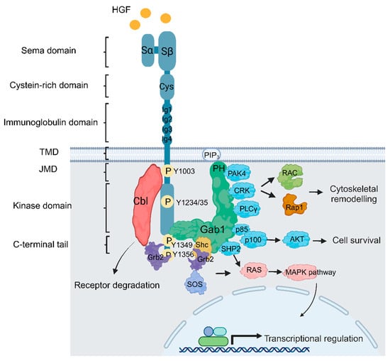
2. Met Structure and Function

The Met RTK was first identified as a chromosomal rearrangement induced by exposure to the carcinogen N-methyl-N′-nitronitrosoguanidine in a human osteogenic sarcoma cell line. This event resulted in the formation of a fusion protein known as Tpr-Met, wherein a leucine zipper dimerization domain was fused with the Met cytoplasmic domain. Consequently, this structural alteration led to the constitutive activation of the kinase domain [29][30][31][32]. This groundbreaking discovery sheds light on the role of RTK fusions and Met in oncogenic activities. The Met receptor is a single-pass transmembrane protein. The Met extracellular domain comprises four immunoglobulin-like (Ig-like) domains, a sema domain critical for binding hepatocyte growth factor (HGF), as well as a heparin-binding domain that enhances biological response [33]. The intracellular domain contains a tyrosine kinase domain, activated following ligand binding, which results in tyrosine phosphorylation of tyrosine residues within the kinase domain, as well as a juxtamembrane domain and a carboxy tail, which act as substrate binding sites and promote downstream signaling pathways (Figure 1) [34][35][36].
Figure 1. Met structure and function. Following HGF binding to MET triggers MET dimerization (Met is illustrated as monomer to simplify the model) and phosphorylation of Met within the activation loops, leading to activation of the receptor and followed by subsequent phosphorylation events in the c-terminal domain (Y1349 and Y1356) that enables Met to engage with a number of downstream signaling pathways, such as cytoskeletal remodelling, cell proliferation, and cell survival through coupling Met with multiple adaptor proteins, such as growth factor receptor-bound protein 2 (Grb2), Src homology 2 domain-containing (Shc), and the p85 subunit of phosphoinositide 3-kinases (PI3K). In addition, Grb2 recruits the docking protein Grb2-associated-binding protein 1 (Gab1), which can recruit other key signaling elements, such as tyrosine phosphatase SRC homology 2 domain-containing phosphatase 2 (SHP2), CRK, and PAK4. Additionally, Grb2 serves the crucial function of recruiting the c-Cbl ubiquitin ligase, which acts as a negative regulator of Met.
HGF plays a vital role in maintaining normal tissue homeostasis and is primarily synthesized in the liver. Its expression is upregulated during liver regeneration, particularly by Kupffer cells, and Met-HGF signaling is critical for full liver regeneration [37][38][39]. The HGF gene is located on chromosome 7q21.1; the protein consists of six distinct structural domains, including a short N-terminal domain, four kringle domains (K1-K4), and a non-catalytic serine proteinase homology (SPH) domain. HGF is initially secreted as an inactive precursor (pro-HGF) and subsequently activated through proteolytic processing in the extracellular environment. Processing leads to the formation of mature HGF, which exists as a heterodimer composed of a 69 kDa alpha chain and a 34 kDa beta chain, linked together by a single disulfide bond [40]. The HGF-Met interaction promotes dimerization and potentially oligomerization of the Met receptor and subsequent trans-phosphorylation of tyrosine residues Y1234 and Y1235 within its kinase domain [41]. This phosphorylation event initiates the trans-phosphorylation of tyrosine residues (Y1349, Y1356) located in the C-terminal tail of the Met receptor, as well as Y1003 in the juxtamembrane domain. The 1349/56 phosphorylated tyrosines serve as docking sites for signaling proteins that contain Src homology 2 (SH2) or phosphotyrosine binding (PTB) domains, including Grb2, which recruits the multisubstrate scaffold protein Gab1, whose tyrosine phosphorylation by Met engages multiple downstream pathways and enhances the activation of the Ras/Raf/MEK/ERK signaling pathway as well as the PI3K/Akt signaling pathway (Figure 1). Collectively, these interactions are tightly regulated and ultimately lead to transmitting downstream cellular responses [42][43][44][45][46][47][48][49].
Moreover, the Met receptor can interact with various cell membrane proteins, including integrins, CD44v6 isoform, and plexin-type receptors, as well as interaction with other RTKs, such as EGFR, Her2, Her3, RET, and IGFR1 [46][49][50][51][52][53][54][55]. These interactions enable the engagement of distinct downstream substrates, which leads to subsequent regulation of diverse cellular processes. It is imperative to prioritize the collective efforts to help identify additional Met-interaction hubs and develop a better understanding of Met signaling pathways. A recent study by Salokas et al. used affinity purification coupled with mass spectrometry (AP-MS) to identify stable binding partners and proximity-dependent biotin identification (BioID) to unveil proximal interactions of Met with other proteins. Notably, this identified key interactions between Met and other receptors, including the insulin receptor (INSR), the TYRO3 tyrosine protein kinase receptor, platelet-derived growth factor receptor beta (PDGFR beta), and neurotrophic receptor tyrosine kinase 3 (NTRK3) [56]. These novel interactions with Met reveal new avenues for exploring potential pathways, broadening our understanding of the diverse cellular processes and signaling networks that drive Met signalling in pathophysiological conditions.

3. MET/HGF Dysregulation and Oncogenic Paradigms in GBM

MET exerts regulatory control over diverse cellular functions, including proliferation, survival, and motility, typically exhibiting low activity in normal cells. However, when MET undergoes abnormal activation in tumor cells, it stimulates enhanced growth, angiogenesis, and invasion, leading to adverse overall survival outcomes [57]. Clinical data from various studies, including The Cancer Genome Atlas (TCGA) consortium, indicated the types of MET aberrations from the GBM cohort, including focal MET amplification, fusion genes, and MET exon 14 skipping mutations (Figure 2) [58][59][60][61][62][63].
Figure 2. Mechanisms of MET oncogenic activation in GBM. (A) Physiologically, HGF-METet interaction promotes dimerization and potentially oligomerization of the Met receptor and subsequent trans-phosphorylation of tyrosine residues Y1234 and Y1235 within its kinase domain, subsequently engaging Met with downstream signaling pathways. (B) Focal MET amplification is associated with high MET expression and enhances ligand-independent oncogenic activity. (C) Gene rearrangements and chromosomal translocations result in constitutive activation of the fusion proteins that typically self-dimerize in a ligand-independent manner, leading to oncogenic activity. (D) Loss of the direct Cbl-TKB-binding site via the loss of exon 14 is associated with reduced ubiquitination, decreased degradation, and sustained Met activation following HGF stimulation, leading to increased oncogenic activity.

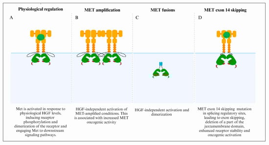
3.1. MET Focal Amplification

Focal MET amplification is the predominant mechanism of wild-type MET alterations in GBM. This amplification of MET at chromosome 7 is defined clinically by fluorescent in situ hybridization (FISH), which measures the ratio between the number of MET copies and the copies of the chromosome 7 centromere (CEP7). In particular, the cut-off of the MET/CEP7 ratio is classified into low (<1.8), medium (1.8 to <4), or high (≥4) [64]. Recent breakthroughs in molecular research have significantly deepened our comprehension of the properties and functions of focal oncogene amplification and rearrangements by introducing innovative methodologies and techniques for their identification, such as copy number variations (CNVs) and whole-genome sequencing (WGS)-based tools [65]. This understanding will help identify the level of amplification and rearrangements, which may predict responses to Met inhibitors and provide an understanding of resistance to other therapeutic options. MET amplification is associated with increased Met activity and/ or constitutive kinase activation in the absence of ligand, as observed in higher-grade GBMs with worse clinical outcomes [66][67]. Data from Lal et al. demonstrated that targeting the Met/HGF axis potentiates the response to γ-radiation synergistically, increases apoptosis, and attenuates cell viability in U87 MG human glioma cell lines and glioma xenograft models [68]. These data support the clinical need to evaluate MET-targeted therapies in combination with other radiotherapeutic or chemotherapeutic agents. Another independent study by Chi and colleagues showed a rapid and efficient clinical and radiographic response when a GBM patient with confirmed MET-amplified status was treated with crizotinib (PF-02341066), a dual ATP competitive inhibitor of Met (cellular IC50, 8 nM) and anaplastic lymphoma kinase (ALK) (cellular IC50, 20 nM) [69]. These data suggest that a subset of GBMs is potentially dependent on MET amplification and, therefore, sensitive to MET inhibition. Such clinical responses will pave the way to stratifying patients with GBM tumors harbouring MET amplification and help advance further clinical investigation on MET aberrations as a therapeutic companion target in GBMs. Additional evidence of the involvement of MET in the landscape of drug resistance is supported by a study conducted by Min and colleagues, which demonstrated that MET signaling activation is essential for GBM stem cells and that METet inhibition suppresses tumor growth and invasiveness [70]. This mechanism was further delineated by De Bacco and colleagues, who identified a subset of radioresistant glioblastoma stem cells (GSCs) driven by the sustained activation of several protein kinases, such as Aurora kinase A, ATM kinase, and the downstream effectors of DNA repair and the phosphorylation and cytoplasmic retention of p21 [71]. The application of Met inhibitors caused DNA damage accumulation in irradiated GSCs and their depletion using in vitro and in vivo xenotransplant models. Regrettably, it is worth noting that the Met inhibitor, JNJ-38877605 used in this study has been precluded from phase 1 clinical trials due to renal toxicity via the formation of species-specific insoluble metabolites [72]. These data suggest that MET contributes to GBM cancer stemness and tumour-initiating cells, potentially in a similar manner to its role in a subset of triple-negative breast cancer [73], and is a promising therapeutic target for this disease.

3.2. Fusion Genes

Chromosomal translocations that result in constitutive activation of RTK are increasingly detected using the latest deep sequencing technologies. MET was first identified as an oncogenic fusion between the TPR locus on chromosome 1 and the MET intracellular kinase domain on chromosome 7. TPR encodes a leucine zipper domain that constitutively dimerizes the Met kinase domain in the absence of ligand [31]. This fusion protein is cytosolic and constitutively activates downstream signalling pathways [74]. Multiple fusions have been detected in GBM. The FIG-ROS1 RTK fusion was the first gene fusion characterized in GBM [75]. This fusion event was identified as an intra-chromosomal homozygous deletion spanning 240 kilobases on chromosome 6q21. Consequently, extensive evidence has confirmed the constitutive activation of the resultant fusion protein, establishing it as an oncogenic entity. Following this discovery, multiple fusion proteins were identified, many of them involving RTKs [76]. The International Cancer Genome Consortium PedBrain Tumor Project identified PTPRZ1-MET (ZM) gene fusions in approximately 10% of cases of primary pediatric GBMs [77], whereas another study detected 15% of the ZM fusion gene in secondary GBM cases [78]. To further explore the oncogenic nature of ZM, Huang and colleagues analyzed a larger cohort of 485 glioma patients [79]. This demonstrated that ZM fusions were predominant in lower-grade and secondary GBMs but were not common in primary GBMs. This fusion transcript indicated a worse prognosis in these patients. The PTPRZ1 gene, located on chromosome 7q31.32, encodes the tyrosine phosphatase receptor type Z1 protein, which is closely located to MET (location 7q31.2). The fusion of both genes caused by intron insertion and tandem duplication can result in both in-frame and out-of-frame transcripts [78]. Unlike ZM, which contains full-length MET, another fusion transcript detected in GBMs is CLIP2-MET, which maintains only the kinase domain [77]. MET fusions result in the upregulation of the mitogen-activated protein kinase (MAPK) signaling pathway, which is associated with aggressive glial tumors in vivo. These tumor formations have been effectively suppressed using Met inhibitors, supporting the advancement of the use of a MET inhibitor in the clinic. However, although initial responses to MET inhibitors have been observed, the development of resistance can be rapid [80]. To further examine the pathologic role of MET fusion genes, Zeng and colleagues characterized exosomes from GBM cells harbouring ZM fusion compared with fusion-free exosomes. This data demonstrated that the internalization of ZM exosomes induced a migratory and invasive phenotype in GBM cells, enhanced neurosphere growth, prompted angiogenesis, and was associated with resistance to temozolomide in GBM cells [81]. Several studies showed inconsistent efficacy when treating pediatric patients with a MET-fusion-expressing GBM using crizotinib. Hu et al. characterized a new MET inhibitor, PLB-10011, which demonstrated remarkable potency in selectively inhibiting MET-altered tumor cells in preclinical models. Molecular dynamic simulation analyses demonstrated that this compound could bind to the conventional ATP-binding pocket of the tyrosine kinase superfamily but with some distinctive interactions in the ATP-binding pocket. An advantage of this small molecule inhibitor is its permeability across the BBB. Subsequently, it has been applied in a phase I clinical trial that enrolled MET-altered chemo-resistant glioma patients [82]. In most cases, the N-terminal signal peptide, necessary for protein targeting to the plasma membrane, is deleted in the MET fusion genes. This structural rearrangement confers a cytosolic location [31][83]. This cytosolic localization would potentially preclude their entry into the endocytic pathway and, hence, lead to lysosomal degradation, a common pathway for the degradation of cell surface RTKs [84]. The development of genomic and sequencing technologies provides a unique opportunity for systematically characterizing cancer cell transcriptomes, including identifying fusion genes resulting from underlying genomic rearrangements [85]. Consequently, this will pave the way to defining novel therapeutic solutions for GBMs characterized by MET gene fusion.

3.3. MET Exon 14 Skipping

Earlier studies demonstrated that uncoupling the Met receptor from ubiquitination is associated with oncogenic activity in the presence of the HGF ligand, highlighting the significance of negative regulation signals on the Met RTK to suppress its oncogenic activity [86][87]. The intracellular Met juxtamembrane domain is partially encoded by exon 14, which contains Y1003, which, when phosphorylated, is a direct binding site for the TKB (tyrosine kinase binding domain) domain of c-Cbl, an E3 ubiquitin ligase that promotes Met protein ubiquitination and subsequent degradation [86][88]. Consequently, the loss of the direct Cbl TKB-binding site, by loss of exon 14, is associated with reduced ubiquitination, decreased degradation, and sustained Met activation following HGF stimulation, leading to increased oncogenic activity [89][90]. Hundreds of distinct genetic alterations lead to MET exon 14 skipping in cancers. These include base substitutions and insertions or deletions (indels) at the splice acceptor site, at the splice donor site, and in intronic noncoding regions that disrupt consensus sequences such as branch sites, polypyrimidine tracts, splice acceptors, and splice donor sites for RNA splicing [91][92][93]. These mutations have been detected in gastric (4.8–7.1%), colorectal (~0–9.3%), and lung adenocarcinoma (3–4%) in addition to GBM. Interestingly, naturally occurring alternative splicing of exon 14 was characterized in cDNA isolated from normal mouse kidney, liver, and brain tissues [94], highlighting that this may also occur in cancers in the absence of mutations.
Recent reports demonstrated that the frequency of METex14del is 14% in secondary GBM, 1% in low-grade GBM (LGG), and 1.7% in primary GBM [82][95][96][97]. The extensive heterogeneity of MET genomic alterations leading to exon 14 skipping presents a challenge in clinical practice for routine detection. Although these mutations can be detected by NGS, whole-exome sequencing (WGS), and Sanger sequencing of MET exon 14 and its flanking introns [98][99], these are not all applicable for routine clinical testing. A robust, targeted NGS fragment analysis that helps with the systematic identification of patients harboring METex14del mutation has been developed that can be adopted for diagnostic applications in clinical settings [100]. It has been shown that integrating plasma NGS testing into the routine management of cancer patients substantially impacts the detection of therapeutically targetable mutations and improves the design of molecularly guided therapy [101]. Recent reports have also detected MET fusion genes coupled with METex14del mutations, which exhibited a poor prognosis [79]. Two types of MET TKIs were recently approved for the treatment of lung cancer patients harbouring METex14del: capmatinib (USA) and tepotinib (Japan) [102][103]. Further studies are required to assess the efficacy of these molecules to target MET aberrations in GBMs. In this regard, some previous studies and clinical trials evaluated the efficacy of cabozantinib (NCT01639508). This small-molecule tyrosine kinase inhibitor targets multiple tyrosine kinases, including VEGFR2, MET, RET, c-KIT, and AXL, and has been clinically evaluated to cross the BBB and has shown clinical efficacy [104][105]. This study addressed the first detailed brain metastases in MET exon 14-positive NSCLC and provided preliminary proof of concept of cabozantinib’s efficacy intracranially [104].

References

  1. Stupp, R.; Weller, M.; Belanger, K.; Bogdahn, U.; Ludwin, S.K.; Lacombe, D.; Mirimanoff, R.O. Radiotherapy plus Concomitant and Adjuvant Temozolomide for Glioblastoma. N. Engl. J. Med. 2005, 352, 987–996.
  2. Miller, K.D.; Ostrom, Q.T.; Kruchko, C.; Patil, N.; Tihan, T.; Cioffi, G.; Fuchs, H.E.; Waite, K.A.; Jemal, A.; Siegel, R.L.; et al. Brain and other central nervous system tumor statistics, 2021. CA Cancer J. Clin. 2021, 71, 381–406.
  3. Eisenbarth, D.; Wang, Y.A. Glioblastoma heterogeneity at single cell resolution. Oncogene 2023, 42, 2155–2165.
  4. Rong, L.; Li, N.; Zhang, Z. Emerging therapies for glioblastoma: Current state and future directions. J. Exp. Clin. Cancer Res. 2022, 41, 142.
  5. Singh, N.; Miner, A.; Hennis, L.; Mittal, S. Mechanisms of temozolomide resistance in glioblastoma—A comprehensive review. Cancer Drug Resist. 2021, 4, 17–43.
  6. Aldape, K.; Zadeh, G.; Mansouri, S.; Reifenberger, G.; von Deimling, A. Glioblastoma: Pathology, molecular mechanisms and markers. Acta Neuropathol. 2015, 129, 829–848.
  7. Wu, F.; Chai, R.C.; Wang, Z.; Liu, Y.Q.; Zhao, Z.; Li, G.Z.; Jiang, H.Y. Molecular classification of IDH-mutant glioblastomas based on gene expression profiles. Carcinogenesis 2019, 40, 853–860.
  8. Bausart, M.; Preat, V.; Malfanti, A. Immunotherapy for glioblastoma: The promise of combination strategies. J. Exp. Clin. Cancer Res. 2022, 41, 35.
  9. Yao, M.; Li, S.; Wu, X.; Diao, S.; Zhang, G.; He, H.; Bian, L.; Lu, Y. Cellular origin of glioblastoma and its implication in precision therapy. Cell. Mol. Immunol. 2018, 15, 737–739.
  10. Ohgaki, H.; Kleihues, P. The definition of primary and secondary glioblastoma. Clin. Cancer Res. 2013, 19, 764–772.
  11. Hegi, M.E.; Diserens, A.C.; Gorlia, T.; Hamou, M.F.; de Tribolet, N.; Weller, M.; Kros, J.M.; Hainfellner, J.A.; Mason, W.; Mariani, L.; et al. MGMT gene silencing and benefit from temozolomide in glioblastoma. N. Engl. J. Med. 2005, 352, 997–1003.
  12. Kotecha, R.; Odia, Y.; Khosla, A.A.; Ahluwalia, M.S. Key Clinical Principles in the Management of Glioblastoma. JCO Oncol. Pract. 2023, 19, 180–189.
  13. Tan, A.C.; Ashley, D.M.; Lopez, G.Y.; Malinzak, M.; Friedman, H.S.; Khasraw, M. Management of glioblastoma: State of the art and future directions. CA Cancer J. Clin. 2020, 70, 299–312.
  14. Patel, A.P.; Tirosh, I.; Trombetta, J.J.; Shalek, A.K.; Gillespie, S.M.; Wakimoto, H.; Cahill, D.P.; Nahed, B.V.; Curry, W.T.; Martuza, R.L.; et al. Single-cell RNA-seq highlights intratumoral heterogeneity in primary glioblastoma. Science 2014, 344, 1396–1401.
  15. Cohen, A.L.; Holmen, S.L.; Colman, H. IDH1 and IDH2 mutations in gliomas. Curr. Neurol. Neurosci. Rep. 2013, 13, 345.
  16. Brennan, C.W.; Verhaak, R.G.; McKenna, A.; Campos, B.; Noushmehr, H.; Salama, S.R.; Zheng, S.; Chakravarty, D.; Sanborn, J.Z.; Berman, S.H.; et al. The somatic genomic landscape of glioblastoma. Cell 2013, 155, 462–477.
  17. Zhang, P.; Xia, Q.; Liu, L.; Li, S.; Dong, L. Current Opinion on Molecular Characterization for GBM Classification in Guiding Clinical Diagnosis, Prognosis, and Therapy. Front. Mol. Biosci. 2020, 7, 562798.
  18. Parsons, D.W.; Jones, S.; Zhang, X.; Lin, J.C.; Leary, R.J.; Angenendt, P.; Mankoo, P.; Carter, H.; Siu, I.M.; Gallia, G.L.; et al. An integrated genomic analysis of human glioblastoma multiforme. Science 2008, 321, 1807–1812.
  19. Skiriute, D.; Vaitkiene, P.; Saferis, V.; Asmoniene, V.; Skauminas, K.; Deltuva, V.P.; Tamasauskas, A. MGMT, GATA6, CD81, DR4, and CASP8 gene promoter methylation in glioblastoma. BMC Cancer 2012, 12, 218.
  20. Li, E.; Hristova, K. Role of receptor tyrosine kinase transmembrane domains in cell signaling and human pathologies. Biochemistry 2006, 45, 6241–6251.
  21. Wintheiser, G.A.; Silberstein, P. Physiology, Tyrosine Kinase Receptors. In StatPearls; StatPearls Publishing: St. Petersburg, FL, USA, 2023.
  22. Stern, Y.E.; Al-Ghabkari, A.; Monast, A.; Fiset, B.; Aboualizadeh, F.; Yao, Z.; Stagljar, I.; Walsh, L.A.; Duhamel, S.; Park, M. Met-HER3 crosstalk supports proliferation via MPZL3 in MET-amplified cancer cells. Cell. Mol. Life Sci. 2022, 79, 178.
  23. Cheng, F.; Guo, D. MET in glioma: Signaling pathways and targeted therapies. J. Exp. Clin. Cancer Res. 2019, 38, 270.
  24. The Cancer Genome Atlas Research Network. Comprehensive genomic characterization defines human glioblastoma genes and core pathways. Nature 2008, 455, 1061–1068.
  25. Stommel, J.M.; Kimmelman, A.C.; Ying, H.; Nabioullin, R.; Ponugoti, A.H.; Wiedemeyer, R.; Stegh, A.H.; Bradner, J.E.; Ligon, K.L.; Brennan, C.; et al. Coactivation of receptor tyrosine kinases affects the response of tumor cells to targeted therapies. Science 2007, 318, 287–290.
  26. Qazi, M.A.; Vora, P.; Venugopal, C.; Sidhu, S.S.; Moffat, J.; Swanton, C.; Singh, S.K. Intratumoral heterogeneity: Pathways to treatment resistance and relapse in human glioblastoma. Ann. Oncol. 2017, 28, 1448–1456.
  27. Eckerich, C.; Zapf, S.; Fillbrandt, R.; Loges, S.; Westphal, M.; Lamszus, K. Hypoxia can induce c-Met expression in glioma cells and enhance SF/HGF-induced cell migration. Int. J. Cancer 2007, 121, 276–283.
  28. Li, Y.; Li, A.; Glas, M.; Lal, B.; Ying, M.; Sang, Y.; Xia, S.; Trageser, D.; Guerrero-Cazares, H.; Eberhart, C.G.; et al. c-Met signaling induces a reprogramming network and supports the glioblastoma stem-like phenotype. Proc. Natl. Acad. Sci. USA 2011, 108, 9951–9956.
  29. Cooper, C.S.; Park, M.; Blair, D.G.; Tainsky, M.A.; Huebner, K.; Croce, C.M.; Vande Woude, G.F. Molecular cloning of a new transforming gene from a chemically transformed human cell line. Nature 1984, 311, 29–33.
  30. Park, M.; Dean, M.; Cooper, C.S.; Schmidt, M.; O’Brien, S.J.; Blair, D.G.; Vande Woude, G.F. Mechanism of met oncogene activation. Cell 1986, 45, 895–904.
  31. Rodrigues, G.A.; Park, M. Dimerization mediated through a leucine zipper activates the oncogenic potential of the met receptor tyrosine kinase. Mol. Cell Biol. 1993, 13, 6711–6722.
  32. Dean, M.; Park, M.; Vande Woude, G.F. Characterization of the rearranged tpr-met oncogene breakpoint. Mol. Cell Biol. 1987, 7, 921–924.
  33. Cai, K.; Zhang, X.; Bai, X.C. Cryo-electron Microscopic Analysis of Single-Pass Transmembrane Receptors. Chem. Rev. 2022, 122, 13952–13988.
  34. Cecchi, F.; Rabe, D.C.; Bottaro, D.P. The Hepatocyte Growth Factor Receptor: Structure, Function and Pharmacological Targeting in Cancer. Curr. Signal Transduct. Ther. 2011, 6, 146–151.
  35. Maulik, G.; Shrikhande, A.; Kijima, T.; Ma, P.C.; Morrison, P.T.; Salgia, R. Role of the hepatocyte growth factor receptor, c-Met, in oncogenesis and potential for therapeutic inhibition. Cytokine Growth Factor Rev. 2002, 13, 41–59.
  36. Sattler, M.; Ma, P.C.; Salgia, R. Therapeutic targeting of the receptor tyrosine kinase Met. Cancer Treat. Res. 2004, 119, 121–138.
  37. Böhm, F.; Köhler, U.A.; Speicher, T.; Werner, S. Regulation of liver regeneration by growth factors and cytokines. EMBO Mol. Med. 2010, 2, 294–305.
  38. Matsumoto, K.; Nakamura, T. Hepatocyte growth factor: Molecular structure, roles in liver regeneration, and other biological functions. Crit. Rev. Oncog. 1992, 3, 27–54.
  39. Schmidt, C.; Bladt, F.; Goedecke, S.; Brinkmann, V.; Zschiesche, W.; Sharpe, M.; Gherardi, E.; Birchmeier, C. Scatter factor/hepatocyte growth factor is essential for liver development. Nature 1995, 373, 699–702.
  40. Toshikazu, N. Structure and function of hepatocyte growth factor. Prog. Growth Factor Res. 1991, 3, 67–85.
  41. Uchikawa, E.; Chen, Z.; Xiao, G.-Y.; Zhang, X.; Bai, X.-C. Structural basis of the activation of c-MET receptor. Nat. Commun. 2021, 12, 4074.
  42. Frigault, M.M.; Naujokas, M.A.; Park, M. Gab2 requires membrane targeting and the Met binding motif to promote lamellipodia, cell scatter, and epithelial morphogenesis downstream from the Met receptor. J. Cell Physiol. 2008, 214, 694–705.
  43. Lock, L.S.; Maroun, C.R.; Naujokas, M.A.; Park, M. Distinct recruitment and function of Gab1 and Gab2 in Met receptor-mediated epithelial morphogenesis. Mol. Biol. Cell 2002, 13, 2132–2146.
  44. Mood, K.; Saucier, C.; Bong, Y.S.; Lee, H.S.; Park, M.; Daar, I.O. Gab1 is required for cell cycle transition, cell proliferation, and transformation induced by an oncogenic met receptor. Mol. Biol. Cell 2006, 17, 3717–3728.
  45. Lock, L.S.; Frigault, M.M.; Saucier, C.; Park, M. Grb2-independent recruitment of Gab1 requires the C-terminal lobe and structural integrity of the Met receptor kinase domain. J. Biol. Chem. 2003, 278, 30083–30090.
  46. Orian-Rousseau, V.; Morrison, H.; Matzke, A.; Kastilan, T.; Pace, G.; Herrlich, P.; Ponta, H. Hepatocyte growth factor-induced Ras activation requires ERM proteins linked to both CD44v6 and F-actin. Mol. Biol. Cell 2007, 18, 76–83.
  47. Maroun, C.R.; Holgado-Madruga, M.; Royal, I.; Naujokas, M.A.; Fournier, T.M.; Wong, A.J.; Park, M. The Gab1 PH domain is required for localization of Gab1 at sites of cell-cell contact and epithelial morphogenesis downstream from the met receptor tyrosine kinase. Mol. Cell Biol. 1999, 19, 1784–1799.
  48. Rajadurai, C.V.; Havrylov, S.; Zaoui, K.; Vaillancourt, R.; Stuible, M.; Naujokas, M.; Zuo, D.; Tremblay, M.L.; Park, M. Met receptor tyrosine kinase signals through a cortactin-Gab1 scaffold complex, to mediate invadopodia. J. Cell Sci. 2012, 125, 2940–2953.
  49. Lai, A.Z.; Abella, J.V.; Park, M. Crosstalk in Met receptor oncogenesis. Trends Cell Biol. 2009, 19, 542–551.
  50. Chen, S.Y.; Chen, H.C. Direct interaction of focal adhesion kinase (FAK) with Met is required for FAK to promote hepatocyte growth factor-induced cell invasion. Mol. Cell Biol. 2006, 26, 5155–5167.
  51. McCall-Culbreath, K.D.; Li, Z.; Zutter, M.M. Crosstalk between the alpha2beta1 integrin and c-met/HGF-R regulates innate immunity. Blood 2008, 111, 3562–3570.
  52. Yamamoto, N.; Mammadova, G.; Song, R.X.; Fukami, Y.; Sato, K. Tyrosine phosphorylation of p145met mediated by EGFR and Src is required for serum-independent survival of human bladder carcinoma cells. J. Cell Sci. 2006, 119, 4623–4633.
  53. Khoury, H.; Naujokas, M.A.; Zuo, D.; Sangwan, V.; Frigault, M.M.; Petkiewicz, S.; Dankort, D.L.; Muller, W.J.; Park, M. HGF converts ErbB2/Neu epithelial morphogenesis to cell invasion. Mol. Biol. Cell 2005, 16, 550–561.
  54. Engelman, J.A.; Zejnullahu, K.; Mitsudomi, T.; Song, Y.; Hyland, C.; Park, J.O.; Lindeman, N.; Gale, C.M.; Zhao, X.; Christensen, J.; et al. MET amplification leads to gefitinib resistance in lung cancer by activating ERBB3 signaling. Science 2007, 316, 1039–1043.
  55. Bauer, T.W.; Somcio, R.J.; Fan, F.; Liu, W.; Johnson, M.; Lesslie, D.P.; Evans, D.B.; Gallick, G.E.; Ellis, L.M. Regulatory role of c-Met in insulin-like growth factor-I receptor-mediated migration and invasion of human pancreatic carcinoma cells. Mol. Cancer Ther. 2006, 5, 1676–1682.
  56. Salokas, K.; Liu, X.; Ohman, T.; Chowdhury, I.; Gawriyski, L.; Keskitalo, S.; Varjosalo, M. Physical and functional interactome atlas of human receptor tyrosine kinases. EMBO Rep. 2022, 23, e54041.
  57. Cruickshanks, N.; Zhang, Y.; Yuan, F.; Pahuski, M.; Gibert, M.; Abounader, R. Role and Therapeutic Targeting of the HGF/MET Pathway in Glioblastoma. Cancers 2017, 9, 87.
  58. Zhao, J.; Chen, A.X.; Gartrell, R.D.; Silverman, A.M.; Aparicio, L.; Chu, T.; Bordbar, D.; Shan, D.; Samanamud, J.; Mahajan, A.; et al. Immune and genomic correlates of response to anti-PD-1 immunotherapy in glioblastoma. Nat. Med. 2019, 25, 462–469.
  59. Wang, L.B.; Karpova, A.; Gritsenko, M.A.; Kyle, J.E.; Cao, S.; Li, Y.; Rykunov, D.; Colaprico, A.; Rothstein, J.H.; Hong, R.; et al. Proteogenomic and metabolomic characterization of human glioblastoma. Cancer Cell 2021, 39, 509–528.e520.
  60. Vaubel, R.A.; Tian, S.; Remonde, D.; Schroeder, M.A.; Mladek, A.C.; Kitange, G.J.; Caron, A.; Kollmeyer, T.M.; Grove, R.; Peng, S.; et al. Genomic and Phenotypic Characterization of a Broad Panel of Patient-Derived Xenografts Reflects the Diversity of Glioblastoma. Clin. Cancer Res. 2020, 26, 1094–1104.
  61. Miller, A.M.; Shah, R.H.; Pentsova, E.I.; Pourmaleki, M.; Briggs, S.; Distefano, N.; Zheng, Y.; Skakodub, A.; Mehta, S.A.; Campos, C.; et al. Tracking tumour evolution in glioma through liquid biopsies of cerebrospinal fluid. Nature 2019, 565, 654–658.
  62. Jonsson, P.; Lin, A.L.; Young, R.J.; DiStefano, N.M.; Hyman, D.M.; Li, B.T.; Berger, M.F.; Zehir, A.; Ladanyi, M.; Solit, D.B.; et al. Genomic Correlates of Disease Progression and Treatment Response in Prospectively Characterized Gliomas. Clin. Cancer Res. 2019, 25, 5537–5547.
  63. Barthel, F.P.; Johnson, K.C.; Varn, F.S.; Moskalik, A.D.; Tanner, G.; Kocakavuk, E.; Anderson, K.J.; Abiola, O.; Aldape, K.; Alfaro, K.D.; et al. Longitudinal molecular trajectories of diffuse glioma in adults. Nature 2019, 576, 112–120.
  64. Camidge, D.R.; Otterson, G.A.; Clark, J.W.; Ignatius Ou, S.H.; Weiss, J.; Ades, S.; Shapiro, G.I.; Socinski, M.A.; Murphy, D.A.; Conte, U.; et al. Crizotinib in Patients With MET-Amplified NSCLC. J. Thorac. Oncol. 2021, 16, 1017–1029.
  65. Deshpande, V.; Luebeck, J.; Nguyen, N.D.; Bakhtiari, M.; Turner, K.M.; Schwab, R.; Carter, H.; Mischel, P.S.; Bafna, V. Exploring the landscape of focal amplifications in cancer using AmpliconArchitect. Nat. Commun. 2019, 10, 392.
  66. Kong, D.S.; Song, S.Y.; Kim, D.H.; Joo, K.M.; Yoo, J.S.; Koh, J.S.; Dong, S.M.; Suh, Y.L.; Lee, J.I.; Park, K.; et al. Prognostic significance of c-Met expression in glioblastomas. Cancer 2009, 115, 140–148.
  67. Petterson, S.A.; Dahlrot, R.H.; Hermansen, S.K.; KA Munthe, S.; Gundesen, M.T.; Wohlleben, H.; Rasmussen, T.; Beier, C.P.; Hansen, S.; Kristensen, B.W. High levels of c-Met is associated with poor prognosis in glioblastoma. J. Neuro-Oncol. 2015, 122, 517–527.
  68. Lal, B.; Xia, S.; Abounader, R.; Laterra, J. Targeting the c-Met pathway potentiates glioblastoma responses to gamma-radiation. Clin. Cancer Res. 2005, 11, 4479–4486.
  69. Chi, A.S.; Batchelor, T.T.; Kwak, E.L.; Clark, J.W.; Wang, D.L.; Wilner, K.D.; Louis, D.N.; Iafrate, A.J. Rapid radiographic and clinical improvement after treatment of a MET-amplified recurrent glioblastoma with a mesenchymal-epithelial transition inhibitor. J. Clin. Oncol. 2012, 30, e30–e33.
  70. Joo, K.M.; Jin, J.; Kim, E.; Ho Kim, K.; Kim, Y.; Gu Kang, B.; Kang, Y.J.; Lathia, J.D.; Cheong, K.H.; Song, P.H.; et al. MET signaling regulates glioblastoma stem cells. Cancer Res. 2012, 72, 3828–3838.
  71. De Bacco, F.; D’Ambrosio, A.; Casanova, E.; Orzan, F.; Neggia, R.; Albano, R.; Verginelli, F.; Cominelli, M.; Poliani, P.L.; Luraghi, P.; et al. MET inhibition overcomes radiation resistance of glioblastoma stem-like cells. EMBO Mol. Med. 2016, 8, 550–568.
  72. Lolkema, M.P.; Bohets, H.H.; Arkenau, H.T.; Lampo, A.; Barale, E.; de Jonge, M.J.A.; van Doorn, L.; Hellemans, P.; de Bono, J.S.; Eskens, F. The c-Met Tyrosine Kinase Inhibitor JNJ-38877605 Causes Renal Toxicity through Species-Specific Insoluble Metabolite Formation. Clin. Cancer Res. 2015, 21, 2297–2304.
  73. Sung, V.Y.C.; Knight, J.F.; Johnson, R.M.; Stern, Y.E.; Saleh, S.M.; Savage, P.; Monast, A.; Zuo, D.; Duhamel, S.; Park, M. Co-dependency for MET and FGFR1 in basal triple-negative breast cancers. NPJ Breast Cancer 2021, 7, 36.
  74. Kim, P.L. Targeting gene fusions in glioma. Curr. Opin. Neurol. 2021, 34, 840–847.
  75. Charest, A.; Lane, K.; McMahon, K.; Park, J.; Preisinger, E.; Conroy, H.; Housman, D. Fusion of FIG to the receptor tyrosine kinase ROS in a glioblastoma with an interstitial del(6)(q21q21). Genes Chromosomes Cancer 2003, 37, 58–71.
  76. You, G.; Fan, X.; Hu, H.; Jiang, T.; Chen, C.C. Fusion Genes Altered in Adult Malignant Gliomas. Front. Neurol. 2021, 12, 715206.
  77. Recurrent MET fusion genes represent a drug target in pediatric glioblastoma. Nat. Med. 2016, 22, 1314–1320.
  78. Bao, Z.S.; Chen, H.M.; Yang, M.Y.; Zhang, C.B.; Yu, K.; Ye, W.L.; Hu, B.Q.; Yan, W.; Zhang, W.; Akers, J.; et al. RNA-seq of 272 gliomas revealed a novel, recurrent PTPRZ1-MET fusion transcript in secondary glioblastomas. Genome Res. 2014, 24, 1765–1773.
  79. Huang, R.; Liu, Y.; Wang, K.; Wang, Z.; Zhang, C.; Zhang, W.; Zhao, Z.; Li, G.; Huang, L.; Chang, Y.; et al. High-sensitive clinical diagnostic method for PTPRZ1-MET and the characteristic protein structure contributing to ligand-independent MET activation. CNS Neurosci. Ther. 2021, 27, 617–628.
  80. Lai, A.Z.; Cory, S.; Zhao, H.; Gigoux, M.; Monast, A.; Guiot, M.C.; Huang, S.; Tofigh, A.; Thompson, C.; Naujokas, M.; et al. Dynamic reprogramming of signaling upon met inhibition reveals a mechanism of drug resistance in gastric cancer. Sci. Signal 2014, 7, ra38.
  81. Zeng, A.L.; Yan, W.; Liu, Y.W.; Wang, Z.; Hu, Q.; Nie, E.; Zhou, X.; Li, R.; Wang, X.F.; Jiang, T.; et al. Tumour exosomes from cells harbouring PTPRZ1-MET fusion contribute to a malignant phenotype and temozolomide chemoresistance in glioblastoma. Oncogene 2017, 36, 5369–5381.
  82. Hu, H.; Mu, Q.; Bao, Z.; Chen, Y.; Liu, Y.; Chen, J.; Wang, K.; Wang, Z.; Nam, Y.; Jiang, B.; et al. Mutational Landscape of Secondary Glioblastoma Guides MET-Targeted Trial in Brain Tumor. Cell 2018, 175, 1665–1678.e1618.
  83. Lamorte, L.; Park, M. The receptor tyrosine kinases: Role in cancer progression. Surg. Oncol. Clin. N. Am. 2001, 10, 271–288.
  84. Peschard, P.; Park, M. From Tpr-Met to Met, tumorigenesis and tubes. Oncogene 2007, 26, 1276–1285.
  85. Edgren, H.; Murumagi, A.; Kangaspeska, S.; Nicorici, D.; Hongisto, V.; Kleivi, K.; Rye, I.H.; Nyberg, S.; Wolf, M.; Borresen-Dale, A.-L.; et al. Identification of fusion genes in breast cancer by paired-end RNA-sequencing. Genome Biol. 2011, 12, R6.
  86. Peschard, P.; Fournier, T.M.; Lamorte, L.; Naujokas, M.A.; Band, H.; Langdon, W.Y.; Park, M. Mutation of the c-Cbl TKB domain binding site on the Met receptor tyrosine kinase converts it into a transforming protein. Mol. Cell 2001, 8, 995–1004.
  87. Peschard, P.; Park, M. Escape from Cbl-mediated downregulation: A recurrent theme for oncogenic deregulation of receptor tyrosine kinases. Cancer Cell 2003, 3, 519–523.
  88. Hammond, D.E.; Urbé, S.; Vande Woude, G.F.; Clague, M.J. Down-regulation of MET, the receptor for hepatocyte growth factor. Oncogene 2001, 20, 2761–2770.
  89. Frampton, G.M.; Ali, S.M.; Rosenzweig, M.; Chmielecki, J.; Lu, X.; Bauer, T.M.; Akimov, M.; Bufill, J.A.; Lee, C.; Jentz, D.; et al. Activation of MET via diverse exon 14 splicing alterations occurs in multiple tumor types and confers clinical sensitivity to MET inhibitors. Cancer Discov. 2015, 5, 850–859.
  90. Kong-Beltran, M.; Seshagiri, S.; Zha, J.; Zhu, W.; Bhawe, K.; Mendoza, N.; Holcomb, T.; Pujara, K.; Stinson, J.; Fu, L.; et al. Somatic mutations lead to an oncogenic deletion of met in lung cancer. Cancer Res. 2006, 66, 283–289.
  91. Fujino, T.; Suda, K.; Mitsudomi, T. Lung Cancer with MET exon 14 Skipping Mutation: Genetic Feature, Current Treatments, and Future Challenges. Lung Cancer 2021, 12, 35–50.
  92. Socinski, M.A.; Pennell, N.A.; Davies, K.D. MET Exon 14 Skipping Mutations in Non-Small-Cell Lung Cancer: An Overview of Biology, Clinical Outcomes, and Testing Considerations. JCO Precis. Oncol. 2021, 5, PO.20.00516.
  93. Recondo, G.; Che, J.; Jänne, P.A.; Awad, M.M. Targeting MET Dysregulation in Cancer. Cancer Discov. 2020, 10, 922–934.
  94. Lee, C.C.; Yamada, K.M. Identification of a novel type of alternative splicing of a tyrosine kinase receptor. Juxtamembrane deletion of the c-met protein kinase C serine phosphorylation regulatory site. J. Biol. Chem. 1994, 269, 19457–19461.
  95. Lee, J.; Ou, S.H.I.; Lee, J.M.; Kim, H.C.; Hong, M.; Kim, S.Y.; Jang, J.; Ahn, S.; Kang, S.Y.; Lee, S.; et al. Gastrointestinal malignancies harbor actionable MET exon 14 deletions. Oncotarget 2015, 6, 28211–28222.
  96. Salgia, R.; Sattler, M.; Scheele, J.; Stroh, C.; Felip, E. The promise of selective MET inhibitors in non-small cell lung cancer with MET exon 14 skipping. Cancer Treat. Rev. 2020, 87, 102022.
  97. Sohn, S.-H.; Sul, H.J.; Kim, B.J.; Zang, D.Y. Responses to the Tepotinib in Gastric Cancers with MET Amplification or MET Exon 14 Skipping Mutations and High Expression of Both PD-L1 and CD44. Cancers 2022, 14, 3444.
  98. Cancer Genome Atlas Research Network. Comprehensive molecular profiling of lung adenocarcinoma. Nature 2014, 511, 543–550.
  99. Si-Yang, L.; Lan-Ying, G.; An-Na, L.; Na-Na, L.; Hong-Fei, G.; Jian, S.; Jin-Ji, Y.; Xu-Chao, Z.; Yang, S.; Zhong-Yi, D.; et al. The Unique Characteristics of MET Exon 14 Mutation in Chinese Patients with NSCLC. J. Thorac. Oncol. 2016, 11, 1503–1510.
  100. Clotilde, D.; Frédéric, L.; Fabienne, E.; Zoulika, K.; Martin, F.; Shéhérazade, S.; Simon, B.; Valérie, G.; Philippe, J.; Marie-Christine, C.; et al. Optimization of Routine Testing for MET Exon 14 Splice Site Mutations in NSCLC Patients. J. Thorac. Oncol. 2018, 13, 1873–1883.
  101. Aggarwal, C.; Thompson, J.C.; Black, T.A.; Katz, S.I.; Fan, R.; Yee, S.S.; Chien, A.L.; Evans, T.L.; Bauml, J.M.; Alley, E.W.; et al. Clinical Implications of Plasma-Based Genotyping with the Delivery of Personalized Therapy in Metastatic Non-Small Cell Lung Cancer. JAMA Oncol. 2019, 5, 173–180.
  102. Wolf, J.; Seto, T.; Han, J.-Y.; Reguart, N.; Garon, E.B.; Groen, H.J.M.; Tan, D.S.-W.; Hida, T.; De Jonge, M.J.; Orlov, S.V.; et al. Capmatinib (INC280) in METΔex14-mutated advanced non-small cell lung cancer (NSCLC): Efficacy data from the phase II GEOMETRY mono-1 study. J. Clin. Oncol. 2019, 37, 9004.
  103. Paik, P.K.; Felip, E.; Veillon, R.; Sakai, H.; Cortot, A.B.; Garassino, M.C.; Mazieres, J.; Viteri, S.; Senellart, H.; Van Meerbeeck, J.; et al. Tepotinib in Non-Small-Cell Lung Cancer with MET Exon 14 Skipping Mutations. N. Engl. J. Med. 2020, 383, 931–943.
  104. Klempner, S.J.; Borghei, A.; Hakimian, B.; Ali, S.M.; Ou, S.I. Intracranial Activity of Cabozantinib in MET Exon 14-Positive NSCLC with Brain Metastases. J. Thorac. Oncol. 2017, 12, 152–156.
  105. Cierra, H.; Kristen, A.B.; Garland, A.; Luis, V.; Daniel, J.G.; Tian, Z. Control of renal cell carcinoma brain metastases with cabozantinib following progression on immune checkpoint inhibitor therapy. Curr. Probl. Cancer Case Rep. 2021, 3, 100060.
More
Information
Subjects: Cell Biology
Contributors MDPI registered users' name will be linked to their SciProfiles pages. To register with us, please refer to https://encyclopedia.pub/register : , ,
View Times: 63
Revisions: 2 times (View History)
Update Date: 11 Mar 2024
1000/1000