Submitted Successfully!
To reward your contribution, here is a gift for you: A free trial for our video production service.
Thank you for your contribution! You can also upload a video entry or images related to this topic.
Version Summary Created by Modification Content Size Created at Operation
1 -- 3877 2024-02-19 14:19:45 |
2 Reference format revised. + 3 word(s) 3880 2024-02-20 02:17:03 |

Video Upload Options

Do you have a full video?

Confirm

Are you sure to Delete?
Cite
If you have any further questions, please contact Encyclopedia Editorial Office.
Barbagallo, F.; Cannarella, R.; Garofalo, V.; Marino, M.; La Vignera, S.; Condorelli, R.A.; Tiranini, L.; Nappi, R.E.; Calogero, A.E. Irisin in Women’s Life Span. Encyclopedia. Available online: https://encyclopedia.pub/entry/55182 (accessed on 19 May 2024).
Barbagallo F, Cannarella R, Garofalo V, Marino M, La Vignera S, Condorelli RA, et al. Irisin in Women’s Life Span. Encyclopedia. Available at: https://encyclopedia.pub/entry/55182. Accessed May 19, 2024.
Barbagallo, Federica, Rossella Cannarella, Vincenzo Garofalo, Marta Marino, Sandro La Vignera, Rosita A. Condorelli, Lara Tiranini, Rossella E. Nappi, Aldo E. Calogero. "Irisin in Women’s Life Span" Encyclopedia, https://encyclopedia.pub/entry/55182 (accessed May 19, 2024).
Barbagallo, F., Cannarella, R., Garofalo, V., Marino, M., La Vignera, S., Condorelli, R.A., Tiranini, L., Nappi, R.E., & Calogero, A.E. (2024, February 19). Irisin in Women’s Life Span. In Encyclopedia. https://encyclopedia.pub/entry/55182
Barbagallo, Federica, et al. "Irisin in Women’s Life Span." Encyclopedia. Web. 19 February, 2024.
Irisin in Women’s Life Span
Edit

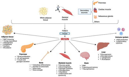
Since its discovery, irisin has attracted much attention for its potential involvement in metabolic and reproductive diseases. It appears to play an important role in different physiological and pathological conditions that can involve women throughout their entire lives. Irisin appears to be an important factor for the hypothalamic–pituitary–gonadal axis activation, and it seems to play a role in the timing of puberty onset. Additionally, serum irisin levels have been proposed as a biomarker for predicting the future development of gestational diabetes (GDM). Its role in polycystic ovary syndrome (PCOS) is still controversial, although an “irisin resistance” mechanism has been hypothesized. Beyond its influence on metabolism, irisin also appears to influence bone health. Irisin levels are inversely correlated with the prevalence of fractures in postmenopausal women. Similar mechanisms have also been postulated in young women with functional hypothalamic amenorrhea (FHA).

irisin bone health metabolism reproduction HPG axis polycystic ovary syndrome (PCOS) functional hypothalamic amenorrhea (FHA) gestational diabetes (GDM) menopause

1. Introduction

Irisin is a 112 amino acid (~12 kDa) hormone, resulting from cleavage of the extracellular portion of the N-terminal membrane protein called fibronectin type III domain containing protein 5 (FNDC5). It was identified for the first time in 2012 by Boström et al. [1] in myocytes of transgenic mice undergoing intense physical activity, where this molecule appeared to be involved in the cross-talk between muscles and other tissues. This peculiarity is expressed by its name, “irisin”, referring to the Greek goddess Iris, the messenger of the Gods [2].
Transgenic mice, in which irisin was first identified, were found to overexpress the Ppargc1a gene encoding for the peroxisome proliferator-activated receptor co-activator-1α (PGC1α). In mouse models of myocytes, irisin was able to promote the “browning processes” of white adipose tissue (WAT) and thermogenesis, mediated by uncoupling protein 1. Subsequent studies have highlighted the pleiotropic actions of irisin [3]. Among its most important effects, irisin is able to promote insulin synthesis and glucose-stimulated insulin secretion, reduce pancreatic ß-cell apoptosis in response to various harmful stimuli, and promote ß-cell proliferation [2][4][5]. Thus, irisin seems to promote the survival and restoration of the normal functional mass of ß-cells, essential for adequate glucose homeostasis [6]. Numerous studies have documented the ability of irisin to act on skeletal muscle, adipose tissue, and liver, improving insulin sensitivity, glucose uptake, reducing gluconeogenesis, glycogenolysis, and lipid accumulation, and increasing glucose storage, lipolysis, and fatty acid oxidation [7]. Therefore, irisin has been proposed as a hormone capable of increasing energy expenditure, promoting weight loss, and decreasing insulin resistance (IR) [8][9]. Irisin has become a potential target for the treatment of metabolic diseases. The main functions of irisin are shown in Figure 1.
Figure 1. Main functions of irisin. The main sources of circulating irisin are the skeletal muscle during physical activity and white adipose tissue. Irisin has multi-spectrum functions, influencing the activity of numerous tissues and organs, mainly skeletal muscle [10], the pancreas [11], the liver [12], the brain [13], the bone [14], and the immune system [15]. Abbreviations: FDNC5, Fibronectin type III domain-containing protein 5; ROS, Reactive oxygen species; WAT, White adipose tissue; BNDF, Brain-derived neurotrophic factor.
Irisin is secreted mainly by skeletal muscle, but also by adipose tissue, the pancreas, sebaceous glands, and cardiac muscle. Irisin immunoreactivity was soon found in several other organs and tissues, including salivary glands, ovaries, testes, the rectum, intracranial arteries, the tongue, optic nerve, stomach, neuronal cells, and sweat glands [8][9] (Figure 1). For these reasons, in recent decades, irisin has been the subject of many studies investigating its possible role in the pathogenesis and development of various diseases.

2. Irisin and Sex Differences: Towards Gender Medicine

There is currently no indicative range of values considered normal for irisin in plasma and serum. Circulating irisin levels appear to vary significantly, with concentrations reported in humans ranging from 0.01 ng/mL to 2000 ng/mL [16]. Although difference in irisin levels between women and men have been hypothesized, the results are still controversial. Previous studies have shown higher irisin levels in girls than boys [17], and in women than men [18].
Al-Daghri et al. [17], in a study conducted on 81 male and 72 female children, found that high circulating levels of irisin are correlated with impaired glucose tolerance, and that this association is more evident in girls. The authors suggest that this sexual dimorphism could be explained by hormonal differences in the two sexes. 17ß-estradiol (E2) could influence irisin circulation through anabolic pathways to increase muscle mass leading to the up-regulation of irisin, and promoting an irisin resistance mechanism [19]
Evidence for the sexual dimorphism of irisin distribution and effects also comes from new insights, which have identified irisin in the central nervous system (CNS). A study conducted on brain samples of marmoset and rhesus monkeys revealed the different distributions of FNDC5 and PGC1A, depending on sex. In females, the pituitary gland and posterior hypothalamus had considerably higher FNDC5 and PGC1A transcript levels than the corresponding male counterparts did. These findings demonstrate for the first time that such genes are expressed in neonatal and adult monkeys in a tissue- and sex-specific manner [20]

3. Irisin and Pubertal Development

Irisin appears to be an important factor activating the function of the hypothalamic-pituitary-gonadal (HPG) axis and reproductive capability [21]. Indeed, irisin and FNDC5 have been identified in the hypothalamus, pituitary gland, ovary, and testis. New insights have led to the identification of irisin in the CNS. Irisin is expressed primarily in the ventromedial nucleus and arcuate nucleus of the hypothalamus, areas known to be involved in the regulation of feeding, energy homeostasis, and reproduction [20]. Furthermore, direct contact between irisin-positive fibers and gonadotropin-releasing hormone (GnRH) neurons was identified, suggesting a direct influence of irisin on GnRH pulses. Furthermore, in mouse models, irisin administration revealed an enhancement of GnRH transcription after one hour of incubation [20]. These findings demonstrated that irisin is involved in GnRH release, and suggested that it might influence GnRH expression and, consequently, reproductive function. However, the role of irisin in the HPG axis is controversial, with opposing evidence. Ulker et al. demonstrated that daily intraperitoneal injections of irisin (100 ng/kg from postnatal day 21 for approximately 10 weeks) delayed the onset of puberty and decreased the expression of GnRH in the brain of female rats [22]. Chronic administration of irisin also affected hormone levels, by lowering serum follicle-stimulating hormone (FSH) levels and increasing serum luteinizing hormone (LH) and E2 levels. In addition to these changes, ovarian tissue from irisin-treated rats showed a reduction in early follicles and an increase in fibrosis. On the other hand, the timing of puberty remained unaltered by the administration of irisin to male rats. However, after prolonged exposure to irisin, male rats’ serum testosterone and LH levels, sperm count, and seminiferous tubule width were significantly increased [22]. Therefore, the authors concluded that the reproductive systems and pubertal maturation of male and female rats respond differently to irisin exposure, and long-term exposure to irisin may alter the female reproductive system. At the same time, it appears to have androgenic effects on the human HPG axis.
The in vitro effects of GnRH on FSH and LH stimulation appear to be compromised when it is administered in combination with irisin [23][24]. The detrimental effects of irisin on GnRH-induced gonadotropin stimulation may be explained by several mechanisms. First, irisin could stimulate FSH and LH secretion directly, and it could decrease their release, exerting a negative feedback on GnRH secretion. Secondly, it can be hypothesized that the effects of irisin are comparable to those of GnRH agonists [16]. Therefore, irisin can bind to GnRH receptors on the cell surface, causing their down-regulation and increasing receptor internalization, thus making them inaccessible to GnRH. This decreases the production of gonadotropins by pituitary desensitization [16].
According to Bastu et al. [25], the irisin precursor protein, FNDC5, can stimulate the growth of mouse ovaries by significantly increasing the number of primary and secondary follicles. Previous research had already revealed that FNDC5 gene knockout mice had fewer antral follicles, in agreement with the findings obtained by these authors [16]. In contrast, some studies have found that irisin has detrimental effects on the ovaries and testes, significantly reducing the number of vegetative cells and Leydig cells, the sperm concentration, and sperm motility in male rats exposed to irisin [26], as well as significantly increasing ovarian fibrosis in female mice [22] and decreasing the number of primary follicles in rats. 
A network of hormones and neuroendocrine pathways, in which GnRH neurons play a major role, complexly regulates the onset of puberty. The balance between inhibitor and stimulatory signals mediated by kisspeptin/neurokinin B/dynorphin (KND), and neuropeptide Y [27] regulates GnRH neurons. The initiation of GnRH pulses confirms the onset of puberty, and allows the increased growth rate, bone maturation, epiphysis fusion, and the development of secondary sexual characteristics. Pulsatile GnRH secretion appears to be linked to body weight (fat/muscle), metabolic condition, and energy reserves during puberty [28]. Energy reserves and the metabolic state of the organism strongly influence the onset of puberty, and it is known that the hypothalamic network can only be activated when the body reaches a critical level of fat and/or muscle mass [21]
Kutlu et al. [29] conducted a study to evaluate irisin levels in 94 girls, including 33 with central precocious puberty (CPP), 31 with precocious puberty (PP), and 30 healthy controls. The authors found that patients with CPP had higher serum irisin levels than the other groups. Furthermore, a positive statistically significant correlation has been reported between irisin levels and BMI standard deviation score (BMI-SDS), height SDS, weight SDS, bone age, uterine long axis, ovarian size, levels of baseline FSH and LH, and peak LH. Therefore, although further prospective studies are needed, the authors hypothesized that increased irisin levels could potentially be used as a marker of CPP.
Irisin levels in 20 children of similar age and pubertal stage were compared with indices of metabolic syndrome, such as impaired glucose metabolism, IR, lipid metabolism, and blood pressure [30]. The results showed that obese children with impaired glucose tolerance had higher irisin levels, followed by obese children with normal glucose tolerance, while the lowest levels were found in normal-weight children. Baseline irisin was strongly correlated with pubertal stage, high-density lipoprotein cholesterol (HDL-c), and a homeostasis model of assessment in a multiple linear regression analysis (HOMA-R), but not with age, gender, BMI, or any other metabolic syndrome parameter. Furthermore, prepubertal children had significantly lower irisin concentrations than pubertal youngsters. Changes in irisin were not substantially correlated with changes in BMI or with any other parameter in longitudinal studies, but they were strongly correlated with the onset of puberty, changes in fasting blood glucose, and with changes in 2 h blood glucose in an oral glucose-tolerance test. Therefore, changes in glucose metabolism characteristics were substantially related to changes in irisin levels.

4. Irisin and Polycystic Ovarian Syndrome (PCOS)

Polycystic ovarian syndrome (PCOS) is the most common endocrinopathy that affects women of reproductive age. It has a prevalence ranging from between 5 and 20%, depending on the different diagnostic criteria used and the population studied [31].
The pathogenesis of the disease is not completely clear, and the clinical manifestations are extremely variable. According to the Rotterdam criteria, at least two of the following three criteria are required to diagnose PCOS: (1) menstrual disorders (oligo-amenorrhea and/or anovulation); (2) clinical or biochemical hyperandrogenism; and (3) polycystic ovarian morphology (PCOM) [32]. Although these diagnostic criteria do not consider the dysmetabolic background of PCOS, it is widely recognized that patients with PCOS commonly have metabolic disorders. The prevalence of obesity is higher in patients with PCOS (relative risk (RR): 2.77; 95% CI: 1.88 to 4.1), compared to those without PCOS [33]. Furthermore, up to 80% of PCOS women exhibit IR, which is independent of BMI and plays an important role in the clinical presentation and development of several metabolic alterations [34]. Genome-wide association studies of single-nucleotide polymorphisms showed that granulosa cells of PCOS patients expressed genes linked to insulin resistance, including protein kinase AMP-activated catalytic subunit alpha 2, matrix metallopeptidase 9, and haptoglobin [35]. As reported above, many studies have shown that irisin improves IR through several mechanisms, including increasing insulin receptor sensitization in skeletal muscle and heart, improving hepatic glucose and lipid metabolism and the functions of pancreatic β cells, and promoting the transformation of WAT into BAT [36]. Therefore, many studies have been conducted to better understand the relationship between irisin and insulin.
The role of irisin in the development of PCOS is controversial. Serum irisin levels in patients with PCOS, first reported by Chang et al., were found to be higher in PCOS compared to controls, suggesting a possible role in the development of the disease [37]. On the other hand, other studies have not reported a significant difference between PCOS and healthy women with respect to irisin levels [38], and even lower irisin levels have been found in women with PCOS [39].
A recent meta-analysis including eight studies, with 918 PCOS patients and 529 controls, demonstrated that irisin levels were at least 45.78 ng/mL (95% confidence interval CI) (12.45, 79.12, p = 0.007) higher in PCOS patients, compared to healthy controls [40]. However, another meta-analysis including the same studies showed that, adjusting for BMI, serum irisin levels in PCOS patients were similar to those of healthy controls [41]. Irisin levels were higher in PCOS patients with higher BMI than in those with lower BMI (d = 0.36, 95% CI 0.15, 6 to 0.56) [41]. Therefore, circulating irisin levels appear to be influenced by BMI [40]. In both PCOS and non-PCOS women, irisin levels were higher in overweight and obese women than in those of normal weight [42][43]. Weight loss led to a significant reduction in irisin levels (15%), while weight regain brought irisin levels back to baseline levels [43].
Given the significant clinical heterogeneity, several classifications have been proposed for distinguishing the different phenotypes of PCOS. A recent study evaluating differences in irisin levels in PCOS women, based on various phenotypes, reported that serum irisin levels were associated with hyperandrogenism, but not with oligo-anovulation or PCOM presence [44]. Li et al. demonstrated that elevated irisin levels in PCOS women were associated with androgen excess, assessed through the free androgen index [45]. Hyperandrogenism is also an important inducer of IR, which highlights the need to further investigate the relationship between irisin, hyperandrogenism, and IR. 
In conclusion, currently, contradictory results link irisin to PCOS. Irisin levels in PCOS patients were found to be higher [37], similar [38], or even lower [39], compared to controls. These differences may be related to a significant heterogeneity of published studies. Irisin levels can be influenced by several factors, particularly BMI. They can also relate to different stages of the diseases. Indeed, irisin could increase in the first phase as a protective mechanism to compensate for reduced insulin sensibility in PCOS patients. However, over time, the excessive increase in irisin can also cause irisin resistance, which in turn leads to a greater risk of metabolic consequences of PCOS. 

5. Irisin and Functional Hypothalamic Amenorrhea

Functional hypothalamic amenorrhea (FHA) is a form of chronic anovulation not related to organic causes, but resulting from various stressors. It is generally associated with three main causes: weight loss, excessive physical exercise, and psychological stress, which can occur alone or, more often, in combination [46]. FHA is one of the most common reproductive disorders in women of childbearing age, and is responsible for 20–35% of secondary cases of amenorrhea and for approximately 3% of cases of women with primary amenorrhea [47].
FHA is characterized by the suppression of GnRH pulsatility, with the consequent reduction in LH and FSH secretion, and, therefore, hypogonadotropic hypogonadism. Although there is no underlying organic cause, some cases of FHA can take a long time to recover, and can lead to major complications, primarily in bone health, due to estrogen deficiency [48]. In young women, estrogens are the most important hormones that regulate bone metabolism, inhibit bone remodeling, decrease bone resorption, and maintain bone formation [48].
In addition to estrogen deficiency, irisin depletion may also contribute to the deterioration in bone health seen in these patients. Indeed, previous studies have reported the potential role of irisin on bone metabolism, through both direct and indirect effects. In mice, irisin enhances osteoblast activation and inhibition of NF-κB ligand activating receptor (RANKL)-mediated osteoclastogenesis and, in turn, increases trabecular and cortical thickness and trabecular density [14]. This mechanism has also been confirmed in humans [49]. The effects of irisin on bone metabolism could also depend on BAT. Indeed, the adipogenesis of this tissue appears to be physiologically related to bone health, and its defective brown adipogenesis has been correlated with bone loss [50]. Irisin levels have also been negative correlated with serum sclerostin, an inhibitor of osteoblast differentiation and bone formation [51]
On the other hand, it has recently been shown that physical activity increases irisin expression in skeletal muscle [1]. Consequently, it must be taken into account that some patients with FHA are athletes who take part in intense physical exercise. 

6. Irisin and Endometriosis

Previous preclinical and clinical studies have suggested that irisin has anti-inflammatory properties [52]. Mechanisms involved in the anti-inflammatory functions of irisin include reducing the production of pro-inflammatory cytokines (such as interleukin-6 (IL-6) and tumor necrosis factor-α), increasing the production of anti-inflammatory cytokines, reducing macrophage proliferation, inducing polarization of alternative-type macrophages, inhibiting pathways of increased vascular permeability, and preventing the formation of inflammasomes (such as toll-like receptor 4/myeloid differentiation factor 88 downstream pathways) [15][53].
It is now known that elevated levels of various pro-inflammatory cytokines play an important role in the pathogenesis of endometriosis. Endometriosis is a common inflammatory disease, characterized by the presence of tissue outside the uterus, which resembles the endometrium, primarily on pelvic organs and tissues. It affects approximately 5–10% of women of reproductive age, and reduces women’s quality of life, due to dysmenorrhea, chronic pelvic pain, irregular uterine bleeding, and infertility [54].

7. Irisin and Gestational Diabetes Mellitus

GDM is a condition that affects pregnant women, and is characterized by hyperglycemia during pregnancy. According to the American Diabetes Association (ADA), GDM is defined as the onset of impaired glucose tolerance during the second or third trimester of pregnancy [55]. According to the data collected and analyzed by the International Diabetes Federation (IDF), the global prevalence of GDM is approximately 14%, although it varies depending on the population studied [56].
Pregnancy is characterized by significant changes in the mother’s metabolic processes, thus facilitating the provision of adequate energy and nutrients to the developing fetus. As a result, pregnant women experience a period of reduced insulin sensitivity during mid-gestation, which intensifies during the third trimester. This subsequently leads to reduced glucose uptake by maternal tissues and increased glucose production through gluconeogenesis [57]. However, in a significant percentage of pregnancies, the IR state is exacerbated, resulting in adverse maternal metabolic conditions and abnormal fetal growth [58]. The mechanisms underlying IR during pregnancy include increased adiposity and the action of placental hormones with diabetogenic effects, such as estrogen, progesterone, cortisol, human chorionic gonadotropin, and prolactin [59][60][61].
GDM has a multifactorial pathogenesis, including both genetic and environmental factors, but the exact mechanism is not fully understood [62]. The presence of GDM during pregnancy increases the risk of perinatal morbidity, pre-eclampsia, fetal macrosomia, and shoulder dystocia, and increases the risk of developing T2DM, cardiovascular disease, and obesity, in both mother and offspring [55]. Early diagnosis and appropriate management of GDM can help mitigate adverse effects in both the mother and fetus, as well as protecting them from potential long-term complications.
The concentrations of irisin increase in a statistically significant manner during pregnancy and particularly in the second and third trimesters, compared to the early stages of gestation. Furthermore, serum irisin levels are significantly higher in pregnant women than in non-pregnant women [63]. Maternal irisin levels are negatively correlated with the HOMA-IR in most studies [64][65], although some of them have not found any correlation [66] or even a positive correlation [67][68]. However, it has been hard for studies to evaluate the association between irisin levels and maternal HOMA-IR, considering potential confounding variables such as age, BMI, physical activity, and nutritional status, which influence circulating irisin levels [69]. To reduce the potential influence of confounding factors, Cai et al. used multiple linear regression analysis, and found an inverse association between maternal serum irisin levels and HOMA-IR [70]. This suggests that the increase in irisin during pregnancy is an adaptive response, to compensate for the increase in IR. Furthermore, maternal irisin showed a negative association with fasting plasma glucose (FPG) [70]
Serum irisin levels have been proposed as a biomarker to predict the future development of GDM. Wang et al. measured maternal irisin levels during the first trimester of pregnancy, and found significantly lower levels in women who subsequently developed GDM compared to those who did not, after adjusting for confounding factors (BMI, insulin, FPG, and lipid metabolism) [71].
Other studies have looked at irisin levels in cord blood. However, currently, the available evidence suggests that levels of this hormone in maternal serum do not appear to correlate with levels in cord blood, either in women with or without GDM [72][73]. This may be because the main source of fetal irisin is the placenta, fetal muscle tissue, and fetal adipose tissue, which is browner and contributes to irisin production [73]

8. Irisin and Menopause

Menopause is a natural biological event that marks the end of a woman’s reproductive age, and typically occurs between her late forties and early fifties. Menopause is associated with a decline in BMD, resulting in an increased risk of osteoporosis and fractures [74]. Irisin has been identified as a potential factor associated with osteoporosis, a condition that causes a significant socioeconomic burden worldwide, particularly in postmenopausal women [75][76].
In in vitro studies, irisin is associated with an increase in bone formation and a decrease in bone resorption, resulting in a reduced risk of osteoporosis in postmenopausal women. At the bone tissue level, irisin acts on target cells, through αV/β5 integrin receptors. This influences the proliferation, differentiation and activity of osteoblasts, osteoclasts, and osteocytes [77]. Irisin treatment of pre-differentiated osteoblasts increases the phosphorylation of Extracellular signal-Regulated Kinase and p38, resulting in higher mRNA levels of osteoblast transcriptional regulators (Runt-related transcription factor 2 (Runx2), Osteoblast-specific Transcription Factor Osterix (Osx)) and early osteoblast differentiation marker genes (alkaline phosphatase (ALP), collagen type 1 alpha 1 gene (ColIa1)), which lead to increased proliferation, differentiation, and mineralization of osteoblasts [78]

9. Conclusions

The positive effects in the regulation of hepatic and pancreatic functions, adipose tissue, and energy expenditure, have made irisin a possible new therapeutic target for the treatment of metabolic diseases. Irisin levels were found to be significantly lower during the first trimester of pregnancy in women who subsequently developed GDM, compared to those who did not develop it in the subsequent months. Therefore, serum irisin levels have been proposed as a biomarker to predict the future development of GDM. Interestingly, irisin could be a marker not only for GDM, but also for macrosomia and placental function. Despite the misnomer of PCOS, this syndrome is not a problem of ovarian cysts, but it is a truly complex disorder, with important metabolic consequences. An “irisin resistance” mechanism, similar to the well-known phenomenon of leptin resistance or IR, has been hypothesized in patients with PCOS. Although the results are still controversial, irisin appears to be involved in the etiology, the development of metabolic comorbidities, and even the treatment of this complex disease.
In contrast to its metabolic effect, the influence of irisin on bone health is still a matter of debate. Preclinical studies have shown that irisin can influence bone metabolism, through both direct and indirect mechanisms. In postmenopausal women, irisin levels are inversely correlated with bone fracture frequency. Similar mechanisms have also been hypothesized in young women with FHA.
Finally, previous preclinical and clinical studies have shown the anti-inflammatory effect of irisin. To date, only one study has reported increased irisin levels in patients with endometriosis, perhaps as an adaptive response to compensate for the increased inflammation underlying this disease.

References

  1. Boström, P.; Wu, J.; Jedrychowski, M.P.; Korde, A.; Ye, L.; Lo, J.C.; Rasbach, K.A.; Boström, E.A.; Choi, J.H.; Long, J.Z.; et al. A PGC1-α-dependent myokine that drives brown-fat-like development of white fat and thermogenesis. Nature 2012, 481, 463–468.
  2. Pukajło, K.; Kolackov, K.; Łaczmański, Ł.; Daroszewski, J. Irisin, a new mediator of energy homeostasis. Adv. Hyg. Exp. Med. 2015, 69, 233–242.
  3. Perakakis, N.; Triantafyllou, G.A.; Fernández-Real, J.M.; Huh, J.Y.; Park, K.H.; Seufert, J.; Mantzoros, C.S. Physiology and role of irisin in glucose homeostasis. Nat. Rev. Endocrinol. 2017, 13, 324–337.
  4. Natalicchio, A.; Marrano, N.; Giorgino, F. Irisina: Ruolo nell’omeostasi del glucosio. L’Endocrinologo 2018, 19, 292.
  5. Norman, D.; Drott, C.J.; Carlsson, P.O.; Espes, D. Irisin—A Pancreatic Islet Hormone. Biomedicines 2022, 10, 258.
  6. Song, R.; Zhao, X.; Zhang, D.Q.; Wang, R.; Feng, Y. Lower levels of irisin in patients with type 2 diabetes mellitus: A meta-analysis. Diabetes Res. Clin. Pract. 2021, 175, 108788.
  7. Marrano, N.; Biondi, G.; Borrelli, A.; Cignarelli, A.; Perrini, S.; Laviola, L.; Giorgino, F.; Natalicchio, A. Irisin and Incretin Hormones: Similarities, Differences, and Implications in Type 2 Diabetes and Obesity. Biomolecules 2021, 11, 286.
  8. Martinez Munoz, I.Y.; Camarillo Romero, E.D.S.; Garduno Garcia, J.J. Irisin a Novel Metabolic Biomarker: Present Knowledge and Future Directions. Int. J. Endocrinol. 2018, 2018, 7816806.
  9. Korta, P.; Pocheć, E.; Mazur-Biały, A. Irisin as a Multifunctional Protein: Implications for Health and Certain Diseases. Medicina 2019, 55, 485.
  10. Ma, C.; Ding, H.; Deng, Y.; Liu, H.; Xiong, X.; Yang, Y. Irisin: A New Code Uncover the Relationship of Skeletal Muscle and Cardiovascular Health During Exercise. Front. Physiol. 2021, 2, 620608.
  11. Yi, P.; Park, J.S.; Melton, D.A. Betatrophin: A hormone that controls pancreatic β cell proliferation. Cell 2013, 53, 747–758.
  12. Park, M.J.; Kim, D.I.; Choi, J.H.; Heo, Y.R.; Park, S.H. New role of irisin in hepatocytes: The protective effect of hepatic steatosis in vitro. Cell Signal 2015, 27, 1831–1839.
  13. Wrann, C.D.; White, J.P.; Salogiannnis, J.; Laznik-Bogoslavski, D.; Wu, J.; Ma, D.; Lin, J.D.; Greenberg, M.E.; Spiegelman, B.M. Exercise induces hippocampal BDNF through a PGC-1α/FNDC5 pathway. Cell Metab. 2013, 18, 649–659.
  14. Colaianni, G.; Cuscito, C.; Mongelli, T.; Oranger, A.; Mori, G.; Brunetti, G.; Colucci, S.; Cinti, S.; Grano, M. Irisin enhances osteoblast differentiation in vitro. Int. J. Endocrinol. 2014, 2014, 902186.
  15. Mazur-Bialy, A.I.; Pocheć, E.; Zarawski, M. Anti-Inflammatory Properties of Irisin, Mediator of Physical Activity, Are Connected with TLR4/MyD88 Signaling Pathway Activation. Int. J. Mol. Sci. 2017, 18, 701.
  16. Luo, Y.; Qiao, X.; Xu, L.; Huang, G. Irisin: Circulating levels in serum and its relation to gonadal axis. Endocrine 2022, 75, 663–671.
  17. Al-Daghri, N.M.; Alkharfy, K.M.; Rahman, S.; Amer, O.E.; Vinodson, B.; Sabico, S.; Piya, M.K.; Harte, A.L.; McTernan, P.G.; Alokail, M.S.; et al. Irisin as a predictor of glucose metabolism in children: Sexually dimorphic effects. Eur. J. Clin. Investig. 2014, 44, 119–124.
  18. Anastasilakis, A.D.; Polyzos, S.A.; Saridakis, Z.G.; Kynigopoulos, G.; Skouvaklidou, E.C.; Molyvas, D.; Vasiloglou, M.F.; Apostolou, A.; Karagiozoglou-Lampoudi, T.; Siopi, A.; et al. Circulating irisin in healthy, young individuals: Day-night rhythm, effects of food intake and exercise, and associations with gender, physical activity, diet, and body composition. J. Clin. Endocrinol. Metab. 2014, 99, 3247–3255.
  19. Moreno-Navarrete, J.M.; Ortega, F.; Serrano, M.; Guerra, E.; Pardo, G.; Tinahones, F.; Ricart, W.; Fernández-Real, J.M. Irisin is expressed and produced by human muscle and adipose tissue in association with obesity and insulin resistance. J. Clin. Endocrinol. Metab. 2013, 98, E769–E778.
  20. Wahab, F.; Khan, I.U.; Polo, I.R.; Zubair, H.; Drummer, C.; Shahab, M.; Behr, R. Irisin in the primate hypothalamus and its effect on GnRH in vitro. J. Endocrinol. 2019, 241, 175–187.
  21. Wahab, F.; Shahab, M.; Behr, R. Hypothesis: Irisin is a metabolic trigger for the activation of the neurohormonal axis governing puberty onset. Med. Hypotheses 2016, 95, 1–4.
  22. Ulker, N.; Yardimci, A.; Kaya Tektemur, N.; Bulmus, O.; Ozer Kaya, S.; Gulcu Bulmus, F.; Turk, G.; Ozcan, M.; Canpolat, S. Irisin may have a role in pubertal development and regulation of reproductive function in rats. Reproduction 2020, 160, 281–292.
  23. Jiang, Q.; Zhang, Q.; Lian, A.; Xu, Y. Irisin stimulates gonadotropins gene expression in tilapia (Oreochromis niloticus) pituitary cells. Anim. Reprod. Sci. 2017, 85, 140–147.
  24. Poretsky, L.; Islam, J.; Avtanski, D.; Lin, Y.K.; Shen, Y.L.; Hirth, Y.; Lesser, M.; Rosenwaks, Z.; Seto-Young, D. Reproductive effects of irisin: Initial in vitro studies. Reprod. Biol. 2017, 17, 285–288.
  25. Bastu, E.; Zeybek, U.; Gurel Gurevin, E.; Yüksel Ozgor, B.; Celik, F.; Okumus, N.; Demiral, I.; Dural, O.; Celik, C.; Bulut, H.; et al. Effects of Irisin and Exercise on Metabolic Parameters and Reproductive Hormone Levels in High-Fat Diet-Induced Obese Female Mice. Reprod. Sci. 2018, 25, 281–291.
  26. Tekin, S.; Beytur, A.; Erden, Y.; Beytur, A.; Cigremis, Y.; Vardi, N.; Turkoz, Y.; Tekedereli, I.; Sandal, S. Effects of intracerebroventricular administration of irisin on the hypothalamus-pituitary-gonadal axis in male rats. J. Cell Physiol. 2019, 234, 8815–8824.
  27. Klenke, U.; Taylor-Burds, C.; Wray, S. Metabolic influences on reproduction: Adiponectin attenuates GnRH neuronal activity in female mice. Endocrinology 2014, 155, 1851–1863.
  28. Castellano, J.M.; Tena-Sempere, M. Metabolic control of female puberty: Potential therapeutic targets. Expert. Opin. Ther. Targets 2016, 20, 1181–1193.
  29. Kutlu, E.; Özgen, I.T.; Bulut, H.; Koçyiǧit, A.; Otçu, H.; Cesur, Y. Serum Irisin Levels in Central Precocious Puberty and Its Variants. J. Clin. Endocrinol. Metab. 2021, 106, E247–E254.
  30. Reinehr, T.; Elfers, C.; Lass, N.; Roth, C.L. Irisin and its relation to insulin resistance and puberty in obese children: A longitudinal analysis. J. Clin. Endocrinol. Metab. 2015, 100, 2123–2130.
  31. Azziz, R.; Carmina, E.; Chen, Z.; Dunaif, A.; Laven, J.S.; Legro, R.S.; Lizneva, D.; Natterson-Horowtiz, B.; Teede, H.J.; Yildiz, B.O. Polycystic ovary syndrome. Nat. Rev. Dis. Primers 2016, 2, 16057.
  32. Rotterdam ESHRE/ASRM-Sponsored PCOS consensus workshop group. Revised 2003 consensus on diagnostic criteria and long-term health risks related to polycystic ovary syndrome (PCOS). Hum. Reprod. 2004, 19, 41–47.
  33. Lim, S.S.; Davies, M.J.; Norman, R.J.; Moran, L.J. Overweight, obesity and central obesity in women with polycystic ovary syndrome: A systematic review and meta-analysis. Hum. Reprod. Update 2012, 18, 618–637.
  34. Amisi, C.A. Markers of insulin resistance in Polycystic ovary syndrome women: An update. World J. Diabetes 2022, 13, 129–149.
  35. Pant, P.; Chitme, H.; Sircar, R.; Prasad, R.; Prasad, H.O. Genome-wide association study for single nucleotide polymorphism associated with mural and cumulus granulosa cells of PCOS (polycystic ovary syndrome) and non-PCOS patients. Future J. Pharm. Sci. 2023, 27.
  36. Gizaw, M.; Anandakumar, P.; Debela, T. A Review on the Role of Irisin in Insulin Resistance and Type 2 Diabetes Mellitus. J. Pharmacopunct. 2017, 20, 235–242.
  37. Chang, C.L.; Huang, S.Y.; Soong, Y.K.; Cheng, P.J.; Wang, C.J.; Liang, I.T. Circulating irisin and glucose-dependent insulinotropic peptide are associated with the development of polycystic ovary syndrome. J. Clin. Endocrinol. Metab. 2014, 99, E2539–E2548.
  38. Pukajło, K.; Łaczmański, Ł.; Kolackov, K.; Kuliczkowska-Płaksej, J.; Bolanowski, M.; Milewicz, A.; Daroszewski, J. Irisin plasma concentration in PCOS and healthy subjects is related to body fat content and android fat distribution. Gynecol. Endocrinol. 2015, 31, 907–911.
  39. Abali, R.; Temel Yuksel, I.; Yuksel, M.A.; Bulut, B.; Imamoglu, M.; Emirdar, V.; Unal, F.; Guzel, S.; Celik, C. Implications of circulating irisin and Fabp4 levels in patients with polycystic ovary syndrome. J. Obs. Gynaecol. 2016, 36, 897–901.
  40. Wang, C.; Zhang, X.Y.; Sun, Y.; Hou, X.G.; Chen, L. Higher circulating irisin levels in patients with polycystic ovary syndrome: A meta-analysis. Gynecol. Endocrinol. 2018, 34, 290–293.
  41. Cai, X.; Qiu, S.; Li, L.; Zügel, M.; Steinacker, J.M.; Schumann, U. Circulating irisin in patients with polycystic ovary syndrome: A meta-analysis. Reprod. Biomed. Online 2018, 36, 172–180.
  42. Li, Q.; Jia, S.; Xu, L.; Li, B.; Chen, N. Metformin-induced autophagy and irisin improves INS-1 cell function and survival in high-glucose environment via AMPK/SIRT1/PGC-1α signal pathway. Food Sci. Nutr. 2019, 7, 1695–1703.
  43. Crujeiras, A.B.; Pardo, M.; Arturo, R.R.; Navas-Carretero, S.; Zulet, M.A.; Martínez, J.A.; Casanueva, F.F. Longitudinal variation of circulating irisin after an energy restriction-induced weight loss and following weight regain in obese men and women. Am J. Hum. Biol. 2014, 26, 198–207.
  44. Zhang, L.; Fang, X.; Li, L.; Liu, R.; Zhang, C.; Liu, H.; Tan, M.; Yang, G. The association between circulating irisin levels and different phenotypes of polycystic ovary syndrome. J. Endocrinol. Investig. 2018, 41, 1401–1407.
  45. Li, H.; Xu, X.; Wang, X.; Liao, X.; Li, L.; Yang, G.; Gao, L. Free androgen index and Irisin in polycystic ovary syndrome. J. Endocrinol. Investig. 2016, 39, 549–556.
  46. Gordon, C.M.; Ackerman, K.E.; Berga, S.L.; Kaplan, J.R.; Mastorakos, G.; Misra, M.; Murad, M.H.; Santoro, N.F.; Warren, M.P. Functional Hypothalamic Amenorrhea: An Endocrine Society Clinical Practice Guideline. J. Clin. Endocrinol. Metab. 2017, 102, 1413–1439.
  47. Practice Committee of the American Society for Reproductive Medicine. Current evaluation of amenorrhea. Fertil. Steril. 2006, 86, S148–S155.
  48. Indirli, R.; Lanzi, V.; Mantovani, G.; Arosio, M.; Ferrante, E. Bone health in functional hypothalamic amenorrhea: What the endocrinologist needs to know. Front. Endocrinol. 2022, 13, 946695.
  49. Ma, Y.; Qiao, X.; Zeng, R.; Cheng, R.; Zhang, J.; Luo, Y.; Nie, Y.; Hu, Y.; Yang, Z.; Zhang, J.; et al. Irisin promotes proliferation but inhibits differentiation in osteoclast precursor cells. FASEB J. 2018, 32, 5813–5823.
  50. Lee, P.; Brychta, R.J.; Collins, M.T.; Linderman, J.; Smith, S.; Herscovitch, P.; Millo, C.; Chen, K.Y.; Celi, F.S. Cold-activated brown adipose tissue is an independent predictor of higher bone mineral density in women. Osteoporos. Int. 2013, 4, 1513–1518.
  51. Klangjareonchai, T.; Nimitphong, H.; Saetung, S.; Bhirommuang, N.; Samittarucksa, R.; Chanprasertyothin, S.; Sudatip, R.; Ongphiphadhanakul, B. Circulating sclerostin and irisin are related and interact with gender to influence adiposity in adults with prediabetes. Int. J. Endocrinol. 2014, 2014, 261545.
  52. Gamal, R.M.; Mohamed, M.E.; Hammam, N.; El Fetoh, N.A.; Rashed, A.M.; Furst, D.E. Preliminary study of the association of serum irisin levels with poor sleep quality in rheumatoid arthritis patients. Sleep Med. 2020, 67, 71–76.
  53. Slate-Romano, J.J.; Yano, N.; Zhao, T.C. Irisin reduces inflammatory signaling pathways in inflammation-mediated metabolic syndrome. Mol. Cell Endocrinol. 2022, 552, 111676.
  54. Zondervan, K.T.; Becker, C.M.; Koga, K.; Missmer, S.A.; Taylor, R.N.; Viganò, P. Endometriosis. Nat. Rev. Dis. Primers 2018, 4, 9.
  55. American Diabetes Association. Classification and diagnosis of diabetes: Standards of medical care in diabetes-2021. Diabetes Care 2021, 44, S15–S33.
  56. Wang, H.; Li, N.; Chivese, T.; Werfalli, M.; Sun, H.; Yuen, L.; Hoegfeldt, C.A.; Elise Powe, C.; Immanuel, J.; Karuranga, S.; et al. IDF Diabetes Atlas: Estimation of Global and Regional Gestational Diabetes Mellitus Prevalence for 2021 by International Association of Diabetes in Pregnancy Study Group’s Criteria. Diabetes Res. Clin. Pract. 2022, 183, 109050.
  57. Catalano, P.M.; Tyzbir, E.D.; Roman, N.M.; Amini, S.B.; Sims, E.A.H. Longitudinal changes in insulin release and insulin resistance in nonobese pregnant women. Am. J. Obstet. Gynecol. 1991, 165, 1667–1672.
  58. Vohr, B.R.; Boney, C.M. Gestational diabetes: The forerunner for the development of maternal and childhood obesity and metabolic syndrome? J. Matern.-Fetal Neonatal Med. 2008, 21, 149–157.
  59. Lain, K.Y.; Catalano, P.M. Factors That Affect Maternal Insulin Resistance and Modify Fetal Growth and Body Composition. Metab. Syndr. Relat. Disord. 2006, 4, 91–100.
  60. Briana, D.D.; Malamitsi-Puchner, A. Adipocytokines in Normal and Complicated Pregnancies. Reprod. Sci. 2009, 16, 921–937.
  61. Lowe, W.L.; Karban, J. Genetics, genomics and metabolomics: New insights into maternal metabolism during pregnancy. Diabet. Med. 2014, 31, 254–262.
  62. Al-Ghazali, M.J.; Ali, H.A.; Al-Rufaie, M.M. Serum irisin levels as a potential marker for diagnosis of gestational diabetes mellitus. Acta Biomed. 2020, 91, 56–63.
  63. Garcés, M.F.; Peralta, J.J.; Ruiz-Linares, C.E.; Lozano, A.R.; Poveda, N.E.; Torres-Sierra, A.L.; Eslava-Schmalbach, J.H.; Alzate, J.P.; Sánchez, Á.Y.; Sanchez, E.; et al. Irisin Levels During Pregnancy and Changes Associated With the Development of Preeclampsia. J. Clin. Endocrinol. Metab. 2014, 99, 2113–2119.
  64. Kuzmicki, M.; Telejko, B.; Lipinska, D.; Pliszka, J.; Szamatowicz, M.; Wilk, J.; Zbucka-Kretowska, M.; Laudanski, P.; Kretowski, A.; Gorska, M.; et al. Serum irisin concentration in women with gestational diabetes. Gynecol. Endocrinol. 2014, 30, 636–639.
  65. Yuksel, M.A.; Oncul, M.; Tuten, A.; Imamoglu, M.; Acikgoz, A.S.; Kucur, M.; Madazli, R. Maternal serum and fetal cord blood irisin levels in gestational diabetes mellitus. Diabetes Res. Clin. Pract. 2014, 104, 171–175.
  66. Erol, O.; Erkal, N.; Ellidağ, H.Y.; İsenlik, B.S.; Aydın, Ö.; Derbent, A.U.; Yılmaz, N. Irisin as an early marker for predicting gestational diabetes mellitus: A prospective study. J. Matern.-Fetal Neonatal Med. 2016, 29, 3590–3595.
  67. Ebert, T.; Stepan, H.; Schrey, S.; Kralisch, S.; Hindricks, J.; Hopf, L.; Platz, M.; Lossner, U.; Jessnitzer, B.; Drewlo, S.; et al. Serum levels of irisin in gestational diabetes mellitus during pregnancy and after delivery. Cytokine 2014, 65, 153–158.
  68. Piya, M.K.; Harte, A.L.; Sivakumar, K.; Tripathi, G.; Voyias, P.D.; James, S.; Sabico, S.; Al-Daghri, N.M.; Saravanan, P.; Barber, T.M.; et al. The identification of irisin in human cerebrospinal fluid: Influence of adiposity, metabolic markers, and gestational diabetes. Am. J. Physiol. Endocrinol. Metab. 2014, 306, 512–518.
  69. Roca-Rivada, A.; Castelao, C.; Senin, L.L.; Landrove, M.O.; Baltar, J.; Crujeiras, A.B.; Seoane, L.M.; Casanueva, F.F.; Pardo, M. FNDC5/Irisin Is Not Only a Myokine but Also an Adipokine. PLoS ONE 2013, 8, e60563.
  70. Cai, L.; Wu, W.; Lin, L.; Chen, Y.; Gao, R.; Shi, B.; Ma, B.; Chen, Y.; Jing, J. Association between plasma irisin and glucose metabolism in pregnant women is modified by dietary n-3 polyunsaturated fatty acid intake. J. Diabetes Investig. 2020, 11, 1326–1335.
  71. Wang, P.; Ma, H.H.; Hou, X.Z.; Song, L.L.; Song, X.L.; Zhang, J.F. Reduced plasma level of irisin in first trimester as a risk factor for the development of gestational diabetes mellitus. Diabetes Res. Clin. Pract. 2018, 142, 130–138.
  72. Cui, L.; Qiao, T.; Xu, F.; Li, Z.; Chen, T.; Su, H.; Chen, G.; Zhang, L.; Xu, D.; Zhang, X. Circulating irisin levels of prenatal and postnatal patients with gestational diabetes mellitus: A systematic review and meta-analysis. Cytokine 2020, 126, 154924.
  73. Ersahin, S.S.; Yurci, A. Cord blood and maternal serum preptin and irisin concentrations are regulated independently in GDM. Eur. Rev. Med. Pharmacol. Sci. 2021, 25, 1954–1958.
  74. Yong, E.; Logan, S. Menopausal osteoporosis: Screening, prevention and treatment. Singap. Med. J. 2021, 62, 159–166.
  75. Zhou, K.; Qiao, X.; Cai, Y.; Li, A.; Shan, D. Lower circulating irisin in middle-aged and older adults with osteoporosis: A systematic review and meta-analysis. Menopause 2019, 26, 1302–1310.
  76. Akkawi, I.; Zmerly, H. Osteoporosis: Current Concepts. Joints 2018, 6, 122–127.
  77. Kornel, A.; Den Hartogh, D.J.; Klentrou, P.; Tsiani, E. Role of the myokine irisin on bone homeostasis: Review of the current evidence. Int. J. Mol. Sci. 2021, 22, 9136.
  78. Qiao, X.; Nie, Y.; Ma, Y.; Chen, Y.; Cheng, R.; Yin, W.; Hu, Y.; Xu, W.; Xu, L. Irisin promotes osteoblast proliferation and differentiation via activating the MAP kinase signaling pathways. Sci. Rep. 2016, 6, 18732.
More
Information
Contributors MDPI registered users' name will be linked to their SciProfiles pages. To register with us, please refer to https://encyclopedia.pub/register : , , , , , , , ,
View Times: 62
Revisions: 2 times (View History)
Update Date: 20 Feb 2024
1000/1000