Submitted Successfully!
To reward your contribution, here is a gift for you: A free trial for our video production service.
Thank you for your contribution! You can also upload a video entry or images related to this topic.
Version Summary Created by Modification Content Size Created at Operation
1 -- 3800 2024-02-07 12:37:43 |
2 format correct Meta information modification 3800 2024-02-08 01:42:38 | |
3 format correct Meta information modification 3800 2024-02-08 07:29:08 |

Video Upload Options

Do you have a full video?

Confirm

Are you sure to Delete?
Cite
If you have any further questions, please contact Encyclopedia Editorial Office.
D’otreppe, J.; Patino-García, D.; Piekos, P.; De Codt, M.; Manavella, D.D.; Courtoy, G.E.; Orellana, R. Endocrine Pathogenic Mediators and Molecular Mechanisms. Encyclopedia. Available online: https://encyclopedia.pub/entry/54848 (accessed on 27 April 2024).
D’otreppe J, Patino-García D, Piekos P, De Codt M, Manavella DD, Courtoy GE, et al. Endocrine Pathogenic Mediators and Molecular Mechanisms. Encyclopedia. Available at: https://encyclopedia.pub/entry/54848. Accessed April 27, 2024.
D’otreppe, Juliette, Daniel Patino-García, Patryk Piekos, Matthieu De Codt, Diego D. Manavella, Guillaume E. Courtoy, Renan Orellana. "Endocrine Pathogenic Mediators and Molecular Mechanisms" Encyclopedia, https://encyclopedia.pub/entry/54848 (accessed April 27, 2024).
D’otreppe, J., Patino-García, D., Piekos, P., De Codt, M., Manavella, D.D., Courtoy, G.E., & Orellana, R. (2024, February 07). Endocrine Pathogenic Mediators and Molecular Mechanisms. In Encyclopedia. https://encyclopedia.pub/entry/54848
D’otreppe, Juliette, et al. "Endocrine Pathogenic Mediators and Molecular Mechanisms." Encyclopedia. Web. 07 February, 2024.
Endocrine Pathogenic Mediators and Molecular Mechanisms
Edit

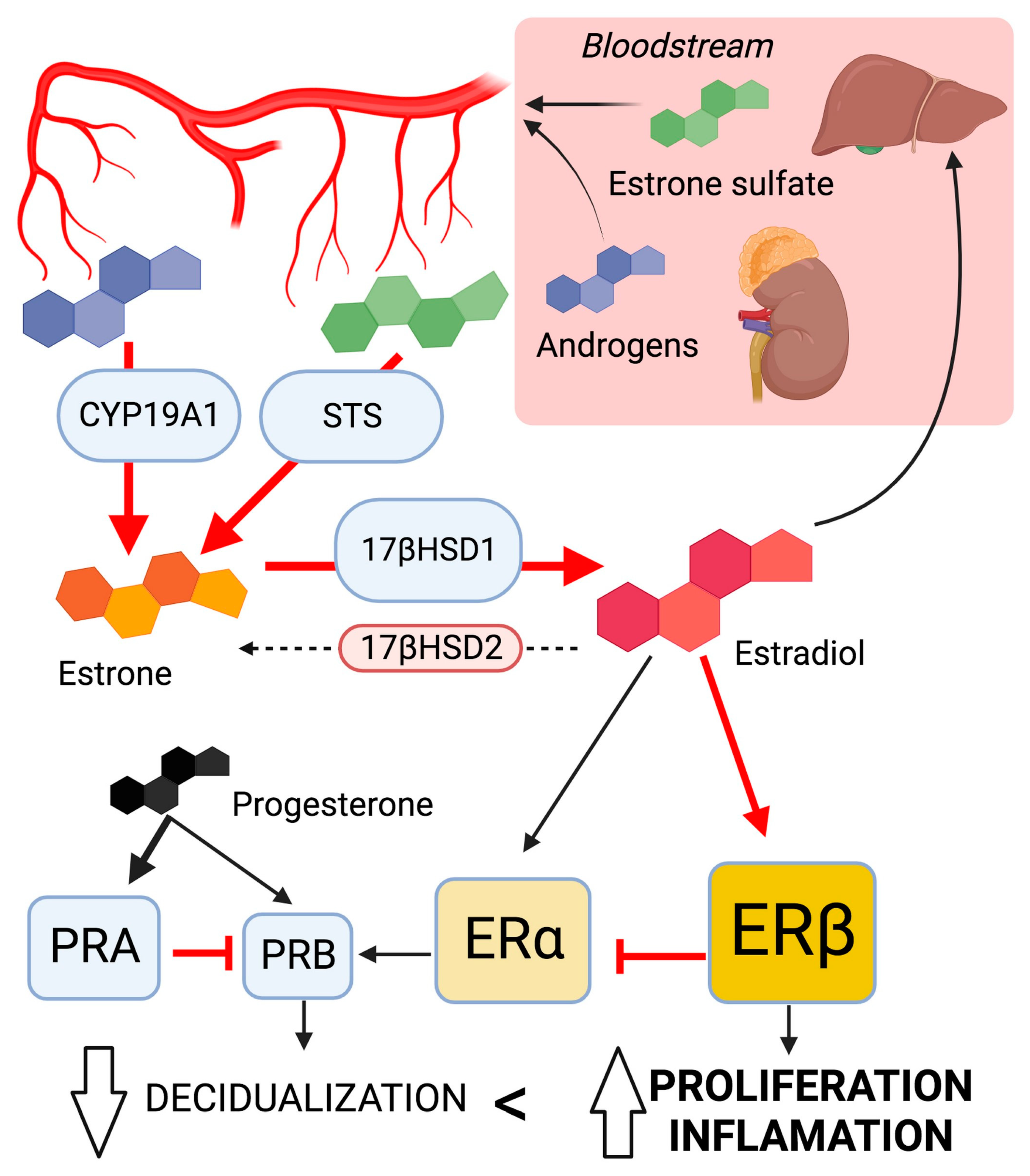
Adenomyosis (ADM) is a multifaceted uterine pathology characterized by the ectopic infiltration of endometrial tissue into the myometrium, affecting approximately 20% of women in the reproductive age group seeking gynecological care. This condition manifests as a range of debilitating symptoms, including dysmenorrhea, menorrhagia, impaired fertility, and heightened susceptibility to miscarriage and obstetric complications. The essential dependence of ADM on estrogen and the impact of endocrine disruptors in its pathogenesis warrant further investigation, and present therapeutic opportunities.

adenomyosis estrogen progesterone endocrine disruptors therapy

1. Intrinsic Hormonal Dysregulation

1.1. The Imbalance of Sex Steroid Hormones

Pronounced estrogen dependency is a unique characteristic of the disease and is central to its development. Indeed, the lesions develop in a hyperestrogenic environment, exhibit distinct patterns of ERs, and manifest signs of localized estrogenic effects, namely an estrogen-responsive uterine contractility. It has been three decades since elevated E2 levels were observed in the menstrual blood of a limited cohort of ADM patients, showing notably higher concentrations compared to patients with endometriosis [1]. Interestingly, increased E2 was limited to the lesions, while circulating levels were unaffected. Consequently, the localized estrogen synthesis in ectopic endometrial cells has been attributed to high estrone (E1) sulfatase levels, which activates circulating sulfated steroids and local activity of aromatase, an enzyme that converts androgens into estrogens [2]. In the context of ADM, sulfatases and sulfotransferases also contribute to the activation of steroids, particularly in the modulation of estrogen levels. Sulfatases, such as estrone sulfatase, play a role in local estrogen synthesis in ectopic endometrial cells by activating circulating sulfated steroids. Additionally, sulfotransferases are involved in the conjugation of sulfate groups onto steroids, affecting their bioavailability and enhancing their activity [3][4]. This explanation was further supported by a specific polymorphism of aromatase cytochrome P450 found in the eutopic endometrium of ADM [5][6]. The role of aromatase in ADM remains a topic of debate, with another group failing to identify a significant contribution of aromatase in endometriosis [7]. Most recently, however, clinical evidence has demonstrated the effectiveness of low-dose aromatase inhibitors in improving symptoms of ADM, such as menorrhagia, hemoglobin levels, and lesion size [8]. Using a recent transgenic murine model, Heinosalo et al. demonstrated that overexpression of the human estrogen biosynthetic enzyme hydroxysteroid, 17-beta-hydroxysteroid dehydrogenase type 1 (17β-HSD1), catalyzing the last step in estrogen activation, leads to the development of an ADM-like phenotype [9]. Conversely, the activity of 17β-HSD2, which deactivates E2 to E1, is downregulated in their eutopic and ectopic endometrium [10][11]. Two components are hypothesized to be responsible for the hyperestrogenic status observed in patients with ADM. An increased local aromatization process and a decreased local estrogen metabolism within both the eutopic and ectopic endometrium are thought to give rise to this occurrence.

Effects of High Estrogen Concentration

The local increase in estrogen concentration with normal peripheral E2 levels may cause hyperperistalsis of the uterus [12]. These specific steroids seem to create a paracrine effect, presumably mediated by the endometrial oxytocin (OT) signaling. This estrogen dominance is considered the “primum movens” in the chain of key events [13].

Hypersensitivity to Estrogen

The higher risk of developing ADM is associated with hypersensitivity to estrogen via specific polymorphism and relatively increased expression of ERα. A decreased expression of progesterone receptor isoform B (PR-B) is also understood to intensify the risk of developing ADM. Concerning the cognate receptors, ERα (NR3A1) and ERβ (NR3A2), membrane ERα and β, and G protein-coupled ER (GPER) were expressed significantly more in ADM than in normal myometrium. These findings are the same as those in endometriosis. The ERα isoform plays dominant roles in uterine development and estrogen sensitivity during the early proliferative phase and differential subtype expression in later phases [14][15]. Accordingly, ERα mediates the E2-induced uterine epithelial cell proliferation of human endometrial cells [16]. Given the central role of this isoform, an in vivo study discovered specific PvuII restriction fragment-length polymorphisms (PP, Pp, pp genotypes) of the ERα gene in ADM patients. The findings suggested a protective characteristic of the P allele, as well as how the local estrogenic effect is more potent with the P allele than with the p allele. Even if it is still unclear how the ERα gene polymorphisms influence its protein function, the authors suggested a possible explanation by a modulation of the ligand estrogen [17]. The ERβ isoform was described as upregulated in adenomyotic lesions and was proposed to be responsible for inflammation in ADM [18]. In regard to the GPER receptor, it enhances contractile responses to OT in the myometrium, seemingly supporting the TIAR theory in ADM [18][19].

Progesterone Resistance

Also proposed is the notion that progesterone resistance may contribute to the hormonal imbalance theory in ADM, as with endometriosis [20]. With this in mind, the proliferative effect driven by hyperestrogenism is not enough when counteracted by progesterone during the secretory phase of the cycle. As a result, hyperproliferation of the endometrium is promoted [21].
The predominance of ERβ over ERα leads to the suppression of PR-B expression and thus the development of progesterone resistance. PR-B and PR-A, two isoforms of the nuclear receptor PR, have dynamic cellular localization, influencing the effect of progesterone. PR-B can promote uterine epithelial cell proliferation but only when not repressed by PR-A. In ADM, PR-B was reportedly suppressed by DNA hypermethylation. Conversely to endometriosis, however, a recent study did not find decreased expression of progesterone membrane receptors in ADM, suggesting molecular differences between ADM and endometriosis [18].

Summary on Sex Steroid Dysregulation

In short, the highly localized concentrations of estrogen combined with altered expression of steroid receptors are central mechanisms leading to ADM (Figure 1). The local conversion of androgens and estrone sulfate into estradiol is catalyzed by increased activity of CYP19A1, STS (steroid sulfatase), and 17βHSD1 enzymes. Additionally, an altered expression pattern of steroid receptors (Erβ >> Erα > PRA > PRB) contributes to ERβ hyperactivation and progesterone resistance. These characteristics lead to reduced decidualization, increased proliferation of endometrial and myometrial smooth muscle cells, and endometrial angiogenesis. Together, these are key elements in the onset and progression of ADM lesions [10].
Figure 1. The dysregulation of sex steroid signaling; involvement in the development of ADM lesions leads to local hyperestrogenism. Changes in key enzyme activity promote estradiol synthesis. The altered pattern of steroid receptor expression enhances estrogen response while suppressing progesterone response. Ellipses represent enzymes; object size correlates with activity. Squares represent sex steroid receptor; object size correlates with expression levels. Arrows indicate the predominant direction in ADM lesions. Created with biorender.com.

1.2. The Pituitary Gland Influence

Prolactin

Several in vivo studies identified the role of prolactin (PRL) in ADM. In 1981, an ectopic anterior pituitary gland transplantation into the uterine lumen was sufficient in inducing ADM in mice. Circulating levels of PRL were consistently higher in pituitary isograft mice than in control mice with submaxillary gland grafts. This emphasizes the significant role of PRL in synergy with ovarian steroid hormones. Indeed, steroid supplements were necessary in pituitary grafts of ovariectomized mice to induce ADM [22]. Similar results were obtained after the administration of PRL or dopamine antagonists, inducing hyperprolactinemia. Moreover, induced ADM mice exhibited a significant upregulation of the messenger ribonucleic acid (mRNA) coding for PRL receptors [22][23]. These early findings were confirmed when observed in spontaneously occurring bovine ADM, where the levels of PRL and its receptors were found to be abnormally high during necropsies. When cells were isolated in vitro, E2 decreased the expression levels of PRL receptors in non-adenomyotic stromal cells and adenomyotic myometrial cells while increasing the secretion of PRL in adenomyotic myometrial cells [24]. In a murine model of ADM exposed to a potent suppressor of pituitary prolactin secretion, the treatment resulted in the absence of adenomyosis in all 39 experimental mice at 12 months of age, while 46.9% of 32 control mice developed the condition. These findings indicate a potential protective role of dopamine agonists within the context of ADM [24][25]. Validating this preclinical data, serum PRL is reported to be higher in patients with ADM than controls. A correlation was even suggested between the rise of PRL levels as an adverse effect of serotonin reuptake inhibiting antidepressants and the development of ADM [26]. The hyperprolactinemic state leads to the invasion of endometrial stromal cells and then glands into the myometrium. This coincides with the overall loosening and disruption of the myometrial layer and the disintegration of individual muscle cells [27]. Moreover, PRL enhances E2 actions in the uterus and stimulates ER expression in the endometrium. This initiates a vicious cycle within the ADM myometrial cells [24]. If not a sufficient explanation for the whole process by itself, the surgical act of grafting mice may cause mechanical disruption, aiding the invasion of endometrial tissue [28]. The intravaginal administration of bromocriptine, a dopamine agonist reducing PRL level, proved significant in decreasing menstrual bleeding and pain [29].

Oxytocin

Through estrogen-mediated contractions in the inner myometrium, OT is suspected to intervene in the microtrauma of the JZ event. It provokes hyperperistalsis of the myometrium via the cognate OT receptor (OTR), resulting in the TIAR effect. OTRs are expressed both in normal endometrium and myometrium, but their expression fluctuates with the cycle phase [30]. However, high non-cyclic expression was observed in a histological biopsy from ADM patients [31]. OTR expression at the JZ is higher in the fundal region of ADM uteri compared to control during the proliferative phase. Moreover, expression of the OTR was lower in the isthmic region than in the fundus region of ADM uteri during the proliferative phase, which is the opposite of control uteri. The dysperistalsis event can be explained by the overexpression of the fundal myometrial OTR in pathological uteri. The opposite expression pattern between fundus and isthmus may even interfere with fertility and the sperm track by disturbing the direction of the JZ contractions [32]. Furthermore, relative overexpression of the OTR in the myometrial cells of ADM patients combined with high-amplitude muscle cell contractility was positively correlated with the severity of dysmenorrhea in patients with ADM. Thus, treatments known to reduce OTR expression, like deacetylase inhibitors and andrographolides, hold potential in treating ADM [33].

Insulin-like Growth Factor 1

Insulin-like growth factor 1 (IGF1) is a multifaceted factor that plays a pivotal role in regulating growth and development. In the uterus, IGF1 is essential for adequate decidualization of the endometrium and directly affects fertility. Significant hormonal crosstalk exists between estrogen and IGF1. E2 enhances IGF1 synthesis and IGF1 potentiates the effects of estrogen [34]. In the endometrium, IGF1 expression and secretion are prominent in stromal cells, while IGF1 receptors are expressed in epithelial cells. In patients with ADM, the expression of IGF1 receptors is significantly elevated, contributing to the aberrant growth of endometrial tissue within the uterine wall [35]. IGF1 may exacerbate inflammation and fibrosis. Furthermore, the interplay between IGF1 and estrogen could further promote lesion formation. These findings warrant greater research to fully elucidate the precise mechanisms by which IGF1 contributes to the development and progression of ADM.

1.3. Genetics and Epigenetics Alteration of Endocrine Signaling

Genetic variants influence enzyme activity and increase the risk of estrogen dependency in ADM. In particular, cytochrome P450 (CYP) and catechol-O-methyltransferase (COMT) gene variants are involved [36]. When comparing ADM to disease-free patients, a recent study found an increased frequency of the C allele in T/C and C/C genotypes of the CYP1A1 gene (CYP1A1 M1 polymorphism), the A allele in C/A and A/A genotypes of the CYP1A2 gene (CYP1A2*1F polymorphism), and the T allele in C/T and C/C genotypes of the CYP19 gene (Arg264Cys polymorphism). The study also found a decreased frequency of the mutant allele and heterozygous and mutant homozygous genotype of the CYP1A2 gene in ADM patients. These results suggest that ADM is triggered by the active hormones and their metabolites’ higher concentration due to enzymes under these polymorphisms’ influence [36].
Another presumption is that the KRAS gene mutation is part of the genetic predisposition to ADM. Among micro-dissected eutopic samples, KRAS mutations were observed in 55.6% of those with ADM, 50% of those with endometriosis, and only 29.1% of the disease-free cohort [37]. KRAS-activating mutations stimulate signaling pathways to enhance cell survival and proliferation and are associated with progesterone resistance in ADM [38]. Recent findings support genetics as a driver in the pathogenesis of ADM through alterations in gene functions, governing not only the steroid function but also the extracellular cell matrix dysregulation, angiogenesis, TIAR mechanism, and inflammatory mediators.
Evidence also reports several types of epigenetic alterations in ADM. In relation to the deoxyribonucleic acid, increased expression of deoxyribonucleic acid methyltransferases DNMT1 and DNMT3B were found in the ectopic endometrium of ADM patients [39]. These enzymes catalyze the transfer of a methyl group to DNA for gene silencing [39][40]. Therefore, they are a candidate for explaining the hypermethylated status of the PR gene observed in ADM and its progesterone resistance [41]. Histone epigenetic modification is a second potential mechanism of the disease. Aberrant expression and localization of class I histone deacetylases (HDACs) was demonstrated in the endometrium, and the immunoreactivity of HDAC1 and HDAC3 was elevated in the adenomyotic endometrium (both ectopic and eutopic) [42]. Hence, the use of valproic acid, a well-known HDAC inhibitor, has been proposed as an effective treatment in refractory disease based on murine observations [43]. In patients with ADM, alterations in epigenetic modifications were detected in RNA molecules. A decrease in RNA methyl regulators and, specifically, lower levels of N6-methyladenosine was observed in the pathological endometrium compared to controls. This contributes to an increase in the expression of various factors, including IGF1 [44]. Finally, the expression of regulatory microRNAs was dysregulated in the eutopic endometrium of ADM patients, including namely members of the miRNA-200 family pivotal in EMT, and Let-7 involved in cell cycle control. The detailed discussion of those examples can be found in the review by Khan et al. and will not be included here [45]. Although there is continuing evidence from several studies that support the involvement of the epigenetic system in ADM, additional research is needed to conclusively pinpoint epigenetic aberrations as a mechanism of disease upgrowth.

2. Extrinsic Factors

Various environmental factors have been associated with ADM development, particularly regarding endocrine disruptors (Figure 2).

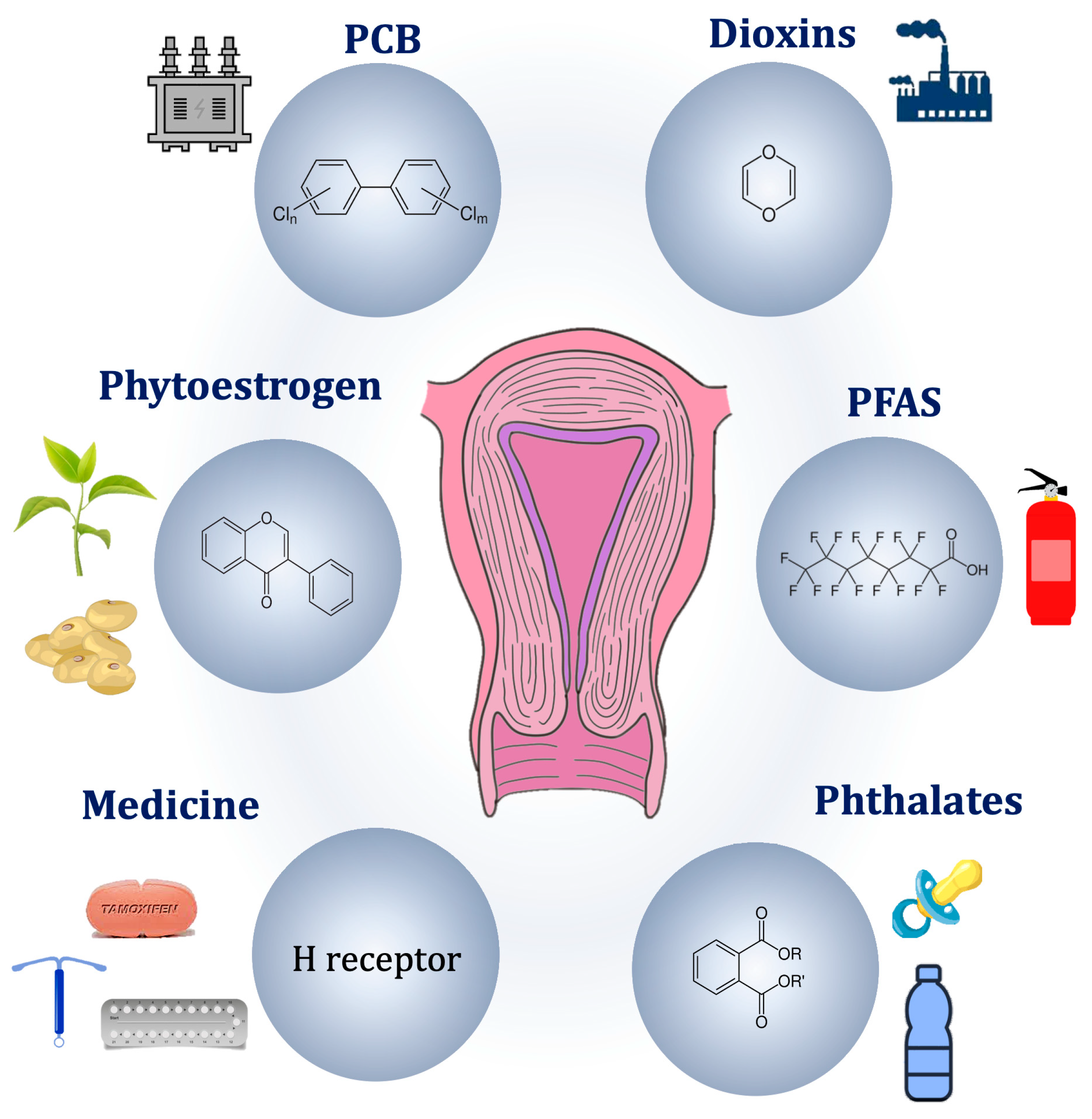
Figure 2. Potential environmental pathogenic causes of adenomyosis.

2.1. Medical Therapies

A possible correlation between the use of hormonal contraceptives and the occurrence of ADM has been considered, but a consensus has not been reached yet. Templeman et al. proposed a positive association between past hormonal contraceptive use and ADM onset, yet ambiguity persists regarding whether subjects primarily employed contraception for birth control or symptom management [46]. It is plausible that the use of the hormonal contraceptive was preferred by the patients already experiencing ADM, rather than the contraceptives being a risk factor for the development of the condition. Conversely, Parazzini et al. could not establish significant association between ADM and a history of hormonal contraceptive use [47][48]. While heightened exposure to exogenous estrogen through hormonal contraceptives may contribute to the development of lesions, such exposure could lead to a reduction in endogenous estrogen production, thereby lowering the risk of ADM [49]. Continued research is necessary to clarify the specific relationship between hormonal contraceptive use and ADM, considering the various reasons for contraceptive use among individuals.
Tamoxifen (TAM) is the prototypical selective ER modulator (SERM), a class of non-steroidal drugs exhibiting agonist and/or antagonist effects given the target tissue. TAM displays anti-estrogenic effects in breast tissue and pro-estrogenic activity in uterine tissue. Due to its demonstrated impact on endometrial tissue, the use of TAM has been identified as a risk factor for ADM [49]. Clinical findings showed that women undergoing treatment for breast cancer with involving TAM are at a higher likelihood of developing ADM than control patients (53% compared to 18%) [50]. Mice studies complemented the human data by revealing an association between TAM exposure and ADM development and progression as mice aged. Interestingly, the study uncovered significant contributions of platelets in the development of TAM-induced lesions, affirming their involvement in the disease and indicating that TAM induces ADM through the TIAR mechanism [51][52].
Boizet-Bonhoure et al. recently demonstrated the effects of chronic exposure to environment-relevant doses of non-steroidal anti-inflammatory drugs (NSAIDs, such as ibuprofen and diclofenac) combined with 17β-ethinylestradiol, in causing ADM development in mice. The research findings revealed impact in both the F1 and F2 generations, implying the occurrence of epigenetic changes [53].

2.2. Endocrine Disrupting Chemicals

Endocrine disrupting chemicals (EDCs) are exogenous compounds that interfere with the endocrine functions, potentially affecting health and promotion of disease [54]. EDCs can be divided into persistent and non-persistent organic pollutants (POPs, nonPOPs), based on their lipid solubility [55]. Both groups are lipophilic, but nonPOPs have a lower lipid solubility, resulting in a short half-life in humans. On the other hand, POPs are not readily biodegradable. Their bioaccumulation in the adipose tissue and slow release in the bloodstream account for the long-term effects [56]. Unfortunately, industrialization has made exposure to EDCs inevitable, either through packaged consumer products or contaminated foods. Since the end of 20th century, warnings have been issued against the harmful effects of EDCs, prompting the ban of polychlorinated biphenyls (PCBs), dichloro-diphenyl-trichloroethane (DDT), and diethylstilbestrol (DES) [57].
Animal studies conclusively demonstrated that EDCs are sufficient to induce ADM or endometriosis. Specifically for ADM, murine studies contributed to the understanding of the role and toxicity of EDCs [58][59][60]. The significant clinical coexistence of both conditions within a patient strongly supports a high probability of a causal relationship between EDC exposure and the initiation of endometrium-related diseases [56]. The probable association of early life exposure to EDCs with ADM offers valuable insights for therapeutic approaches and essentially its prevention [61].

Persistent Organic Pollutants

POPs are a group of synthetic chemicals resistant to environmental degradation, such as PCBs (mainly used as insulant), perfluoroalkyl substances (PFAS, notably in firefighting foams and non-stick cookware), tetrachlorodibenzo-p-dioxin (TCDD, present in Agent Orange), and dichlorodiphenyltrichloroethane (DTT, insecticide). They persist in the environment for decades, or even centuries, and can bioaccumulate in trophic networks. The hydroxylated metabolites of POPs can also have estrogenic activity.
Multiple epidemiological studies investigated the involvement of PCBs in the context of endometriosis [61]. Reflecting potential prenatal exposure, a Chinese study reported a notable correlation between a shorter anogenital distance amongst patients diagnosed with endometriosis or ADM [61]. Moreover, the sum of PCB levels was significantly higher in patients with rectovaginal ADM compared to patients with endometriosis and controls.
The ENDO study found that two types of PFAS were associated with increased incidence of endometriosis diagnosis. No association has been reported for ADM yet [62].
Association studies established a link between TCDD and endometriosis [63][64]. TCDD has the capacity to modulate signaling pathways mediated by the steroid hormones in the normal uterine physiology [65]. Bruner-Tran et al. conducted a retrospective investigation to identify mice with ADM-like lesions resulting from any type of exposure to TCDD over multiple generations [58][66]. Deep adenomyotic lesions were detected in more than half of the mice with a history of either direct (F1-F2) or indirect (F3) exposure [58]. Nonetheless, further studies are needed to assess a potential link between TCDD and ADM.

Non-Persistent Organic Pollutants

NonPOPs, such as glyphosate, phtalates, bisphenol, and pyrethroids, can degrade and break down relatively quickly in the environment, often within days to months. They are typically less likely to bioaccumulate in organisms than POPs and have lower potential for long-range transport through air and water.
Clinical studies address the potential role of gene–environment interactions in the context of ADM. Huang et al. revealed an increased risk for ADM in individuals who carry the glutathione S-transferase M1 polymorphism (GSTM1) and are exposed to high levels of phthalates compared to those unexposed [67]. Although rare cases of EDCs induce genetic mutations, most EDCs are unable to alter DNA sequences. Conversely, an association between epigenetic modifications and EDC exposure has been expressed. Some toxic agents in the environment were also proven to generate epigenetic modifications within the germline, hence causing multi- and transgenerational repercussions [68]. Despite a lack of evidence regarding the exact bond between EDCs and epigenetics, it seems like they act with two mechanisms: gene-specific and global action [69].
Phthalates are EDCs characterized by an anti-androgenic and pro-estrogenic effect [70]. The concentration of phthalates in endometriosis patients’ blood is significantly higher than in control patients [71][72]. Other studies showed an increase in urinary phthalates in patients suffering from endometriosis [67][73]. Similar results were seen in an additional study from this group that identified a modest increase in urinary phthalates in patients with either endometriosis or ADM [74]. In a case–control study, urinary levels of phthalates, particularly MEHP (the primary metabolite of DEHP), were higher and strongly associated with significantly increase in risk for ADM (OR = 10.4; 95% CI, 1.26–85.0) [67].
Bisphenols are estrogen-mimicking molecules that maintain a low concentration of PRs, eventually leading to uterine cyclicity disruption [75]. Using a murine model, Newbold et al. identified a correlation between BPA neonatal exposure and suggested a link between parental EDC exposure and the onset of endometriosis and ADM in the female offspring [76]. In 2010, another research group demonstrated that prenatal exposure to BPA in mice caused development of endometrial glands and stroma within adipose tissue neighboring the reproductive tract, accompanied by the expression of ERs and HOX-A10 [59]. However, the substantiating evidence linking BPA to ADM remains relatively scant in comparison to its association with endometriosis.
Diethylstillbestrol (DES) is a synthetic potent estrogen that was given to mitigate the risk of pregnancy loss but was prohibited in the 1970s following the disclosure of significant morbidities to females and their female offspring [77]. Further studies highlighted a link between in utero exposure to DES and the risk of endometriosis [78]. Although epidemiology studies have not identified a link between DES exposure and ADM in humans, mice studies suggested a positive correlation between DES and ADM [79][80].

2.3. Natural Endocrine Disruptors

Phytoestrogens can function as endocrine disruptors by binding to ERs and either mimicking or blocking the effects of natural estrogen. These actions can result in hormonal imbalances. They can exert ER-independent mechanisms of action, such as altering hormone-binding globulin levels. Furthermore, some phytoestrogens inhibit aromatase and other enzymes involved in the synthesis of steroid hormones. Several studies have suggested that phytoestrogens may be involved in the development or progression of uterine diseases, such as endometrial cancer [81][82].

2.4. Mode of Action of Endocrine Disruption

Most endocrine disruptors act gene-specifically by interfering with NR function, but global action is considered. NRs regulate gene-specific chromatin states by engaging histone modifiers and recruiting DNMTs and thymine DNA glycosylases (TDGs) to specified genomic loci [83]. This is supported by different studies in mice, though still not demonstrated in humans [60][84]. EDCs act globally on DNMTs by downstream regulation of messenger RNA and/or microRNA expression by defective receptors [85][86]. Moreover, studies showed a dysregulation of DNA demethylases and histone-modifying enzymes by EDC exposure [87][88].
Based on current scientific knowledge, endocrine disruptors can have an agonist or antagonist effect on hormone receptors or alter hormone receptor expression, as described with the TCDD and decreased PR expression in mice uteri [89]. EDCs, with the help of EDC co-factors, are also linked to a perturbation of both nuclear steroid receptors and cell surface membrane receptors, causing a dysregulation of downstream signal transduction [90][91]. Furthermore, they can alter the activity of hormone-responsive cells by interfering with hormonal transport [90].

References

  1. Takahashi, K.; Nagata, H.; Kitao, M. Clinical Usefulness of Determination of Estradiol Level in the Menstrual Blood for Patients with Endometriosis. Nihon Sanka Fujinka Gakkai Zasshi 1989, 41, 1849–1850.
  2. Yamamoto, T.; Noguchi, T.; Tamura, T.; Kitawaki, J.; Okada, H. Evidence for Estrogen Synthesis in Adenomyotic Tissues. Am. J. Obstet. Gynecol. 1993, 169, 734–738.
  3. Bulun, S.E.; Lin, Z.; Imir, G.; Amin, S.; Demura, M.; Yilmaz, B.; Martin, R.; Utsunomiya, H.; Thung, S.; Gurates, B.; et al. Regulation of Aromatase Expression in Estrogen-Responsive Breast and Uterine Disease: From Bench to Treatment. Pharmacol. Rev. 2005, 57, 359–383.
  4. Purohit, A.; Reed, M.J. Regulation of Estrogen Synthesis in Postmenopausal Women. Steroids 2002, 67, 979–983.
  5. Kitawaki, J. Adenomyosis: The Pathophysiology of an Oestrogen-Dependent Disease. Best. Pract. Res. Clin. Obstet. Gynaecol. 2006, 20, 493–502.
  6. Kitawaki, J.; Noguchi, T.; Amatsu, T.; Maeda, K.; Tsukamoto, K.; Yamamoto, T.; Fushiki, S.; Osawa, Y.; Honjo, H. Expression of Aromatase Cytochrome P450 Protein and Messenger Ribonucleic Acid in Human Endometriotic and Adenomyotic Tissues but Not in Normal Endometrium. Biol. Reprod. 1997, 57, 514–519.
  7. Colette, S.; Lousse, J.C.; Defrère, S.; Curaba, M.; Heilier, J.F.; Van Langendonckt, A.; Mestdagt, M.; Foidart, J.M.; Loumaye, E.; Donnez, J. Absence of Aromatase Protein and MRNA Expression in Endometriosis. Hum. Reprod. 2009, 24, 2133–2141.
  8. Sharma, S.; Roychoudhury, S.; Padmaja Bhattacharya, M.; Hazra, S.; Majhi, A.K.; Oswal, K.C.; Chattopadhyay, R. Low-Dose Letrozole À an Effective Option for Women with Symptomatic Adenomyosis Awaiting IVF: A Pilot Randomized Controlled Trial. Reprod. BioMed. Online 2023, 47, 84–93.
  9. Heinosalo, T.; Rytkönen, K.T.; Saarinen, N.; Järvensivu, P.; Damdimopoulou, P.; Strauss, L.; Orasniemi, S.; Horshauge, P.; Gabriel, M.; Koskimies, P.; et al. Overexpression of Human Estrogen Biosynthetic Enzyme Hydroxysteroid (17beta) Dehydrogenase Type 1 Induces Adenomyosis-like Phenotype in Transgenic Mice. Int. J. Mol. Sci. 2022, 23, 4815.
  10. Zhai, J.; Vannuccini, S.; Petraglia, F.; Giudice, L.C. Adenomyosis: Mechanisms and Pathogenesis. Semin. Reprod. Med. 2020, 38, 129–143.
  11. Kitawaki, J.O.; Koshiba, H.; Ishihara, H.; Kusuki, I.; Tsukamoto, K.; Honjo, H. Progesterone Induction of 17-Hydroxysteroid Dehydrogenase Type 2 during the Secretory Phase Occurs in the Endometrium of Estrogen-Dependent Benign Diseases But Not in Normal Endometrium. J. Clin. Endocrinol. Metab. 2000, 85, 3292–3296.
  12. Urabe, M.; Yamamoto, T.; Kitawaki, J.; Honjo, H.; Okada, H. Estrogen Biosynthesis in Human Uterine Adenomyosis. Acta Endocrinol. 1989, 121, 259–264.
  13. Vannuccini, S.; Tosti, C.; Carmona, F.; Huang, S.J.; Chapron, C.; Guo, S.W.; Petraglia, F. Pathogenesis of Adenomyosis: An Update on Molecular Mechanisms. Reprod. BioMed. Online 2017, 35, 592–601.
  14. Hewitt, S.C.; Winuthayanon, W.; Korach, K.S. What’s New in Estrogen Receptor Action in the Female Reproductive Tract. J. Mol. Endocrinol. 2016, 56, 55–71.
  15. Oehler, M.K.; Greschik, H.; Fischer, D.C.; Tong, X.; Schuele, R.; Kieback, D.G. Functional Characterization of Somatic Point Mutations of the Human Estrogen Receptor α (HERα) in Adenomyosis Uteri. Mol. Hum. Reprod. 2004, 10, 853–860.
  16. Yu, K.; Huang, Z.Y.; Xu, X.L.; Li, J.; Fu, X.W.; Deng, S.L. Estrogen Receptor Function: Impact on the Human Endometrium. Front. Endocrinol. 2022, 13, 827724.
  17. Kitawaki, J.; Obayashi, H.; Ishihara, H.; Koshiba, H.; Kusuki, I.; Kado, N.; Tsukamoto, K.; Hasegawa, G.; Nakamura, N.; Honjo, H. Oestrogen Receptor-Alpha Gene Polymorphism Is Associated with Endometriosis, Adenomyosis and Leiomyomata. Hum. Reprod. 2001, 16, 51–55.
  18. Sztachelska, M.; Ponikwicka-Tyszko, D.; Martínez-Rodrigo, L.; Bernaczyk, P.; Palak, E.; Półchłopek, W.; Bielawski, T.; Wołczyński, S. Functional Implications of Estrogen and Progesterone Receptors Expression in Adenomyosis, Potential Targets for Endocrinological Therapy. J. Clin. Med. 2022, 11, 4407.
  19. Hong, D.G.; Park, J.Y.; Chong, G.O.; Lee, Y.H.; Lee, H.J.; Shinn, J.U.; Lee, Y.S.; Seong, W.J. Transmembrane G Protein-Coupled Receptor 30 Gene Polymorphisms and Uterine Adenomyosis in Korean Women. Gynecol. Endocrinol. 2019, 35, 498–501.
  20. Nisolle, M.; Donnez, J. Peritoneal Endometriosis, Ovarian Endometriosis, and Adenomyotic Nodules of the Rectovaginal Septum Are Three Different Entities. Fertil. Steril. 1997, 68, 585–596.
  21. Mehasseb, M.K.; Panchal, R.; Taylor, A.H.; Brown, L.; Bell, S.C.; Habiba, M. Estrogen and Progesterone Receptor Isoform Distribution through the Menstrual Cycle in Uteri with and without Adenomyosis. Fertil. Steril. 2011, 95, 2228–2235.e1.
  22. Mori, T.; Nagasawa, H.; Takahashi, S. The induction of adenomyosis in mice by intrauterine pituitary isografts. Life Sci. 1981, 29, 1277–1282.
  23. Singtripop, T.; Mori, T.; Park, M.K.; Sakamoto, S.; Kawashima, S. Development of Uterine Adenomyosis after Treatment with Dopamine Antagonists in Mice. Life Sci. 1991, 49, 201–206.
  24. Łupicka, M.; Socha, B.M.; Szczepańska, A.A.; Korzekwa, A.J. Prolactin Role in the Bovine Uterus during Adenomyosis. Domest. Anim. Endocrinol. 2017, 58, 1–13.
  25. Nagasawa, H.; Mori, T. Stimulation of mammary tumorigenesis and suppression of uterine adenomyosis by temporary inhibition of pituitary prolactin secretion during youth in mice (41492). Proc. Soc. Exp. Biol. Med. 1982, 171, 164–167.
  26. Sengupta, P.; Sharma, A.; Mazumdar, G.; Banerjee, I.; Tripathi, S.K.; Bagchi, C.; Das, N. The Possible Role of Fluoxetine in Adenomyosis: An Animal Experiment with Clinical Correlations. J. Clin. Diagn. Res. 2013, 7, 1530–1534.
  27. Mori, T.; Ohta, Y.; Nagasawa, H. Ultrastructural changes in uterine myometrium of mice with experimentally-induced adenomyosis. Experientia 1984, 40, 1385–1387.
  28. Marquardt, R.M.; Jeong, J.W.; Fazleabas, A.T. Animal Models of Adenomyosis. Semin. Reprod. Med. 2020, 38, 168–178.
  29. Andersson, J.K.; Khan, Z.; Weaver, A.L.; Vaughan, L.E.; Gemzell-Danielsson, K.; Stewart, E.A. Vaginal Bromocriptine Improves Pain, Menstrual Bleeding and Quality of Life in Women with Adenomyosis: A Pilot Study. Acta Obstet. Gynecol. Scand. 2019, 98, 1341–1350.
  30. Takemura, M.; Nomura, S.; Kimura, T.; Inoue, T.; Onoue, H.; Azuma, C.; Saji, F.; Kitamura, Y.; Tanizawa, O. Expression and localization of oxytocin receptor gene in human uterine endometrium in relation to the menstrual cycle. Endocrinology 1993, 132, 1830–1835.
  31. Mechsner, S.; Grum, B.; Gericke, C.; Loddenkemper, C.; Dudenhausen, J.W.; Ebert, A.D. Possible Roles of Oxytocin Receptor and Vasopressin-1α Receptor in the Pathomechanism of Dysperistalsis and Dysmenorrhea in Patients with Adenomyosis Uteri. Fertil. Steril. 2010, 94, 2541–2546.
  32. Zhang, Y.; Yu, P.; Sun, F.; Li, T.C.; Cheng, J.M.; Duan, H. Expression of Oxytocin Receptors in the Uterine Junctional Zone in Women with Adenomyosis. Acta Obstet. Gynecol. Scand. 2015, 94, 412–418.
  33. Guo, S.W.; Mao, X.; Ma, Q.; Liu, X. Dysmenorrhea and Its Severity Are Associated with Increased Uterine Contractility and Overexpression of Oxytocin Receptor (OTR) in Women with Symptomatic Adenomyosis. Fertil. Steril. 2013, 99, 231–240.
  34. Sekulovski, N.; Whorton, A.E.; Shi, M.; Hayashi, K.; MacLean, J.A. Insulin Signaling Is an Essential Regulator of Endometrial Proliferation and Implantation in Mice. FASEB J. 2021, 35, e21440.
  35. Konopka, B.; Skasko, E.; Kluska, A.; Goluda, M.; Janiec-Jankowska, A.; Paszko, Z.; Ujec, M. Changes in the concentrations of receptors of insulin-like growth factor-I, epithelial growth factor, oestrogens and progestagens in adenomyosis foci, endometrium and myometrium of women during menstrual cycle. Eur. J. Gynaecol. Oncol. 1998, 19, 93–97.
  36. Artymuk, N.; Zotova, O.; Gulyaeva, L. Adenomyosis: Genetics of Estrogen Metabolism. Horm. Mol. Biol. Clin. Investig. 2019, 37, 20180069.
  37. Inoue, S.; Hirota, Y.; Ueno, T.; Fukui, Y.; Yoshida, E.; Hayashi, T.; Kojima, S.; Takeyama, R.; Hashimoto, T.; Kiyono, T. Uterine Adenomyosis Is an Oligoclonal Disorder Associated with KRAS Mutations. Nat. Commun. 2019, 10, 5785.
  38. Bulun, S.E.; Yildiz, S.; Adli, M.; Wei, J.J. Adenomyosis Pathogenesis: Insights from next-Generation Sequencing. Hum. Reprod. Update 2021, 27, 1086–1097.
  39. Liu, X.; Guo, S.W. Aberrant Immunoreactivity of Deoxyribonucleic Acid Methyltransferases in Adenomyosis. Gynecol. Obstet. Investig. 2012, 74, 100–108.
  40. Cedar, H.; Bergman, Y. Linking DNA Methylation and Histone Modification: Patterns and Paradigms. Nat. Rev. Genet. 2009, 10, 295–304.
  41. Nie, J.; Liu, X.; Guo, S.W. Promoter Hypermethylation of Progesterone Receptor Isoform B (PR-B) in Adenomyosis and Its Rectification by a Histone Deacetylase Inhibitor and a Demethylation Agent. Reprod. Sci. 2010, 17, 995–1005.
  42. Liu, X.; Nie, J.; Guo, S.W. Elevated Immunoreactivity against Class i Histone Deacetylases in Adenomyosis. Gynecol. Obstet. Invest. 2012, 74, 50–55.
  43. Liu, X.; Guo, S.W. Valproic Acid Alleviates Generalized Hyperalgesia in Mice with Induced Adenomyosis. J. Obstet. Gynaecol. Res. 2011, 37, 696–708.
  44. Zhai, J.; Li, S.; Sen, S.; Opoku-Anane, J.; Du, Y.; Chen, Z.J.; Giudice, L.C. M6A RNA Methylation Regulators Contribute to Eutopic Endometrium and Myometrium Dysfunction in Adenomyosis. Front. Genet. 2020, 11, 716.
  45. Khan, K.N.; Fujishita, A.; Mori, T. Pathogenesis of Human Adenomyosis: Current Understanding and Its Association with Infertility. J. Clin. Med. 2022, 11, 4057.
  46. Templeman, C.; Marshall, S.F.; Ursin, G.; Horn-Ross, P.L.; Clarke, C.A.; Allen, M.; Deapen, D.; Ziogas, A.; Reynolds, P.; Cress, R.; et al. Adenomyosis and Endometriosis in the California Teachers Study. Fertil. Steril. 2008, 90, 415–424.
  47. Parazzini, F.; Vercellini, P.; Panazza, S.; Chatenoud, L.; Oldani, S. Crosignani PG. Risk factors for adenomyosis. Hum. Reprod. 1997, 12, 1275–1279.
  48. Parazzini, F.; Mais, V.; Cipriani, S.; Busacca, M.; Venturini, P. Determinants of Adenomyosis in Women Who Underwent Hysterectomy for Benign Gynecological Conditions: Results from a Prospective Multicentric Study in Italy. Eur. J. Obstet. Gynecol. Reprod. Biol. 2009, 143, 103–106.
  49. Upson, K.; Missmer, S.A. Epidemiology of Adenomyosis. Semin. Reprod. Med. 2020, 38, 89–107.
  50. Cohen, I.; Beyth, Y.; Shapira, J.; Tepper, R.; Fishman, A.; Cordoba, M.; Bernheim, J.; Yigael, D.; Altaras, M.M. High frequency of adenomyosis in postmenopausal breast cancer patients treated with tamoxifen. Gynecol. Obstet. Investig. 1997, 44, 200–205.
  51. Jin, Z.; Wu, X.; Liu, H.; Xu, C. Celecoxib, a Selective COX2 Inhibitor, Markedly Reduced the Severity of Tamoxifeninduced Adenomyosis in a Murine Model. Exp. Ther. Med. 2020, 19, 3289–3299.
  52. Shen, M.; Liu, X.; Zhang, H.; Guo, S.W. Transforming Growth Factor Β1 Signaling Coincides with Epithelial-Mesenchymal Transition and Fibroblast-to-Myofibroblast Transdifferentiation in the Development of Adenomyosis in Mice. Hum. Reprod. 2016, 31, 355–369.
  53. Brigitte Boizet-Bonhoure; Stéphanie Déjardin; Mélissa Girard; Quentin Durix; Francis Poulat; Pascal Philibert; Adenomyotic Lesions Are Induced in the Mouse Uterus after Exposure to NSAID and EE2 Mixtures at Environmental Doses. Int. J. Mol. Sci. 2024, 25, 2003.
  54. Rumph, J.T.; Stephens, V.R.; Archibong, A.E.; Osteen, K.G.; Bruner-Tran, K.L. Environmental Endocrine Disruptors and Endometriosis. In Advances in Anatomy Embryology and Cell Biology; Springer Science and Business Media Deutschland GmbH: Berlin/Heidelberg, Germany, 2020; Volume 232, pp. 57–78.
  55. Cho, Y.J.; Yun, J.H.; Kim, S.J.; Kwon, H.Y. Nonpersistent Endocrine Disrupting Chemicals and Reproductive Health of Women. Obstet. Gynecol. Sci. 2020, 63, 1–12.
  56. Stephens, V.R.; Rumph, J.T.; Ameli, S.; Bruner-Tran, K.L.; Osteen, K.G. The Potential Relationship Between Environmental Endocrine Disruptor Exposure and the Development of Endometriosis and Adenomyosis. Front. Physiol. 2022, 28, 807685.
  57. Schug, T.T.; Johnson, A.F.; Birnbaum, L.S.; Colborn, T.; Guillette, L.J.; Crews, D.P.; Collins, T.; Soto, A.M.; Vom Saal, F.S.; McLachlan, J.A.; et al. Minireview: Endocrine Disruptors: Past Lessons and Future Directions. Mol. Endocrinol. 2016, 30, 833–847.
  58. Bruner-Tran, K.L.; Duleba, A.J.; Taylor, H.S.; Osteen, K.G. Developmental Toxicant Exposure Is Associated with Transgenerational Adenomyosis in a Murine Model. Biol. Reprod. 2016, 95, 73.
  59. Signorile, P.G.; Spugnini, E.P.; Mita, L.; Mellone, P.; D’Avino, A.; Bianco, M.; Diano, N.; Caputo, L.; Rea, F.; Viceconte, R.; et al. Pre-Natal Exposure of Mice to Bisphenol A Elicits an Endometriosis-like Phenotype in Female Offspring. Gen. Comp. Endocrinol. 2010, 168, 318–325.
  60. Jefferson, W.N.; Karimi Kinyamu, H.; Wang, T.; Miranda, A.X.; Padilla-Banks, E.; Suen, A.A.; Williams, C.J. Widespread Enhancer Activation via ERα Mediates Estrogen Response in Vivo during Uterine Development. Nucleic Acids Res. 2018, 46, 5487–5503.
  61. Liu, X.; Ding, D.; Shen, M.; Yan, D.; Guo, S.W. Shorter Anogenital Distance in Women with Ovarian Endometriomas and Adenomyosis, but Not Uterine Leiomyomas. Biomedicines 2023, 11, 2618.
  62. Louis, G.M.; Peterson, C.M.; Chen, Z.; Hediger, M.L.; Croughan, M.S.; Sundaram, R.; Stanford, J.B.; Fujimoto, V.Y.; Varner, M.W.; Giudice, L.C.; et al. Perfluorochemicals and endometriosis: The ENDO study. Epidemiology 2012, 23, 799–805.
  63. Matta, K.; Lefebvre, T.; Vigneau, E.; Cariou, V.; Marchand, P.; Guitton, Y.; Royer, A.L.; Ploteau, S.; Le Bizec, B.; Antignac, J.P.; et al. Associations between Persistent Organic Pollutants and Endometriosis: A Multiblock Approach Integrating Metabolic and Cytokine Profiling. Environ. Int. 2022, 158, 106926.
  64. Rier, S.E.; Martin, D.C.; Bowman, R.E.; Dmowski, W.P.; Becker, J.L. Endometriosis in rhesus monkeys (Macaca mulatta) following chronic exposure to 2,3,7,8-tetrachlorodibenzo-p-dioxin. Fundam. Appl. Toxicol. 1993, 21, 433–441.
  65. Szántó, M.; Gupte, R.; Kraus, W.L.; Pacher, P.; Bai, P. PARPs in lipid metabolism and related diseases. Prog. Lipid Res. 2021, 84, 101117.
  66. Bruner-Tran, K.L.; Gnecco, J.; Ding, T.; Glore, D.R.; Pensabene, V.; Osteen, K.G. Exposure to the Environmental Endocrine Disruptor TCDD and Human Reproductive Dysfunction: Translating Lessons from Murine Models. Reprod. Toxicol. 2017, 68, 59–71.
  67. Huang, P.C.; Tsai, E.M.; Li, W.F.; Liao, P.C.; Chung, M.C.; Wang, Y.H.; Wang, S.L. Association between Phthalate Exposure and Glutathione S-Transferase M1 Polymorphism in Adenomyosis. Hum. Reprod. 2010, 25, 986–994.
  68. Skinner, M.K.; Manikkam, M.; Guerrero-Bosagna, C. Epigenetic Transgenerational Actions of Environmental Factors in Disease Etiology. Trends Endocrinol. Metab. 2010, 21, 214–222.
  69. Alavian-Ghavanini, A.; Rüegg, J. Understanding Epigenetic Effects of Endocrine Disrupting Chemicals: From Mechanisms to Novel Test Methods. Basic. Clin. Pharmacol. Toxicol. 2018, 122, 38–45.
  70. Czernych, R.; Chraniuk, M.; Zagożdżon, P.; Wolska, L. Characterization of Estrogenic and Androgenic Activity of Phthalates by the XenoScreen YES/YAS in Vitro Assay. Environ. Toxicol. Pharmacol. 2017, 53, 95–104.
  71. Reddy, B.S.; Rozati, R.; Reddy, S.; Kodampur, S.; Reddy, P.; Reddy, R. High Plasma Concentrations of Polychlorinated Biphenyls and Phthalate Esters in Women with Endometriosis: A Prospective Case Control Study. Fertil. Steril. 2006, 85, 775–779.
  72. Reddy, B.S.; Rozati, R.; Reddy, B.V.R.; Raman, N.V.V.S.S. Association of Phthalate Esters with Endometriosis in Indian Women. BJOG 2006, 113, 515–520.
  73. Upson, K.; Sathyanarayana, S.; De Roos, A.J.; Thompson, M.L.; Scholes, D.; Dills, R.; Holt, V.L. Phthalates and Risk of Endometriosis. Environ. Res. 2013, 126, 91–97.
  74. Huang, P.C.; Li, W.F.; Liao, P.C.; Sun, C.W.; Tsai, E.M.; Wang, S.L. Risk for Estrogen-Dependent Diseases in Relation to Phthalate Exposure and Polymorphisms of CYP17A1 and Estrogen Receptor Genes. Environ. Sci. Pollut. Res. 2014, 21, 13964–13973.
  75. Aldad, T.S.; Rahmani, N.; Leranth, C.; Taylor, H.S. Bisphenol-A Exposure Alters Endometrial Progesterone Receptor Expression in the Nonhuman Primate. Fertil. Steril. 2011, 96, 175–179.
  76. Newbold, R.R.; Jefferson, W.N.; Padilla-Banks, E. Long-Term Adverse Effects of Neonatal Exposure to Bisphenol A on the Murine Female Reproductive Tract. Reprod. Toxicol. 2007, 24, 253–258.
  77. Giusti, R.M.; Iwamoto, K.; Hatch, E.E. Diethylstilbestrol revisited: A review of the long-term health effects. Ann. Intern. Med. 1995, 122, 778–788.
  78. Upson, K.; Sathyanarayana, S.; Scholes, D.; Holt, V.L. Early-Life Factors and Endometriosis Risk. Fertil. Steril. 2015, 104, 964–971.e5.
  79. Huseby, R.A.; Thurlow, S. Effects of Prenatal Exposure of Mice to “Low-Dose” Diethylstilbestrol and the Development of Adenomyosis Associated with Evidence of Hyperprolactinemia. Am. J. Obstet. Gynecol. 1982, 144, 939–949.
  80. McLachlan, J.A.; Newbold, R.R.; Bullock, B.C. Long-term effects on the female mouse genital tract associated with prenatal exposure to diethylstilbestrol. Cancer Res. 1980, 40, 3988–3999.
  81. Domínguez-López, I.; Yago-Aragón, M.; Salas-Huetos, A.; Tresserra-Rimbau, A.; Hurtado-Barroso, S. Effects of Dietary Phytoestrogens on Hormones throughout a Human Lifespan: A Review. Nutrients 2020, 12, 2456.
  82. Youseflu, S.; Sadatmahalleh, S.J.; Mottaghi, A.; Kazemnejad, A. Dietary Phytoestrogen Intake and the Risk of Endometriosis in Iranian Women: A Case-Control Study. Int. J. Fertil. Steril. 2020, 13, 296–300.
  83. Kouzmenko, A.; Ohtake, F.; Fujiki, R.; Kato, S. Hormonal Gene Regulation through DNA Methylation and Demethylation. Epigenomics 2010, 2, 765–774.
  84. Bruner-Tran, K.L.; Resuehr, D.; Ding, T.; Lucas, J.A.; Osteen, K.G. The Role of Endocrine Disruptors in the Epigenetics of Reproductive Disease and Dysfunction: Potential Relevance to Humans. Curr. Obstet. Gynecol. Rep. 2012, 1, 116–123.
  85. Derghal, A.; Djelloul, M.; Trouslard, J.; Mounien, L. An Emerging Role of Micro-RNA in the Effect of the Endocrine Disruptors. Front. Neurosci. 2016, 10, 318.
  86. Liu, J.; Zhang, L.; Winterroth, L.C.; Garcia, M.; Weiman, S.; Wong, J.W.; Sunwoo, J.B.; Nadeau, K.C. Epigenetically Mediated Pathogenic Effects of Phenanthrene on Regulatory T Cells. J. Toxicol. 2013, 2013, 967029.
  87. Liu, Y.; Zhang, Y.; Tao, S.; Guan, Y.; Zhang, T.; Wang, Z. Global DNA Methylation in Gonads of Adult Zebrafish Danio Rerio under Bisphenol A Exposure. Ecotoxicol. Environ. Saf. 2016, 130, 124–132.
  88. Jefferson, W.N.; Chevalier, D.M.; Phelps, J.Y.; Cantor, A.M.; Padilla-Banks, E.; Newbold, R.R.; Archer, T.K.; Karimi Kinyamu, H.; Williams, C.J. Persistently Altered Epigenetic Marks in the Mouse Uterus after Neonatal Estrogen Exposure. Mol. Endocrinol. 2013, 27, 1666–1677.
  89. Nayyar, T.; Bruner-Tran, K.L.; Piestrzeniewicz-Ulanska, D.; Osteen, K.G. Developmental Exposure of Mice to TCDD Elicits a Similar Uterine Phenotype in Adult Animals as Observed in Women with Endometriosis. Reprod. Toxicol. 2007, 23, 326–336.
  90. La Merrill, M.A.; Vandenberg, L.N.; Smith, M.T.; Goodson, W.; Browne, P.; Patisaul, H.B.; Guyton, K.Z.; Kortenkamp, A.; Cogliano, V.J.; Woodruff, T.J.; et al. Consensus on the Key Characteristics of Endocrine-Disrupting Chemicals as a Basis for Hazard Identification. Nat. Rev. Endocrinol. 2020, 16, 45–57.
  91. Routledge, E.J.; White, R.; Parker, M.G.; Sumpter, J.P. Differential Effects of Xenoestrogens on Coactivator Recruitment by Estrogen Receptor (ER) α and ERβ. J. Biol. Chem. 2000, 275, 35986–35993.
More
Information
Contributors MDPI registered users' name will be linked to their SciProfiles pages. To register with us, please refer to https://encyclopedia.pub/register : , , , , , ,
View Times: 158
Revisions: 3 times (View History)
Update Date: 08 Feb 2024
1000/1000