Submitted Successfully!
To reward your contribution, here is a gift for you: A free trial for our video production service.
Thank you for your contribution! You can also upload a video entry or images related to this topic.
Version Summary Created by Modification Content Size Created at Operation
1 -- 4383 2024-01-25 23:45:41 |
2 format correct -3 word(s) 4380 2024-01-26 10:27:53 |

Video Upload Options

Do you have a full video?

Confirm

Are you sure to Delete?
Cite
If you have any further questions, please contact Encyclopedia Editorial Office.
Waseem, M.; Wang, B. Organoids in Prostate Cancer. Encyclopedia. Available online: https://encyclopedia.pub/entry/54369 (accessed on 29 April 2024).
Waseem M, Wang B. Organoids in Prostate Cancer. Encyclopedia. Available at: https://encyclopedia.pub/entry/54369. Accessed April 29, 2024.
Waseem, Mohammad, Bi-Dar Wang. "Organoids in Prostate Cancer" Encyclopedia, https://encyclopedia.pub/entry/54369 (accessed April 29, 2024).
Waseem, M., & Wang, B. (2024, January 25). Organoids in Prostate Cancer. In Encyclopedia. https://encyclopedia.pub/entry/54369
Waseem, Mohammad and Bi-Dar Wang. "Organoids in Prostate Cancer." Encyclopedia. Web. 25 January, 2024.
Organoids in Prostate Cancer
Edit

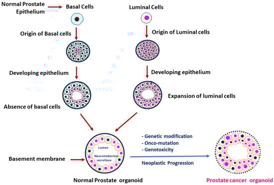
Prostate cancer (PCa) has been known as the most prevalent cancer disease and the second leading cause of cancer mortality in men almost all over the globe. There is an urgent need for establishment of PCa models that can recapitulate the progress of genomic landscapes and molecular alterations during development and progression of this disease. Notably, several organoid models have been developed for assessing the complex interaction between PCa and its surrounding microenvironment. PCa organoids have been emerged as powerful in vitro 3D model systems that recapitulate the molecular features (such as genomic/epigenomic changes and tumor microenvironment) of PCa metastatic tumors. In addition, application of organoid technology in mechanistic studies (i.e., for understanding cellular/subcellular and molecular alterations) and translational medicine has been recognized as a promising approach for facilitating the development of potential biomarkers and novel therapeutic strategies.

prostate cancer organoids 3D model precision medicine in vitro and in vivo models castration resistant prostate cancer (CRPC) neuroendocrine prostate cancer (NEPC)

1. Background and Recent Stage of Prostate Organoid Culture

The earlier development of prostate 3D culture technology was achieved by establishing mouse-derived spheroid cultures. Such methodology was performed by culturing whole or fractionated mouse prostate epithelium in commercially available serum-free medium coupled with a 3D extracellular matrix (ECM), finally facilitating the development of spheroids with self-renewing and self-organizing differentiation potentials [1][2]. The usage of Matrigel in the development of organoid culture has been pinpointed as a key component for early success for developing organoids. These early strategies allowed scientists to develop 3D prostate cultures by continuing the propagation of luminal cells followed by the addition of androgens such as dihydrotestosterone (DHT) to the medium. This approach resulted in a limited luminal differentiation to intermediate phenotypes with AR expression; however, the secretory phenotype of prostate luminal epithelium was still missing in the established spheroid models [3]. Later, the 3D normal prostate organoids consisting of both basal and luminal cells were developed [2][4], and 3D PCa organoids were developed from metastatic PCa biopsies and circulating PCa cells [2]. These 3D models (Figure 1) of normal prostate organoids and PCa organoids (developed from PCa tissues/circulating cells, or through neoplastic transformation of normal prostate organoids) can be utilized to address the key molecular mechanisms underlying PCa development and/or progression.
Figure 1. The majority of PCas are defined by abnormal luminal cell development and lack of basal cells when compared to prostate tissues with normal basal and luminal cells. During transformation to neoplasm, impairment of prostatic epithelium was characterized. The normal prostatic cluster of cells (normal prostate organoid, Left) consists of an epithelial compartment comprising of basal (sky blue) and luminal (light orange) cells, and a limited population of neuroendocrine cells that act as stem cells in response to repairing the cellular damage. During neoplastic progression, a normal prostate organoid is transformed into a PCa organoid through multiple genetic alterations and onco-mutations. A PCa organoid (Right) is characterized by luminal hyperproliferation, breakdown of basement membrane, loss of basal cells, prominent nucleoli (blue/pink), and nuclear enlargement.
In recent years, Julio and her colleagues developed a novel strategy for generation of PCa organoids to evaluate therapeutic responses [5]. This group optimized the protocol for efficiently generating patient-derived organoids (PDOs), and detailed characterization of PCa organoids was provided. Furthermore, in collaboration with the NEXUS Personalized Health Technologies, Julio et al. further developed a PDO for medium-throughput screening for drug efficacies. In this study, the authors demonstrated that PDOs maintain the molecular and cellular features of the original prostate carcinoma that the PDOs were derived from. For instance, the PDOs share comparable genomic landscapes and gene expression profiles when compared to parental prostate carcinoma [5].
In 2009, Sato et al. [6] defined the organoid culture medium by including stem cell niche components, including Wnt pathway agonist R-spondin-1, epidermal growth factor (EGF), and the bone morphogenetic protein (BMP) antagonist Noggin. This formulation efficiently improved the long-term self-renewal and differentiation potential of mouse intestinal crypt stem cells into a 3D ECM. Using basal medium containing nicotinamide, prostaglandin E2 (PGE2), and components that inhibit TGF-β and MAPK signaling, Jung et al. successfully established human intestinal organoids. These intestinal organoids were developed from cocultures containing intact intestinal crypts and isolated stem cells derived from normal or tumor tissues [7]. Employing various stem cell-promoting culture conditions, several epithelial tumor organoids were established. For example, after optimization of tissue-specific factors (such as adding estrogen or testosterone), subtypes of breast cancer organoids or PCa organoids were established [8].
Earlier studies conducted by Clevers and colleagues demonstrated a PCa organoid model that was developed through continuous growth and differentiation of prostate luminal epithelial stem cells. This organoid model has been used to study the role of R-spondin, Noggin, and testosterone [9][10]. Prostate-specific modification of intestinal medium and other changes in 3D culture conditions [11] have led to a robust procedure for developing mouse-derived prostate organoids [12].
Gao and colleagues reported the development of PCa organoid culture from metastatic CRPC patient tissues [8]. However, various success rates of developing sustainable CRPC organoids from patient tumor needle biopsies have been reported. An average of ≤10% success rates of developing CRPC organoids are observed, which is significantly lower than the rates for many other epithelial tumor organoids (such as pancreatic, colorectal, and breast cancer organoids). These results reflect the complexity of tumor heterogeneity of CRPC and also suggest a need to further optimize the culture conditions for establishing fidelity CRPC organoid models [9].
In recent years, an increasing number of new PCa organoid models have been established from metastatic PCa tissue biopsies. These new models are highlighted to represent the most important PCa subtypes, such as AR-driven (AR+) adenocarcinoma, AR-independent neuroendocrine positive (AR NE+), or double-negative (AR NE) PCa [8][13][14][15]. In contrast, an organoid model derived from castration-sensitive PCa has also been established [5]. Furthermore, modified culture conditions have been applied to develop PCa organoids from patient-derived xenograft (PDX), aiming to establish novel PCa organoid models mimicking the PCa heterogeneity [13].

2. Tumor Model System in PCa

PCa represents the most common cancer type in males, and it has gained an increasing attention all over the world. In male population, it has also been ranked among the highest level of cancer-associated mortality [16]. Several studies have utilized organoids as ex vivo models to evaluate tumor microenvironment (TME) and translational medicine in tumor biology [2][11][14][17][18][19][20][21][22][23][24]. Heninger et al. established patient-derived organoids (PDOs) from locally advanced PCa [25]. According to their orthogonal analyses, these organoids reserved and maintained the complexity of TME as observed in parental PCa. According to orthogonal flow cytometry analysis, PCa organoids retained a distinct subpopulation of epithelial cells and reserved the expression signature of AR and AR-related genes as parental PCa cells [25]. Using mouse prostate organoids, Grbesa et al. revealed that mutations in SPOP (the most frequently mutated tumor suppressor gene in human primary PCa) contribute to AR accessibility and binding patterns that are similar to those in primary PCa [26].
Gao and his group developed an optimized condition wherein the continuous propagation of normal basal and luminal prostate epithelial cells was considered in the development of PCa organoid models. In this study, PCa organoids were derived from different PCa sources, including cell lines (DU-145, PC-3, LAPC4, 22Rv1, LNCaP, and VCaP), PCa tissues, iPSCs, CDXs, PDXs and circulating tumor cells. These PCa organoids have been reported to recapitulate the different molecular characterization of PCa subpopulations from different sources. Notably, the PCa organoids derived from cell lines exhibited similar molecular signatures to both primary and metastatic PCa. For instance, mutations in SPOP, loss/deletion of PTEN, TMPRSS2-ERG expression, or mutations in TP53, PIK3R1 and/or FOXO1 (frequently occurred in CRPCs) were retained in these PCa organoid models. In addition, these organoid models represent a series of diverse subgroups of CRPCs, including AR-dependent adenocarcinoma, AR-negative adenocarcinoma, and neuroendocrine carcinoma [8].
In addition, Clevers et al. developed a mini-gut-based 3D culture procedure for the development of primary mouse and human PCa organoids consisting of differentiated basal and luminal cells [27]. Compared to other PCa organoids, luminal cell-derived organoids seem to be better resembled to mimic the prostate gland. Moreover, a previous report showed that long-term cultured organoids are genetically stable, and they can reconstitute prostate glands through tissue recombination approaches [4]. Furthermore, this organoid system, developed in Matrigel/EGF-based culture supplemented with androgens, has been reported as a feasible model system for PCa study [11].

2.1. In Vitro and In Vivo 3D PCa Models

Current in vitro tumor models, such as PCa cell lines, show the limitations on featuring the genetic signature and molecular alterations compared to PCa tissue samples and PCa development/progression. Instead, PCa organoids are considered as 3D models with context of tumor microenvironment and heterogeneity, thereby serving as ideal model systems for mechanistic studies and drug screening assays [28]. Several 3D models were introduced described below.

2.1.1. Spheroids

Tumor spheroids and tumorspheres are two common spherical cancer models. Formation of tumorspheres is achieved by clonal proliferation of tumor cells in low-adherent conditions with stem cell medium, while tumor spheroids are formed by aggregation and compaction of multiple tumor cells in nonadherent conditions [29]. Tumorsphere models have been used in several PCa studies. For example, co-inhibition of glucocorticoid receptor (GR) and β-catenin (by CORT-108297 and MSAB) resulted in reducing/suppressing PCa tumorsphere formation and stemness while sensitizing the resistant PCa to docetaxel [30]. In addition, PCa tumorspheres, derived from VCaP cells, were used to assess the inhibitory effects of Stattic and Napabucasin (STAT3 inhibitors) in PCa metastasis [31].
Spheroids are categorized as 3D cell models derived from cancer cell lines or patient-derived cancer samples, and they are cultured in suspension by using a scaffold and/or a hydrogel-based approach [32]. Compared to the limitations of 2D models in in vitro studies, spheroids share the advantage of mimicking several in vivo tumor phenotypes including cell–cell and cell–ECM interactions. These features allow for scientists to utilize spheroids to study cell proliferation, metabolism, hypoxia, and tumor heterogeneity in PCa. Prostate tumor-derived spheroids (also named “prostaspheres”) were utilized to examine the genetic/molecular characteristics under in vitro conditions [33]. Spheroids have been described as cost-effective 3D cell models for screening drug responses. However, spheroids have also been shown to have a lack of organized form and uniformity in culture states. In recent years, microfluidic systems have further revolutionized the process for developing PCa spheroids. However, hypoxia-induced necrosis remains a challenge for maintaining tumor spheroids. Fluidic systems (such as Microwell Flow Device, MFD) allow in-well laminar flow around the spheroids, consequently reducing necrosis and increasing integrity of cellular structures. These flow-cultured spheroids demonstrate advantages for studying hypoxia adoption, metabolism and drug efficacies in a 3D condition [34].
The process of establishing spheroids starts from long-term culturing of CRC cells and healthy intestinal stem cells in the presence of Wnt, R-spondin1, EGF, and noggin [6][27][35][36]. It is known that the organoids developed from healthy intestinal stem cells in Matrigel are able to maintain their normal genome over a period of time [37]. Medium containing Matrigel, cocktail of stem cell growth factors, TGF-β receptor inhibitor (A83-01), and p38 MAPK inhibitor (SB202190) have been used as growth media for the coculturing of healthy human intestinal cells with colon cancer cells, ultimately developing CRC organoids [35]. Similar culture protocols have been adopted to develop other types of cancer organoids, including pancreatic cancer [38] and PCa [8] organoids.
As described above, organoid models have the potential to reserve the tumor heterogeneity of parental PCa. Spheroid culture is a 3D model with a higher success rate compared to organoid culture. These spheroid models have been established by using ultra-low-attachment culture plates. Furthermore, Rho-associated protein kinase inhibitor Y-27632 has been applied to substantially support the development of spheroid culture models [39][40]. In 2018, Linxweiler et al. reported spheroids as efficient 3D models generated from radical prostatectomy specimens of PCa patients [41]. This type of 3D PCa culture has been considered as a promising model for drug development and clinical applications.
In 2019, the Saarland University developed spheroid models from radical prostatectomy of PCa patients [41]. Specifically, more than 100 spheroid models were developed using the tissues collected from 173 PCa patients. These models were demonstrated to have higher viability after maintenance of for several months, and were well-adopted to cryopreservation procedures under in vitro conditions. Among the established spheroid models, most of the tumor spheroids expressed high levels of AR, CK8, and AMACR proteins, similar to their parental PCa cells. In summary, the advance in culture technology and tissue engineering has greatly improved the success rates for the development of patient-derived spheroid, featuring the characteristics of parental tumor phenotypes and tumor heterogeneity in PCa.

2.1.2. Patient-Derived Organoids (PDOs)

In the past decade, the Memorial Sloan Kettering Cancer Center has particularly highlighted the development of “MSK-PCa models”, which are patient-derived organoid (PDO) models of PCa [8]. These PCa models were primarily established from the biopsy of PCa metastases and circulating tumor cells. In this study, the success rates of the established PDO models were ranging between 15 and 20%. These PDO models could successfully form tumors after these organoids were transplanted into SCID mice. Therefore, the MSK-PCa organoid models (PDOs) are considered ideal 3D models that recapitulate the molecular diversity of different PCa subtypes and can be applied to in vivo studies.
Likewise, Weil Cornell Medicine has developed “ORG WCM organoid models” from metastatic PCa biopsies of NEPC patients [14]. Specifically, fresh tumor tissues from 25 PCa patients at metastatic stages were employed for establishing organoid models, with a success rate of 16% (4 patients out of a total of 25). The organoids were further implemented into NOD/SCID mice to establish patient-derived organoid xenografts (PDOXs). Moreover, the PDOXs were re-passaged, sub-cultured, and employed in in vitro functional assays. The metastatic tumors collected from NEPC patients were used for the development of organoids and PDOXs. These organoids and PDOXs were found to highly express CRPC-NEPC marker genes, such as MYCN, PEG10, SRRM4, EZH2, SOX2, BRN2, and FOXA2 and low levels of AR and its associated signaling genes. Additionally, these organoid models were effectively utilized as 3D models for assessing drug efficacies (i.e., efficacies of EZH2 inhibitors) in CRPC and/or NEPC.
Under the optimal culture condition, the organoids were developed from basal and luminal cells. These PCa organoids maintain multipotent progenitor cells and hold up intact AR signaling [4]. In addition, PDOs were developed from metastatic and circulating tumor cells, which were shown to maintain both histological and molecular features of patient tumors. These PDOs recapitulate the genetic features of the PCa, such as expression of the TMPRSS2-ERG fusion gene, mutations in SPOP, loss of TP53, PTEN and CHD1 [8]. In other words, drug screening assays were conducted in PDO models for assessing the drug sensitivities in the context of patient genotypes. Taken together, these results suggest PDO as a promising 3D model for PCa research [13].

2.1.3. Induced-Pluripotent Stem Cell (iPSC)-Derived Organoids

Similar to PDOs, iPSCs have also been highlighted for the establishment of human prostate organoids [42][43]. Using co-culture strategies with urogenital mesenchyme, prostate iPSC-derived organoids were developed. These 3D cultures were shown to retain the features of prostate epithelial differentiations. It was reported that iPSC-derived organoids could form glandular morphology, recapitulate prostate tissue histology and express key genes such as AR, NKX3.1 (prostate specific homeobox protein) and prostate specific antigen (PSA) [44]. iPSC-derived organoids, after transformation to PCa organoids due to multiple genetic/oncogenic alterations, have been proposed as promising 3D models for examining molecular mechanisms underlying PCa disease and evaluating drug efficacies for personalized medicine. These iPSC-derived organoids have also shed light on the development of novel models for genome editing using CRISPR/Cas9 technology. According to previous studies, iPSC-derived organoids have been successfully established as 3D models for studying glioblastoma, pancreatic and prostate cancer. Ultimately, these iPSC-derived organoids represent a promising model system mimicking patient phenotypes/characteristics in vitro, thereby leading to future discovery of new therapeutic targets in different types of cancers, including PCa [45][46][47].
Organoids derived from pluripotent stem cells (PSCs) and adult stem cells (ASCs) are considered self-organized 3D models that prominently mimic and depict both biochemical and metabolic signals in PCa. These 3D cultures have also characterized the in vivo epithelial architecture, genetics, and key functions of the organ of origin [48]. PSC-mediated organoids are developed from either induced pluripotent stem cells (iPSCs, capable to generate reprogramming of adult somatic cells) or embryonic stem cells (ESCs, pluripotent, self-renewing cells that differentiate into specialized cell types in response to developmental cues). In contrast to PSC-derived organoids that recapitulate in vivo organ development, ASC-derived organoids are utilized to model adult tissue regeneration. Despite the advantage of recapitulating organ/tissue features in iPSC/ASC-derived organoids, these organoids are not capable of completely recapitulating the in vivo microenvironment. Through numerous efforts from earlier investigations, 3D culture conditions have been optimized for more efficient establishment of organoids from both healthy and cancerous prostate tissues [4][6][8][11].

2.1.4. Patient-Derived Xenograft (PDX)-Derived Organoids

PDX-derived organoids are defined as 3D organoid cultures, developed from PDXs in animal models. PDX lines from various cancers have been recently demonstrated as excellent models for cancer research due to their similar genomic and phenotypic features, and tumor heterogeneity to human cancer diseases [13][49][50]. Previous studies have also shown that differential drug responses may occur in patients with distinct genomic makeups. PDX-derived organoids represent 3D model systems carrying the genetic/genomic features in different PCa patients, therefore serving as ideal model systems for studying PCa diseases [4][8][51]. In 2018, Beshiri et al. first reported the establishment of a PDX-derived organoid from the LuCaP-PDX mouse model. These PDX-derived organoids were further used for drug screening/development purposes [10][13]. Notably, the LuCaP-PDXs have been well characterized and clinically considered as a cohort of advanced PCa PDXs. Although PDX-derived organoids share promising potentials for PCa research, it remains a challenge to establish a consistent PDX-derived organoid for in vitro applications [42][43]. For instance, low success rates of developing CRPC organoids with long-term/stable phenotypes were found. Besides the low availabilities of CRPC organoids, considerable variations were observed between different CRPC organoids [43]. To address this issue, LuCaP PDX-derived organoids were developed. These organoids exhibited exponential growth modes in long-term cultures, thereby serving as ideal PDX-derived 3D models for in vitro functional assays and drug efficacy studies for PCa diseases [2][4][8][51].

2.1.5. Three-Dimensional Models for PCa Epigenomic Studies

In the past decade, next-generation sequencing has emerged as a powerful technology for understanding the PCa genome, transcriptome, and epigenome [52]. Epigenetic/epigenomic alterations (i.e., histone modification, DNA methylation, etc.) have been widely investigated during the process of PCa development and progression [53]. Aberrant DNA methylation patterns have been extensively studied in several kinds of cancers, including PCa [54][55]. Hypermethylation occurs at promoter regions of APC (encoding adenomatous polyposis coli), RASSF2 (Ras associated domain family member 2), and GSTP1 (glutathione-S transferase). DNA methylation patterns have been extensively studied in various PCa diseases, and the hypermethylation signatures in tumor suppressor genes/oncogenes were suggested as potential biomarkers for primary PCa and mCRPC [53][54][56].
Previously, Zhao et al. investigated the genome-wide DNA methylation patterns in mCRPC, and the study suggested the alteration of DNA methylation pattern begins in an early stage of PCa progression [57]. Intriguingly, PDOs derived from early and advanced PCa have sustained epigenetic features similar to the original primary and advanced tumors, respectively [14][25]. Additionally, 3D models have been demonstrated as an excellent model for epigenomic studies, such as the study of DNA methylation alterations in PCa. Lu et al. and Stepper et al. have shown that fusion of DNA methyltransferase genes (DNMT1 and DNMT3) to nuclease inactivated CRISPR/Cas9 (dCas9, dead Cas9) resulted in reduced DNA methylation of the target genes, through inhibiting activities of DNMT1 and DNMT3 [58][59]. Likewise, alterations in histone modifications have also been observed during the process of PCa development and progression [60]. It has been suggested that dysregulation of histone demethylases and histone methyltransferases are critically linked to PCa aggressiveness and drug resistance [61][62]. Interestingly, the application of dCas9 fusing to genes encoding histone modification enzymes is thought to be an efficient approach to study histone modifications. Utilization of this CRISPR-dCas9-based technology (a targeted epigenome editing approach) to investigate the epigenomic alterations in 3D organoid models could provide molecular insights into the mechanisms underlying epigenetic/epigenomic alterations in PCa [63][64][65].

2.1.6. Strategies for PDO Development from Tumor Immune Microenvironment (TIME)

Prostate tumors have been considered as ”immunologically cold” due to limited infiltrating cytotoxic lymphocytes and suppressed myeloid populations in prostate tumor immune microenvironment (TIME) [66][67][68]. Single-cell technology has made immunotherapy studies possible at the cellular level, particularly in a coculturing system combining PCa cells with diverse immune cells such as mononuclear phagocytes (monocytes, dendritic cells, and macrophages), T cells, NK cells, B cells and mast cells [69][70][71]. Due to the immunosuppression and immunogenic impairment in prostate TIME, blockade of immune checkpoints has been shown to be ineffective for evoking strong antitumor responses in a majority of the PCa patients [72][73]. Previous studies in mouse models have been conducted with better efficacies of the modulation of immune checkpoints in the context of TIME. Such experimental models, however, still reflected the limitations on the effects of physiological/genetic variations in complex inflammatory and immunological interactions [74]. To elucidate the complex mechanisms underlying immunotherapeutic response in PCa, there is an urgent need to establish novel 3D models with compatible prostate TIME for scientists to further explore the interactions between PCa cells and immune system under in vitro and in vivo conditions.
Extensive studies have been performed to investigate the cross-talk between tumor and immune cells in 2D culture models. These studies have shed light on specific immune cell types either in conditioned media obtained from PCa or directly co-culturing immune cells with PCa cells. To develop these 2D models in the context of TIME, an immortalized lymphocyte line (Jurkat cells, or primary immune cells collected from PBMCs) was co-cultured with PCa cell lines [75]. However, these models were shown to have limited effects on tumor/immune interactions for immunotherapeutic applications. Recently, Lee et al. developed engineered chimeric antigen receptor T (CAR-T) from PBMC-derived T cells for targeting CEACAM5 (a protein upregulated in tumor cells) to provoke cytotoxicity in neuroendocrine PCa (NEPC) cell lines [76].
Several PCa models have been established to explore PCa biology in the context of TIME in the past years. In 2022, Yamaguchi and his research team established an in vitro system by co-culturing PCa cell lines with CAR-T cells and PBMC derived macrophages [77]. The study revealed that M2 macrophages enhanced CAR-T activity against PCa cells under the in vitro condition, indicating the clinical significance of including multiple immune compartments for testing immunotherapeutic effects in vitro. However, these strategies were only favorable at an early stage for developing novel immunomodulatory approaches, and PCa heterogenous TIME need to be established in an improved 3D model for evaluating the clinical significance of novel immunotherapies for PCa patients.
In terms of 3D tumor models for immunotherapy studies, TIME should be considered when a variety of 3D tumor models are established. These 3D models include the 3D cultures from PDOs, PDEs and microfluidic systems [78][79]. To more effectively elucidate the cross-talk between immune cells and non-immune compartments of cancer cells, it is critical to ensure that the immunotherapy study is conducted in a tissue-derived organoid model or a 3D co-culture system combining cancer and immune cells. PDOs and explant cultures have been demonstrated with an advantage of sustainable host-resident immune cells; however, the poor viabilities of such 3D cultures hinder their significance for immunomodulatory studies. To address this, establishment of an optimal culture that maintains epithelial/stromal components with immune cell population is in need for future immunotherapeutic studies in PCa. To establish a workable in vitro 3D culture similar to PDO models, co-culture systems containing PBMC-derived cells (or immune cells isolated from tissues) and tumor organoids have been developed. These in vitro models, with optimization of culture conditions with immune population of interest, have been utilized to explore the molecular mechanisms underlying immunotherapy in PCa [80][81].
Although an organoid system with complex PCa-TIME interaction remains to be developed, recent studies have highlighted the promising progress in the development of tumor–immune interaction systems in other solid tumors. For instance, immune profiling studies on non-small cell lung cancer (NSCLC), renal cancer, and melanoma PDOs with TIME have revealed that the CD8+ and CD4+ T cells, CD14/CD68/CD 69+ macrophages, NK (natural killer), NKT (natural killer T) and B cells were well-maintained in tumor PDO models [82][83][84]. These results suggested that PDO models can be efficiently utilized for testing novel immunotherapeutic strategies for provoking/activation of T cells. Particularly, these models can be used to evaluate the efficacies of immune checkpoint blockers (such as anti-PD-1 antibody) on activating T cell to against PCa cells. Furthermore, it has been reported that human pancreatic organoids could be effectively co-cultured with cancer-associated fibroblast and human peripheral T cells, successfully demonstrating T cell infiltration in a PDO model [78][85][86]. Additionally, previous studies have shown that co-culturing NSCLC or colon organoids with matched PBMC cells resulted in an increase in the CD8+ anti-tumor T cell population [87]. Notably, it has been reported that a metabolic shift in tissue microenvironment contributes to the recruitment and activation of immune cells, particularly when pembrolizumab (anti-PD-1 antibody) was applied to NSCLC model containing TIME [88][89].
In summary, development of PCa organoid systems considering prostate TIME holds promise for scientists to further develop personalized immunotherapeutic strategies for PCa patients.

2.1.7. In Vivo Implantation of PCa Organoids

In recent years, several studies have been conducted by implanting organoids in mouse models [90][91][92]. Park et al. [92] developed a PCa organoid model using oncogene-transduced human primary prostate basal and luminal cells. Their study suggested that the luminal cells are involved in later stages of oncogenic transformation during the development of PCa. These oncogene-transformed organoids could further be developed into atypical and/or glandular architecture once these organoids were used to develop xenografts in the immunodeficient NOD-SCID mice [93]. Specifically, PCa organoids, derived from basal or luminal organoids, were collected and subcutaneously implanted into immunodeficient mice. The xenografts derived from c-Myc/AKT-transduced basal organoids exhibited histological features of poorly differentiated adenocarcinoma, while the xenografts derived from luminal organoids displayed characteristics of low-grade adenocarcinoma.
A previous study also showed that PCa organoids were orthotopically implanted into C57BL/6J mice for investigating intra-epithelial Activin A signaling, a signaling pathway critical for progenitor proliferative potential in response to the microenvironment in prostate epithelium. In the mouse model implemented with prostate organoids, it was shown that the intra-epithelial Activin A signaling inhibits cell proliferation, independent from regulation by Smad. This finding implicates a critical role of Activin A signaling in healthy prostate cells, potentially paving an alternative avenue for developing novel therapies by targeting quiescent tumor progenitors [94].

References

  1. Shi, X.; Gipp, J.; Bushman, W. Anchorage-independent culture maintains prostate stem cells. Dev. Biol. 2007, 312, 396–406.
  2. Drost, J.; Karthaus, W.R.; Gao, D.; Driehuis, E.; Sawyers, C.L.; Chen, Y.; Clevers, H. Organoid culture systems for prostate epithelial and cancer tissue. Nat. Protoc. 2016, 11, 347–358.
  3. Lukacs, R.U.; Goldstein, A.S.; Lawson, D.A.; Cheng, D.; Witte, O.N. Isolation, cultivation and characterization of adult murine prostate stem cells. Nat. Protoc. 2010, 5, 702–713.
  4. Karthaus, W.R.; Iaquinta, P.J.; Drost, J.; Gracanin, A.; van Boxtel, R.; Wongvipat, J.; Dowling, C.M.; Gao, D.; Begthel, H.; Sachs, N.; et al. Identification of multipotent luminal progenitor cells in human prostate organoid cultures. Cell 2014, 159, 163–175.
  5. Karkampouna, S.; La Manna, F.; Benjak, A.; Kiener, M.; De Menna, M.; Zoni, E.; Grosjean, J.; Klima, I.; Garofoli, A.; Bolis, M.; et al. Patient-derived xenografts and organoids model therapy response in prostate cancer. Nat. Commun. 2021, 12, 1117.
  6. Sato, T.; Vries, R.G.; Snippert, H.J.; van de Wetering, M.; Barker, N.; Stange, D.E.; van Es, J.H.; Abo, A.; Kujala, P.; Peters, P.J.; et al. Single Lgr5 stem cells build crypt-villus structures in vitro without a mesenchymal niche. Nature 2009, 459, 262–265.
  7. Jung, P.; Sato, T.; Merlos-Suarez, A.; Barriga, F.M.; Iglesias, M.; Rossell, D.; Auer, H.; Gallardo, M.; Blasco, M.A.; Sancho, E.; et al. Isolation and in vitro expansion of human colonic stem cells. Nat. Med. 2011, 17, 1225–1227.
  8. Gao, D.; Vela, I.; Sboner, A.; Iaquinta, P.J.; Karthaus, W.R.; Gopalan, A.; Dowling, C.; Wanjala, J.N.; Undvall, E.A.; Arora, V.K.; et al. Organoid cultures derived from patients with advanced prostate cancer. Cell 2014, 159, 176–187.
  9. Drost, J.; Clevers, H. Organoids in cancer research. Nat. Rev. Cancer 2018, 18, 407–418.
  10. Beshiri, M.; Agarwal, S.; Yin, J.J.; Kelly, K. Prostate organoids: Emerging experimental tools for translational research. J. Clin. Investig. 2023, 133, e169616.
  11. Chua, C.W.; Shibata, M.; Lei, M.; Toivanen, R.; Barlow, L.J.; Bergren, S.K.; Badani, K.K.; McKiernan, J.M.; Benson, M.C.; Hibshoosh, H.; et al. Single luminal epithelial progenitors can generate prostate organoids in culture. Nat. Cell Biol. 2014, 16, 951–961.
  12. Agarwal, S.; Hynes, P.G.; Tillman, H.S.; Lake, R.; Abou-Kheir, W.G.; Fang, L.; Casey, O.M.; Ameri, A.H.; Martin, P.L.; Yin, J.J.; et al. Identification of Different Classes of Luminal Progenitor Cells within Prostate Tumors. Cell Rep. 2015, 13, 2147–2158.
  13. Beshiri, M.L.; Tice, C.M.; Tran, C.; Nguyen, H.M.; Sowalsky, A.G.; Agarwal, S.; Jansson, K.H.; Yang, Q.; McGowen, K.M.; Yin, J.; et al. A PDX/Organoid Biobank of Advanced Prostate Cancers Captures Genomic and Phenotypic Heterogeneity for Disease Modeling and Therapeutic Screening. Clin. Cancer Res. 2018, 24, 4332–4345.
  14. Puca, L.; Bareja, R.; Prandi, D.; Shaw, R.; Benelli, M.; Karthaus, W.R.; Hess, J.; Sigouros, M.; Donoghue, A.; Kossai, M.; et al. Patient derived organoids to model rare prostate cancer phenotypes. Nat. Commun. 2018, 9, 2404.
  15. Tang, F.; Xu, D.; Wang, S.; Wong, C.K.; Martinez-Fundichely, A.; Lee, C.J.; Cohen, S.; Park, J.; Hill, C.E.; Eng, K.; et al. Chromatin profiles classify castration-resistant prostate cancers suggesting therapeutic targets. Science 2022, 376, eabe1505.
  16. Sartor, O.; de Bono, J.S. Metastatic Prostate Cancer. N. Engl. J. Med. 2018, 378, 645–657.
  17. Risbridger, G.P.; Toivanen, R.; Taylor, R.A. Preclinical Models of Prostate Cancer: Patient-Derived Xenografts, Organoids, and Other Explant Models. Cold Spring Harb. Perspect. Med. 2018, 8, a030536.
  18. Beltran, H.; Oromendia, C.; Danila, D.C.; Montgomery, B.; Hoimes, C.; Szmulewitz, R.Z.; Vaishampayan, U.; Armstrong, A.J.; Stein, M.; Pinski, J.; et al. A Phase II Trial of the Aurora Kinase A Inhibitor Alisertib for Patients with Castration-resistant and Neuroendocrine Prostate Cancer: Efficacy and Biomarkers. Clin. Cancer Res. 2019, 25, 43–51.
  19. Watt, M.J.; Clark, A.K.; Selth, L.A.; Haynes, V.R.; Lister, N.; Rebello, R.; Porter, L.H.; Niranjan, B.; Whitby, S.T.; Lo, J.; et al. Suppressing fatty acid uptake has therapeutic effects in preclinical models of prostate cancer. Sci. Transl. Med. 2019, 11, eaau5758.
  20. Chakraborty, G.; Armenia, J.; Mazzu, Y.Z.; Nandakumar, S.; Stopsack, K.H.; Atiq, M.O.; Komura, K.; Jehane, L.; Hirani, R.; Chadalavada, K.; et al. Significance of BRCA2 and RB1 Co-loss in Aggressive Prostate Cancer Progression. Clin. Cancer Res. 2020, 26, 2047–2064.
  21. Hwang, J.H.; Seo, J.H.; Beshiri, M.L.; Wankowicz, S.; Liu, D.; Cheung, A.; Li, J.; Qiu, X.; Hong, A.L.; Botta, G.; et al. CREB5 Promotes Resistance to Androgen-Receptor Antagonists and Androgen Deprivation in Prostate Cancer. Cell Rep. 2019, 29, 2355–2370.e6.
  22. Zhang, Z.; Karthaus, W.R.; Lee, Y.S.; Gao, V.R.; Wu, C.; Russo, J.W.; Liu, M.; Mota, J.M.; Abida, W.; Linton, E.; et al. Tumor Microenvironment-Derived NRG1 Promotes Antiandrogen Resistance in Prostate Cancer. Cancer Cell 2020, 38, 279–296.e9.
  23. Joshi, A.; Roberts, M.J.; Alinezhad, S.; Williams, E.D.; Vela, I. Challenges, applications and future directions of precision medicine in prostate cancer—The role of organoids and patient-derived xenografts. BJU Int. 2020, 126, 65–72.
  24. Gil, V.; Miranda, S.; Riisnaes, R.; Gurel, B.; D’Ambrosio, M.; Vasciaveo, A.; Crespo, M.; Ferreira, A.; Brina, D.; Troiani, M.; et al. HER3 Is an Actionable Target in Advanced Prostate Cancer. Cancer Res. 2021, 81, 6207–6218.
  25. Heninger, E.; Kosoff, D.; Rodems, T.S.; Sethakorn, N.; Singh, A.; Gungurthi, H.; Carlson, K.N.; Yang, B.; Gilsdorf, C.; Pasch, C.A.; et al. Live cell molecular analysis of primary prostate cancer organoids identifies persistent androgen receptor signaling. Med. Oncol. 2021, 38, 135.
  26. Grbesa, I.; Augello, M.A.; Liu, D.; McNally, D.R.; Gaffney, C.D.; Huang, D.; Lin, K.; Ivenitsky, D.; Goueli, R.; Robinson, B.D.; et al. Reshaping of the androgen-driven chromatin landscape in normal prostate cells by early cancer drivers and effect on therapeutic sensitivity. Cell Rep. 2021, 36, 109625.
  27. Clevers, H. Modeling Development and Disease with Organoids. Cell 2016, 165, 1586–1597.
  28. Pamarthy, S.; Sabaawy, H.E. Patient derived organoids in prostate cancer: Improving therapeutic efficacy in precision medicine. Mol. Cancer 2021, 20, 125.
  29. Weiswald, L.B.; Bellet, D.; Dangles-Marie, V. Spherical cancer models in tumor biology. Neoplasia 2015, 17, 1–15.
  30. Martinez, S.R.; Elix, C.C.; Ochoa, P.T.; Sanchez-Hernandez, E.S.; Alkashgari, H.R.; Ortiz-Hernandez, G.L.; Zhang, L.; Casiano, C.A. Glucocorticoid Receptor and beta-Catenin Interact in Prostate Cancer Cells and Their Co-Inhibition Attenuates Tumorsphere Formation, Stemness, and Docetaxel Resistance. Int. J. Mol. Sci. 2023, 24, 7130.
  31. Thulin, M.H.; Maatta, J.; Linder, A.; Sterbova, S.; Ohlsson, C.; Damber, J.E.; Widmark, A.; Persson, E. Inhibition of STAT3 prevents bone metastatic progression of prostate cancer in vivo. Prostate 2021, 81, 452–462.
  32. Bialkowska, K.; Komorowski, P.; Bryszewska, M.; Milowska, K. Spheroids as a Type of Three-Dimensional Cell Cultures-Examples of Methods of Preparation and the Most Important Application. Int. J. Mol. Sci. 2020, 21, 6225.
  33. Ellem, S.J.; De-Juan-Pardo, E.M.; Risbridger, G.P. In vitro modeling of the prostate cancer microenvironment. Adv. Drug Deliv. Rev. 2014, 79–80, 214–221.
  34. Payne, M.C.; Ho, S.; Hashimoto, T.; Imboden, S.; Diaz, J.A.; Lee, B.S.; Rupert, M.J.; Cai, N.Y.; Goldstein, A.S.; Lin, N.Y.C. Microwell-based flow culture increases viability and restores drug response in prostate cancer spheroids. Biotechnol. J. 2023, 18, e2200434.
  35. Sato, T.; Stange, D.E.; Ferrante, M.; Vries, R.G.; Van Es, J.H.; Van den Brink, S.; Van Houdt, W.J.; Pronk, A.; Van Gorp, J.; Siersema, P.D.; et al. Long-term expansion of epithelial organoids from human colon, adenoma, adenocarcinoma, and Barrett’s epithelium. Gastroenterology 2011, 141, 1762–1772.
  36. Kashfi, S.M.H.; Almozyan, S.; Jinks, N.; Koo, B.K.; Nateri, A.S. Morphological alterations of cultured human colorectal matched tumour and healthy organoids. Oncotarget 2018, 9, 10572–10584.
  37. Behjati, S.; Huch, M.; van Boxtel, R.; Karthaus, W.; Wedge, D.C.; Tamuri, A.U.; Martincorena, I.; Petljak, M.; Alexandrov, L.B.; Gundem, G.; et al. Genome sequencing of normal cells reveals developmental lineages and mutational processes. Nature 2014, 513, 422–425.
  38. Boj, S.F.; Hwang, C.I.; Baker, L.A.; Chio, I.I.; Engle, D.D.; Corbo, V.; Jager, M.; Ponz-Sarvise, M.; Tiriac, H.; Spector, M.S.; et al. Organoid models of human and mouse ductal pancreatic cancer. Cell 2015, 160, 324–338.
  39. Ishiguro, T.; Sato, A.; Ohata, H.; Ikarashi, Y.; Takahashi, R.U.; Ochiya, T.; Yoshida, M.; Tsuda, H.; Onda, T.; Kato, T.; et al. Establishment and Characterization of an In Vitro Model of Ovarian Cancer Stem-like Cells with an Enhanced Proliferative Capacity. Cancer Res. 2016, 76, 150–160.
  40. Ohata, H.; Ishiguro, T.; Aihara, Y.; Sato, A.; Sakai, H.; Sekine, S.; Taniguchi, H.; Akasu, T.; Fujita, S.; Nakagama, H.; et al. Induction of the stem-like cell regulator CD44 by Rho kinase inhibition contributes to the maintenance of colon cancer-initiating cells. Cancer Res. 2012, 72, 5101–5110.
  41. Linxweiler, J.; Hammer, M.; Muhs, S.; Kohn, M.; Pryalukhin, A.; Veith, C.; Bohle, R.M.; Stockle, M.; Junker, K.; Saar, M. Patient-derived, three-dimensional spheroid cultures provide a versatile translational model for the study of organ-confined prostate cancer. J. Cancer Res. Clin. Oncol. 2019, 145, 551–559.
  42. Moad, M.; Pal, D.; Hepburn, A.C.; Williamson, S.C.; Wilson, L.; Lako, M.; Armstrong, L.; Hayward, S.W.; Franco, O.E.; Cates, J.M.; et al. A novel model of urinary tract differentiation, tissue regeneration, and disease: Reprogramming human prostate and bladder cells into induced pluripotent stem cells. Eur. Urol. 2013, 64, 753–761.
  43. Hepburn, A.C.; Curry, E.L.; Moad, M.; Steele, R.E.; Franco, O.E.; Wilson, L.; Singh, P.; Buskin, A.; Crawford, S.E.; Gaughan, L.; et al. Propagation of human prostate tissue from induced pluripotent stem cells. Stem Cells Transl. Med. 2020, 9, 734–745.
  44. Hepburn, A.C.; Curry, E.L.; Moad, M.; Steele, R.E.; Franco, O.E.; Wilson, L.; Singh, P.; Crawford, S.; Gaughan, L.; Mills, I.; et al. High-throughput propagation of human prostate tissue from induced-pluripotent stem cells. bioRxiv 2019, bioRxiv:637876.
  45. Breunig, M.; Merkle, J.; Wagner, M.; Melzer, M.K.; Barth, T.F.E.; Engleitner, T.; Krumm, J.; Wiedenmann, S.; Cohrs, C.M.; Perkhofer, L.; et al. Modeling plasticity and dysplasia of pancreatic ductal organoids derived from human pluripotent stem cells. Cell Stem Cell 2021, 28, 1105–1124.e19.
  46. Sancho-Martinez, I.; Nivet, E.; Xia, Y.; Hishida, T.; Aguirre, A.; Ocampo, A.; Ma, L.; Morey, R.; Krause, M.N.; Zembrzycki, A.; et al. Establishment of human iPSC-based models for the study and targeting of glioma initiating cells. Nat. Commun. 2016, 7, 10743.
  47. Feng, W.; Cao, Z.; Lim, P.X.; Zhao, H.; Luo, H.; Mao, N.; Lee, Y.S.; Rivera, A.A.; Choi, D.; Wu, C.; et al. Rapid interrogation of cancer cell of origin through CRISPR editing. Proc. Natl. Acad. Sci. USA 2021, 118, e2110344118.
  48. Schutgens, F.; Clevers, H. Human Organoids: Tools for Understanding Biology and Treating Diseases. Annu. Rev. Pathol. 2020, 15, 211–234.
  49. Shi, C.; Chen, X.; Tan, D. Development of patient-derived xenograft models of prostate cancer for maintaining tumor heterogeneity. Transl. Androl. Urol. 2019, 8, 519–528.
  50. Basak, D.; Gregori, L.; Johora, F.; Deb, S. Preclinical and Clinical Research Models of Prostate Cancer: A Brief Overview. Life 2022, 12, 1607.
  51. Aparicio, S.; Hidalgo, M.; Kung, A.L. Examining the utility of patient-derived xenograft mouse models. Nat. Rev. Cancer 2015, 15, 311–316.
  52. Stevens, C.; Hightower, A.; Buxbaum, S.G.; Falzarano, S.M.; Rhie, S.K. Genomic, epigenomic, and transcriptomic signatures of prostate cancer between African American and European American patients. Front. Oncol. 2023, 13, 1079037.
  53. Conteduca, V.; Hess, J.; Yamada, Y.; Ku, S.Y.; Beltran, H. Epigenetics in prostate cancer: Clinical implications. Transl. Androl. Urol. 2021, 10, 3104–3116.
  54. Saghafinia, S.; Mina, M.; Riggi, N.; Hanahan, D.; Ciriello, G. Pan-Cancer Landscape of Aberrant DNA Methylation across Human Tumors. Cell Rep. 2018, 25, 1066–1080.e8.
  55. Cancer Genome Atlas Research, N. The Molecular Taxonomy of Primary Prostate Cancer. Cell 2015, 163, 1011–1025.
  56. Zavridou, M.; Strati, A.; Bournakis, E.; Smilkou, S.; Tserpeli, V.; Lianidou, E. Prognostic Significance of Gene Expression and DNA Methylation Markers in Circulating Tumor Cells and Paired Plasma Derived Exosomes in Metastatic Castration Resistant Prostate Cancer. Cancers 2021, 13, 780.
  57. Zhao, S.G.; Chen, W.S.; Li, H.; Foye, A.; Zhang, M.; Sjostrom, M.; Aggarwal, R.; Playdle, D.; Liao, A.; Alumkal, J.J.; et al. The DNA methylation landscape of advanced prostate cancer. Nat. Genet. 2020, 52, 778–789.
  58. Lu, A.; Wang, J.; Sun, W.; Huang, W.; Cai, Z.; Zhao, G.; Wang, J. Reprogrammable CRISPR/dCas9-based recruitment of DNMT1 for site-specific DNA demethylation and gene regulation. Cell Discov. 2019, 5, 22.
  59. Stepper, P.; Kungulovski, G.; Jurkowska, R.Z.; Chandra, T.; Krueger, F.; Reinhardt, R.; Reik, W.; Jeltsch, A.; Jurkowski, T.P. Efficient targeted DNA methylation with chimeric dCas9-Dnmt3a-Dnmt3L methyltransferase. Nucleic Acids Res. 2017, 45, 1703–1713.
  60. Pomerantz, M.M.; Qiu, X.; Zhu, Y.; Takeda, D.Y.; Pan, W.; Baca, S.C.; Gusev, A.; Korthauer, K.D.; Severson, T.M.; Ha, G.; et al. Prostate cancer reactivates developmental epigenomic programs during metastatic progression. Nat. Genet. 2020, 52, 790–799.
  61. Duan, L.; Chen, Z.; Lu, J.; Liang, Y.; Wang, M.; Roggero, C.M.; Zhang, Q.J.; Gao, J.; Fang, Y.; Cao, J.; et al. Histone lysine demethylase KDM4B regulates the alternative splicing of the androgen receptor in response to androgen deprivation. Nucleic Acids Res. 2019, 47, 11623–11636.
  62. Wang, N.; Ma, T.; Yu, B. Targeting epigenetic regulators to overcome drug resistance in cancers. Signal Transduct. Target. Ther. 2023, 8, 69.
  63. Hilton, I.B.; D’Ippolito, A.M.; Vockley, C.M.; Thakore, P.I.; Crawford, G.E.; Reddy, T.E.; Gersbach, C.A. Epigenome editing by a CRISPR-Cas9-based acetyltransferase activates genes from promoters and enhancers. Nat. Biotechnol. 2015, 33, 510–517.
  64. Elbadawy, M.; Abugomaa, A.; Yamawaki, H.; Usui, T.; Sasaki, K. Development of Prostate Cancer Organoid Culture Models in Basic Medicine and Translational Research. Cancers 2020, 12, 777.
  65. Driehuis, E.; Clevers, H. CRISPR/Cas 9 genome editing and its applications in organoids. Am. J. Physiol. Gastrointest. Liver Physiol. 2017, 312, G257–G265.
  66. Davidsson, S.; Ohlson, A.L.; Andersson, S.O.; Fall, K.; Meisner, A.; Fiorentino, M.; Andren, O.; Rider, J.R. CD4 helper T cells, CD8 cytotoxic T cells, and FOXP3+ regulatory T cells with respect to lethal prostate cancer. Mod. Pathol. 2013, 26, 448–455.
  67. Vitkin, N.; Nersesian, S.; Siemens, D.R.; Koti, M. The Tumor Immune Contexture of Prostate Cancer. Front. Immunol. 2019, 10, 603.
  68. Movassaghi, M.; Chung, R.; Anderson, C.B.; Stein, M.; Saenger, Y.; Faiena, I. Overcoming Immune Resistance in Prostate Cancer: Challenges and Advances. Cancers 2021, 13, 4757.
  69. Tuong, Z.K.; Loudon, K.W.; Berry, B.; Richoz, N.; Jones, J.; Tan, X.; Nguyen, Q.; George, A.; Hori, S.; Field, S.; et al. Resolving the immune landscape of human prostate at a single-cell level in health and cancer. Cell Rep. 2021, 37, 110132.
  70. Guan, X.; Polesso, F.; Wang, C.; Sehrawat, A.; Hawkins, R.M.; Murray, S.E.; Thomas, G.V.; Caruso, B.; Thompson, R.F.; Wood, M.A.; et al. Androgen receptor activity in T cells limits checkpoint blockade efficacy. Nature 2022, 606, 791–796.
  71. Nair, S.S.; Weil, R.; Dovey, Z.; Davis, A.; Tewari, A.K. The Tumor Microenvironment and Immunotherapy in Prostate and Bladder Cancer. Urol. Clin. N. Am. 2020, 47, e17–e54.
  72. Antonarakis, E.S.; Piulats, J.M.; Gross-Goupil, M.; Goh, J.; Ojamaa, K.; Hoimes, C.J.; Vaishampayan, U.; Berger, R.; Sezer, A.; Alanko, T.; et al. Pembrolizumab for Treatment-Refractory Metastatic Castration-Resistant Prostate Cancer: Multicohort, Open-Label Phase II KEYNOTE-199 Study. J. Clin. Oncol. 2020, 38, 395–405.
  73. Cha, H.R.; Lee, J.H.; Ponnazhagan, S. Revisiting Immunotherapy: A Focus on Prostate Cancer. Cancer Res. 2020, 80, 1615–1623.
  74. Masopust, D.; Sivula, C.P.; Jameson, S.C. Of Mice, Dirty Mice, and Men: Using Mice To Understand Human Immunology. J. Immunol. 2017, 199, 383–388.
  75. Sun, L.; Li, C.W.; Chung, E.M.; Yang, R.; Kim, Y.S.; Park, A.H.; Lai, Y.J.; Yang, Y.; Wang, Y.H.; Liu, J.; et al. Targeting Glycosylated PD-1 Induces Potent Antitumor Immunity. Cancer Res. 2020, 80, 2298–2310.
  76. Lee, J.K.; Bangayan, N.J.; Chai, T.; Smith, B.A.; Pariva, T.E.; Yun, S.; Vashisht, A.; Zhang, Q.; Park, J.W.; Corey, E.; et al. Systemic surfaceome profiling identifies target antigens for immune-based therapy in subtypes of advanced prostate cancer. Proc. Natl. Acad. Sci. USA 2018, 115, E4473–E4482.
  77. Yamaguchi, Y.; Gibson, J.; Ou, K.; Lopez, L.S.; Ng, R.H.; Leggett, N.; Jonsson, V.D.; Zarif, J.C.; Lee, P.P.; Wang, X.; et al. PD-L1 blockade restores CAR T cell activity through IFN-gamma-regulation of CD163+ M2 macrophages. J. Immunother. Cancer 2022, 10, e004400.
  78. Rodrigues, J.; Heinrich, M.A.; Teixeira, L.M.; Prakash, J. 3D In Vitro Model (R)evolution: Unveiling Tumor-Stroma Interactions. Trends Cancer 2021, 7, 249–264.
  79. Colombo, E.; Cattaneo, M.G. Multicellular 3D Models to Study Tumour-Stroma Interactions. Int. J. Mol. Sci. 2021, 22, 1633.
  80. Jeong, S.R.; Kang, M. Exploring Tumor-Immune Interactions in Co-Culture Models of T Cells and Tumor Organoids Derived from Patients. Int. J. Mol. Sci. 2023, 24, 14609.
  81. Sun, C.P.; Lan, H.R.; Fang, X.L.; Yang, X.Y.; Jin, K.T. Organoid Models for Precision Cancer Immunotherapy. Front. Immunol. 2022, 13, 770465.
  82. Yuan, J.; Li, X.; Yu, S. Cancer organoid co-culture model system: Novel approach to guide precision medicine. Front. Immunol. 2022, 13, 1061388.
  83. Neal, J.T.; Li, X.; Zhu, J.; Giangarra, V.; Grzeskowiak, C.L.; Ju, J.; Liu, I.H.; Chiou, S.H.; Salahudeen, A.A.; Smith, A.R.; et al. Organoid Modeling of the Tumor Immune Microenvironment. Cell 2018, 175, 1972–1988.e16.
  84. Ou, L.; Liu, S.; Wang, H.; Guo, Y.; Guan, L.; Shen, L.; Luo, R.; Elder, D.E.; Huang, A.C.; Karakousis, G.; et al. Patient-derived melanoma organoid models facilitate the assessment of immunotherapies. EBioMedicine 2023, 92, 104614.
  85. Tsai, S.; McOlash, L.; Palen, K.; Johnson, B.; Duris, C.; Yang, Q.; Dwinell, M.B.; Hunt, B.; Evans, D.B.; Gershan, J.; et al. Development of primary human pancreatic cancer organoids, matched stromal and immune cells and 3D tumor microenvironment models. BMC Cancer 2018, 18, 335.
  86. Piro, G.; Agostini, A.; Larghi, A.; Quero, G.; Carbone, C.; Esposito, A.; Rizzatti, G.; Attili, F.; Alfieri, S.; Costamagna, G.; et al. Pancreatic Cancer Patient-Derived Organoid Platforms: A Clinical Tool to Study Cell- and Non-Cell-Autonomous Mechanisms of Treatment Response. Front. Med. 2021, 8, 793144.
  87. Dijkstra, K.K.; Cattaneo, C.M.; Weeber, F.; Chalabi, M.; van de Haar, J.; Fanchi, L.F.; Slagter, M.; van der Velden, D.L.; Kaing, S.; Kelderman, S.; et al. Generation of Tumor-Reactive T Cells by Co-culture of Peripheral Blood Lymphocytes and Tumor Organoids. Cell 2018, 174, 1586–1598.e12.
  88. Fan, T.W.; Higashi, R.M.; Song, H.; Daneshmandi, S.; Mahan, A.L.; Purdom, M.S.; Bocklage, T.J.; Pittman, T.A.; He, D.; Wang, C.; et al. Innate immune activation by checkpoint inhibition in human patient-derived lung cancer tissues. Elife 2021, 10, e69578.
  89. Zhou, Y.; Lin, Z.; Zhang, X.; Chen, C.; Zhao, H.; Hong, S.; Zhang, L. First-line treatment for patients with advanced non-small cell lung carcinoma and high PD-L1 expression: Pembrolizumab or pembrolizumab plus chemotherapy. J. Immunother. Cancer 2019, 7, 120.
  90. Tanaka, C.; Furihata, K.; Naganuma, S.; Ogasawara, M.; Yoshioka, R.; Taniguchi, H.; Furihata, M.; Taniuchi, K. Establishment of a mouse model of pancreatic cancer using human pancreatic cancer cell line S2-013-derived organoid. Hum. Cell 2022, 35, 735–744.
  91. Roper, J.; Tammela, T.; Cetinbas, N.M.; Akkad, A.; Roghanian, A.; Rickelt, S.; Almeqdadi, M.; Wu, K.; Oberli, M.A.; Sanchez-Rivera, F.J.; et al. In vivo genome editing and organoid transplantation models of colorectal cancer and metastasis. Nat. Biotechnol. 2017, 35, 569–576.
  92. Park, J.W.; Lee, J.K.; Phillips, J.W.; Huang, P.; Cheng, D.; Huang, J.; Witte, O.N. Prostate epithelial cell of origin determines cancer differentiation state in an organoid transformation assay. Proc. Natl. Acad. Sci. USA 2016, 113, 4482–4487.
  93. Shultz, L.D.; Ishikawa, F.; Greiner, D.L. Humanized mice in translational biomedical research. Nat. Rev. Immunol. 2007, 7, 118–130.
  94. Cambuli, F.; Foletto, V.; Alaimo, A.; De Felice, D.; Gandolfi, F.; Palumbieri, M.D.; Zaffagni, M.; Genovesi, S.; Lorenzoni, M.; Celotti, M.; et al. Intra-epithelial non-canonical Activin A signaling safeguards prostate progenitor quiescence. EMBO Rep. 2022, 23, e54049.
More
Information
Contributors MDPI registered users' name will be linked to their SciProfiles pages. To register with us, please refer to https://encyclopedia.pub/register : ,
View Times: 96
Revisions: 2 times (View History)
Update Date: 26 Jan 2024
1000/1000