Submitted Successfully!
To reward your contribution, here is a gift for you: A free trial for our video production service.
Thank you for your contribution! You can also upload a video entry or images related to this topic.
Version Summary Created by Modification Content Size Created at Operation
1 -- 4344 2024-01-25 10:54:23 |
2 format change Meta information modification 4344 2024-01-26 06:14:07 |

Video Upload Options

Do you have a full video?

Confirm

Are you sure to Delete?
Cite
If you have any further questions, please contact Encyclopedia Editorial Office.
Liu, L.; Xie, Y.; Yahaya, B.S.; Wu, F. Enigmatic Role of GIGANTEA in Plant Biology. Encyclopedia. Available online: https://encyclopedia.pub/entry/54339 (accessed on 06 July 2024).
Liu L, Xie Y, Yahaya BS, Wu F. Enigmatic Role of GIGANTEA in Plant Biology. Encyclopedia. Available at: https://encyclopedia.pub/entry/54339. Accessed July 06, 2024.
Liu, Ling, Yuxin Xie, Baba Salifu Yahaya, Fengkai Wu. "Enigmatic Role of GIGANTEA in Plant Biology" Encyclopedia, https://encyclopedia.pub/entry/54339 (accessed July 06, 2024).
Liu, L., Xie, Y., Yahaya, B.S., & Wu, F. (2024, January 25). Enigmatic Role of GIGANTEA in Plant Biology. In Encyclopedia. https://encyclopedia.pub/entry/54339
Liu, Ling, et al. "Enigmatic Role of GIGANTEA in Plant Biology." Encyclopedia. Web. 25 January, 2024.
Enigmatic Role of GIGANTEA in Plant Biology
Edit

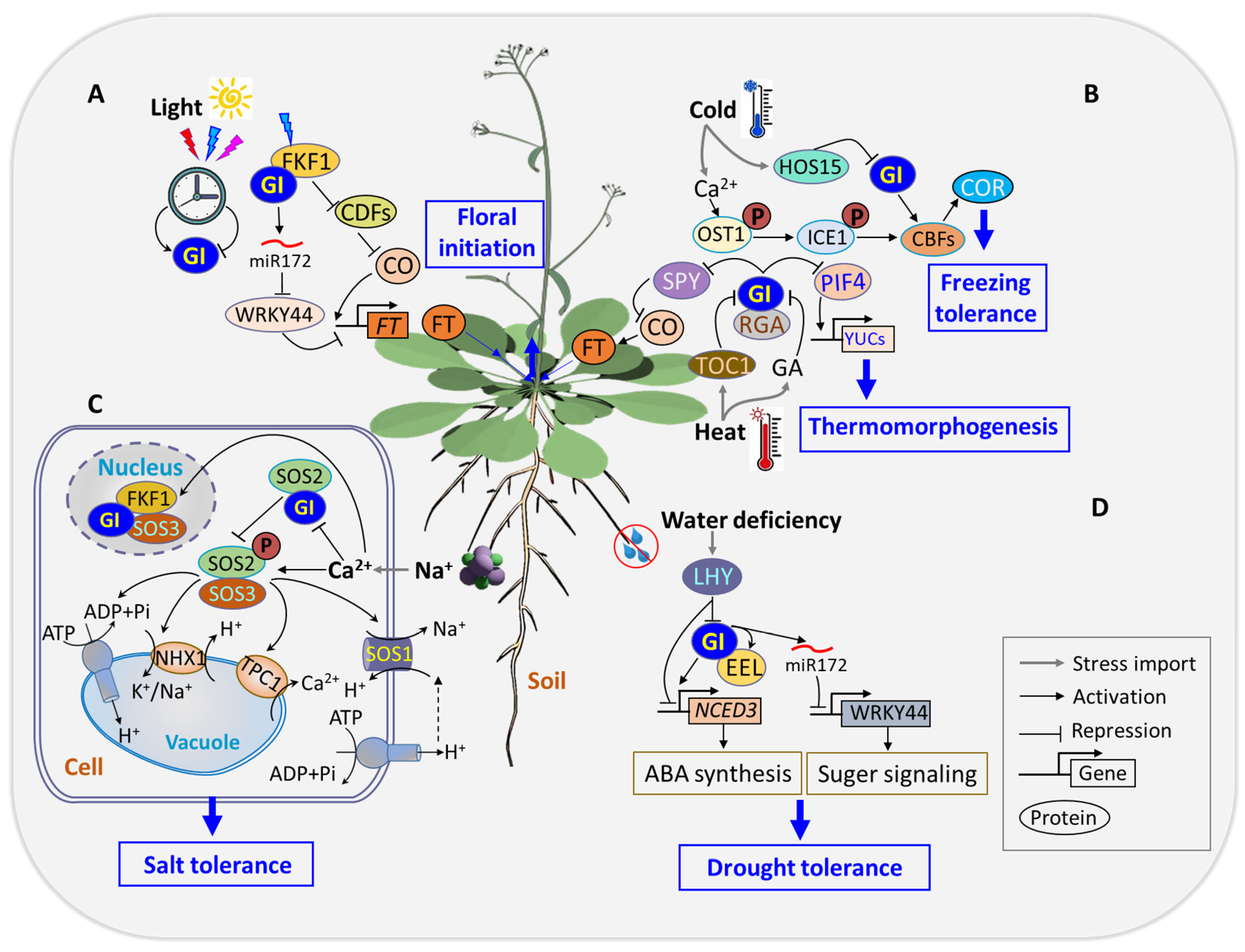
GIGANTEA (GI) is a conserved nuclear protein crucial for orchestrating the clock-associated feedback loop in the circadian system by integrating light input, modulating gating mechanisms, and regulating circadian clock resetting. It serves as a core component which transmits blue light signals for circadian rhythm resetting and overseeing floral initiation. Beyond circadian functions, GI influences various aspects of plant development (chlorophyll accumulation, hypocotyl elongation, stomatal opening, and anthocyanin metabolism). GI has also been implicated to play a pivotal role in response to stresses such as freezing, thermomorphogenic stresses, salinity, drought, and osmotic stresses. Positioned at the hub of complex genetic networks, GI interacts with hormonal signaling pathways like abscisic acid (ABA), gibberellin (GA), salicylic acid (SA), and brassinosteroids (BRs) at multiple regulatory levels. This intricate interplay enables GI to balance stress responses, promoting growth and flowering, and optimize plant productivity.

GI circadian clock flowering time stress tolerance stimulus response

1. Introduction

Agricultural productivity is limited by multiple abiotic stresses, which affects plant growth and development. These stresses induce intricate alterations in cellular metabolism, necessitating adjustments in the central metabolic network of plants to maintain cellular and metabolic homeostasis [1]. The limiting effect of these stresses on crops is intensified by climate change, emphasizing the need for cultivars with enhanced adaptability to ensure global food security [2][3][4]. Plants have evolved intricate mechanisms to cope with abiotic stresses, including regulatory pathways involved in stress signal perception, transduction, transcriptional regulation, and protein modifications [5][6]. Functional genomic approaches, such as high-throughput transcriptomics and proteomics, have contributed to identifying stress-responsive genes and proteins, which have enhanced our understanding of plant responses to environmental challenges [7][8].
Stress-responsive genes fall into two functional categories, playing crucial roles in plant adaptation to abiotic stress [9]. The first category comprises regulatory proteins, including transcription factors, protein kinases, phosphatases, and calcium receptors. These regulators participate in signal transduction pathways by influencing the expression of downstream stress-inducible genes. The second category encompasses diverse protein molecules, such as water channel proteins, chaperones, sugar and proline transporters, osmotin, detoxification enzymes, anti-freezing proteins, and late embryogenesis abundant (LEA) proteins [10][11][12][13]. These proteins act together and contribute to the multifaceted responses employed by plants to mitigate the adverse effects of abiotic stress.
The phenotype of GI was initially identified as a late-flowering mutant (gi) in Arabidopsis thaliana [14][15]. The dynamic responsiveness of GI across various developmental stages underscores its active involvement in physiological processes such as seed dormancy, germination, hypocotyl emergence, circadian clock regulation, flower initiation, carbohydrate metabolism, and stress responses [16][17][18][19][20][21]. The intricate temporal regulation of GI during diurnal cycles highlights its interconnection with the circadian clock [22], showcasing its pivotal role in coordinating plant temporal responses. GI’s influence extends from breaking seed dormancy to circadian clock regulation and stress responses. Its significant role in carbohydrate metabolism underscores its versatility in regulating adaption to environmental challenges [19][20][21]. The regulation and stability of GI’s protein are paramount to normal functioning of the circadian clock system [23][24]. Transcriptomic studies reveal GI’s widespread impact on nearly 80% of all genes in plants species like rice (Oryza sativa), poplar (Populus trichocarpa), and Arabidopsis (Arabidopsis thaliana), emphasizing its central position in temporal coordination in plants [25].
Despite its discovery over six decades ago, the biochemical function of GI has largely remained elusive [26]. While extensive evidence supports the exclusive regulation of GI transcripts and proteins within the circadian clock system, understanding of the molecular-level regulation of GI abundance and its precise biochemical function remains limited [23]. Over the last two decades, GI has emerged as a focal point of research, revealing its involvement in a myriad of biological processes in plants. Serving as a pleiotropic gene, GI’s diverse roles span various aspects of plant physiology, including circadian clock regulation, light sensing and signaling, flowering time regulation, chlorophyll accumulation, hypocotyl elongation, sugar metabolism, abiotic stress tolerance, and even miRNA processing [27][28].

2. Stimulus Response

Light is pivotal to shaping key growth and developmental processes in plants, such as photosynthesis, photomorphogenesis, phototropism, and shading escape [29][30]. Sunlight serves as the natural light source and provides the optimal illumination needed for plants to maximize growth and development. Two critical variables, light intensity and spectral quality, significantly impact various aspects of plant physiology. Light intensity affects crucial plant processes such as food production, stem elongation, leaf color, flowering, and overall plant yield [31][32]. Meanwhile, light spectral quality is essential in activating plant photoreceptors, spanning from UV-B to far-red, and includes blue light receptors such as cryptochromes (CRYs), phototropism, and ZTLs [33][34]. Light signaling involves plants’ ability to perceive both the quantity and quality of light through specialized photoreceptors and transduce this information into transcriptional networks that regulate the expression of specific genes to modulate light responses [35].
GI interacts with various proteins at both transcriptional and post-translational levels to influence several biological processes [23][36]. Red and far-red lights are crucial components of light quality and play significant roles in plant growth and development [37]. Phytochromes are central to light signaling pathways primarily via interacting with PHYtochrome-interacting factors (PIFs), which are members of the bHLH transcription factor family and are known to negatively regulate photomorphogenesis in the dark [37][38]. GI modulates light signaling by regulating PIF activity and accumulation through multiple mechanisms, including transcriptional and post-translational regulations [39][40]. GI influences PIF4 and PIF5 mRNA expression during the early night and interacts with PIF7 to repress transcription in response to shade at dusk, illustrating the broader impact of GI on photoperiodic growth and response to environmental cues [41][42]. The interplay between GI and light signaling pathways (Figure 1) adds a layer of complexity to our understanding of how plants integrate environmental cues to regulate key physiological processes.
Figure 1. GI is implicated in light signaling, contributing to circadian rhythm resetting. In the circadian clock, GI directly activates the expression of LHY and TOC1 to reset the circadian rhythm. GI collaborates with the central clock components in the evening feedback loop, forming a chaperone complex with heat shock protein (HSP90) and ZTL to regulate ZTL stability. This complex promotes the degradation of TOC1, influencing overall clock function. During the evening, TOC1 and evening complex elements reciprocally suppress GI. Under long-day conditions, GI interacts with FKF1 to degrade cycling dof factor (CDF), which is a repressor of CO, leading to elevation of CO transcript abundance and promotion of FT expression to regulate flowering. Additionally, GI integrates light signaling with the circadian clock by regulating PIF proteins (PIF4 and PIF5), which affects output rhythms like hypocotyl elongation.

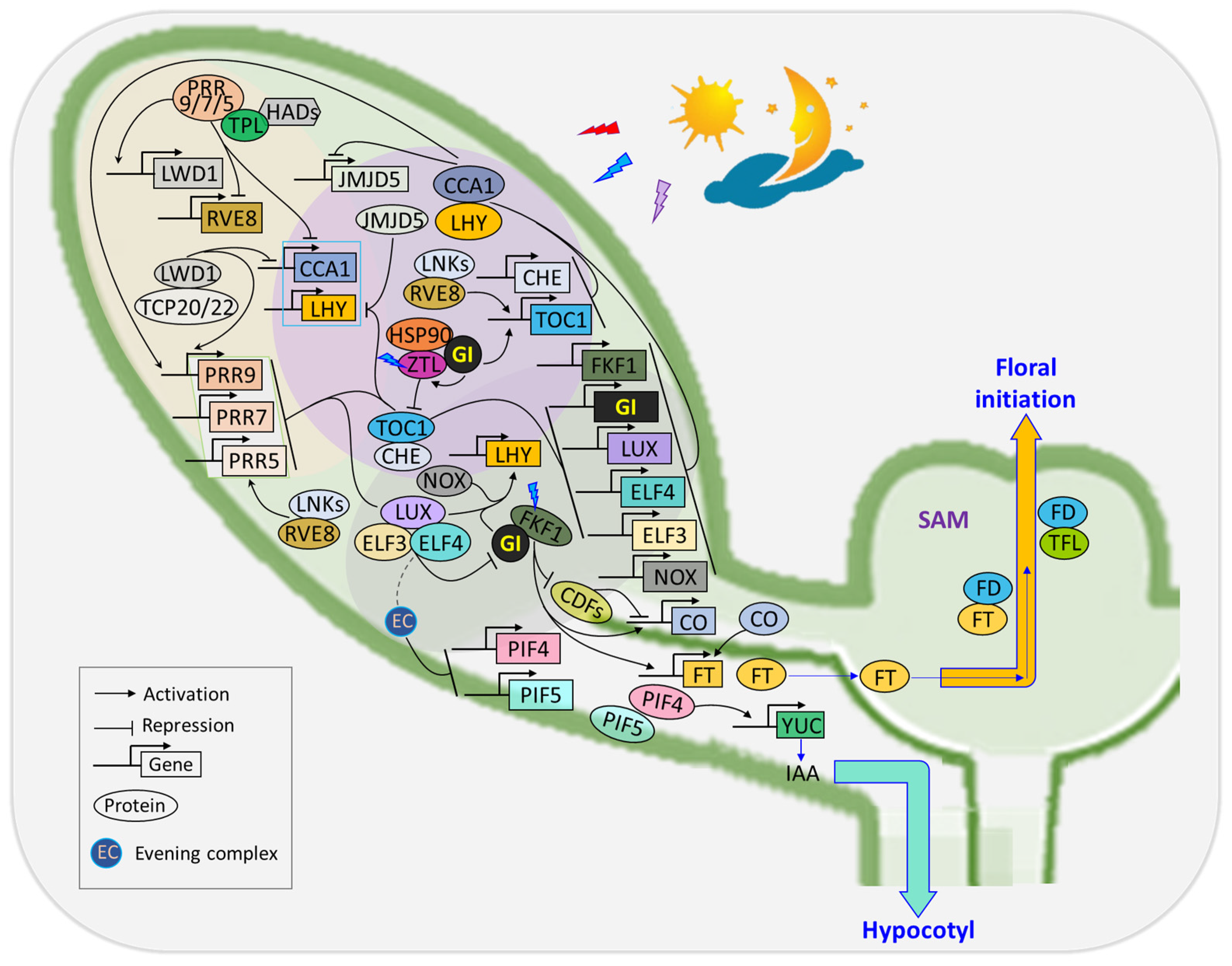
3. Flowering Time Regulation

(1)
Orchestrating floral transition in response to photoperiodic signals
The transition from vegetative growth to flowering is a complex process fine-tuned by environmental signals, with the photoperiodic pathway playing a central role. Such transition is influenced by light, photoperiod, and the circadian clock and revolves around key players such as CO and the florigen hormone FT [43]. Light is perceived by multiple photoreceptors in the leaves, and signal output responses are supervised by the circadian clock. GI acts as a gating factor by regulating the FT expression in CO-dependent and CO-independent pathways. CO, a pivotal component of the photoperiodic pathway, orchestrates the production of FT under long-day photoperiods. Its peak expression in short-day photoperiods is post-dark due to insufficient stabilization by light. CDF1 regulates CO transcription by binding to its promoter at sunrise to repress its expression [44].
Flowering time regulation within the circadian clock involves one of the output pathways mediated by GI, which regulates the amount of CO. Under long-day conditions, GI and FKF1 are co-expressed at ZT10 and form a complex. This complex accumulates and peaks in the middle of the day, leading to the degradation of CDF repressors and an increase in CO transcript abundance. This consequently promotes the expression of FT [21] (Figure 1). Under short-day conditions, where the expression of GI precedes that of FKF1, the formation of the GI-FKF1 repressor complex is disrupted. This disruption results in a reduction in abundance of CO and FT [45]. GI mutants exhibit reduced CO mRNA abundance, further confirming GI’s positive regulatory role in flowering time [46]. In addition to the CO-dependent regulation of FT, GI can independently regulate FT either by directly binding to its promoter or through microRNA-based regulation. In the latter, GI positively regulates miRNA172 [47], leading to the inhibition of TARGET OF EAT1 (TOE1), TOE2, and TOE3 transcriptional repressors, whose functions are crucial in controlling flowering time [48] (Figure 2A). Additionally, GI inhibits SPY expression in a light-dependent manner. SPY, in turn, suppresses CO and FT expression, with spy-4 plants mitigating the late-flowering phenotype of gi-1 plants [49]. In all, GI serves as a key mediator between the circadian clock and the master regulators (CO and FT) in the photoperiodic flowering pathway. The FT transcription was activated in leaf vascular tissue (phloem) [50], and its protein was transported to the shoot apex to induce flowering [51].
Figure 2. GI serves as a central hub protein involved in crosstalk of numerous stress responses and flowering regulation. GI functions as a pleiotropic gene that mediates regulatory pathways, influencing various aspects of flowering (A) and responses to cold or heat (B), salt (C), and drought (D) stresses. The intricate interplay between these pathways enables GI to balance stress responses, promoting both growth and flowering and enhancing plant resilience under adverse conditions.
(2)
Stress tolerance
GI regulates diverse facets of plant growth and development, from flowering time to circadian clock regulation, light signaling, starch accumulation, chlorophyll biogenesis, and miRNA processing. GI also plays a crucial regulatory role in shaping plants’ response to the environment. Drought is one of the most prominent abiotic stresses that induces a cascade of responses in plants, including the production of reactive oxygen species (ROS), oxidative damage, ion toxicity, and nutrient imbalances [52]. GI’s involvement in plant response to drought stress is underscored by its impact on flowering time regulation, which is a crucial mechanism adopted by plants for drought escape [53][54]. Arabidopsis flowering time mutants subjected to conditions triggering drought escape revealed the pivotal roles of GI, FT, and TWIN SISTER OF FT (TSF) genes in orchestrating the plant’s response to water availability changes [55]. GI’s involvement in drought escape is elucidated through an ABA-dependent activation of the SUPPRESSOR OF OVEREXPRESSION OF CONSTANS 1 (SOC1). The collaboration between GI and the bZIP transcription factor enhanced em level (EEL) forms a complex that modulates diurnal ABA biosynthesis, which influences drought tolerance [27]. Further insights into GI’s role in drought escape unveil a regulatory pathway where GI suppresses WRKY44 through miRNA172, contributing to the plant’s ability to cope with drought stress [56]. GI mutants exhibited abnormal drought escape and tolerant phenotypes, emphasizing the intricate network through which GI influences plants’ response to environmental challenges (Figure 2D).
Global climate change has occasioned temperature extremities, posing significant challenges to optimal plant growth, yield, and fruit quality [57][58]. In response to low temperatures, plants activate gene alterations that regulate the production of metabolites, which enhances resistance against damages caused by cold stress [59]. GI plays a crucial role in regulating freeze tolerance, contributing to plants’ ability to withstand cold stress through various regulatory mechanisms. Transcriptome profiling of Arabidopsis exposed to cold stress revealed an upregulation of GI transcripts, facilitating cold stress acclimation independently of C-repeat binding protein (CBP) [60]. Under constitutive cold stress, GI is induced, and the gi-3 mutant exhibits decreased cold tolerance and impaired acclimation compared to the WT [61]. This emphasizes the significance of GI in plants’ adaptive response to cold stress conditions. HOS15, a transcriptional repressor of GI, operates independently of COP1 in mediating GI’s degradation and regulation of flowering time in response to low ambient temperature [62]. The interaction between GI and the CDF module plays a pivotal role in mediating the transcriptional regulation of CDFs, thereby influencing freezing tolerance in Arabidopsis [40]. This intricate module adds another layer to the regulatory network through which GI contributes to plants’ ability to tolerate and acclimate to cold stress (Figure 2B).
Furthermore, temperature signals integrate into the clock transcriptional circuitry through the EC consisting of ELF3/4 and LUX. This regulation extends to the transcription of PRR9/PRR7, GI, LUX, and PIF4 in response to both temperature changes and variations in steady-state growth temperature [63]. Under long-day conditions, elevated temperatures promote the accumulation of GI protein. GI then interacts with and stabilizes the repressor of ga1-3 (RGA), a DELLA protein, which consequently dampens PIF4-mediated thermomorphogenesis [64]. Conversely, under short days with reduced GI accumulation, RGA undergoes rapid degradation through the gibberellic-acid-mediated ubiquitination–proteasome pathway to facilitate thermomorphogenic growth [64]. These findings suggest that the GI–RGA–PIF4 signaling module facilitates day-length-dependent plant thermomorphogenic responses (Figure 2B).

4. Chlorophyll Accumulation Is Regulated by GI in Plants

Plants exhibit the capacity to regulate chlorophyll distribution across tissues, which balances their visibility and functionality. In petals, chlorophyll accumulation is limited to preserve the conspicuousness of flowers, while leaves accumulate substantial amounts crucial for photosynthesis [65]. Chloroplasts house the chlorophyll, which serves as sites for light energy capture and conversion during photosynthesis [66]. GI, modulated by the circadian clock, plays a pivotal role in chloroplast biogenesis in Arabidopsis. The gi-2 mutant displays reduced sensitivity to the chloroplast biogenesis inhibitor lincomycin, maintaining higher photosynthetic protein levels. Conversely, wild-type and GI-overexpressing transgenic lines exhibit lincomycin hypersensitivity, leading to variegated leaves and reduced photosynthetic protein abundance [67]. This underscores GI’s involvement in chloroplast and chlorophyll biogenesis. In a related study, Arabidopsis GI mutant alleles (gi-3, gi-4, gi-5, and gi-6) show significantly higher seedling chlorophyll content than the wild type after paraquat treatment, confirming GI as a negative regulator of chlorophyll biogenesis [68].
Nitric oxide treatment suppresses GI mRNA abundance, resulting in increased chlorophyll content in Arabidopsis [69]. Under the long-day photoperiod, gi-201 and gi-2 mutants maintain green leaves for 32 days post-emergence compared to the wild type. Conversely, gi-2 mutants overexpressing GI exhibit leaf tip yellowing at 24 days post-emergence, which intensified at 28 days post-emergence. Chlorophyll fluorescence (Fv/Fm) was higher in gi-201 and gi-2 mutants than in the wild type, highlighting GI’s dual role as a positive regulator of leaf senescence and a negative regulator of chlorophyll accumulation [70]. Mutation in GI impacts the CAB2 gene, which is a key component of the light-harvesting complex of photosystem II [18]. Silencing of GI’s paralog PhGI1 in Petunia hybrida results in phgi1 with greener apical regions and increased chlorophyll accumulation compared to the wild type [68]. Conversely, loss of function of a rice GI mutant, osgi, displayed significantly reduced leaf chlorophyll content compared to the wild type, indicating species-specific variations in GI-mediated chlorophyll regulation [71].

5. GI Regulates Stomatal Opening in Plants

Stomata are minute pores on the epidermis of leaves and stems which connect internal air spaces with the external atmosphere. Guard cells control the stomatal aperture, modulating gas exchange and water loss through transpiration in response to environmental and exogenous signals [72][73]. Blue light acts as a stomatal opening signal, perceived by receptor kinases phot1 and phot2, which activates the plasma membrane H+-ATPase [74][75]. GI functions in the blue light signaling pathway by directly binding to the LOV motif of ZTL, LKP2, and FKF1 [21]. Core components of the photoperiodic flowering pathway, including cryptochromes (CRY), GI, CO, EFL3, FT, TSF, and suppressor of overexpression of co1 (SOC1), are expressed in guard cells to regulate light-induced stomatal opening [76][77]. Overexpression of these components leads to opened stomata, while knockout mutants exhibit reduced light-induced stomatal opening. CRYs are blue light photoreceptors which promote floral transition [78] by preventing ubiquitination of GI and CO proteins [79][80], thereby controlling stomatal opening through FT and TSF in response to photoperiod [81]. TSF mutants and overexpression in phot1 and phot2 mutants display suppressed and constitutive stomatal opening, respectively [81]. Similarly, gi-1 and co-1 mutants exhibit suppressed blue-light-induced stomatal opening, while GI and CO overexpression results in constitutive open-stomata phenotypes. The GI- EEL complex binds to the promoter of 9-cis-epoxycarotenoid dioxygenase 3 (NCED3) and activates its transcription to mediate stomatal opening. The gi-1, eel, and gi-1eel mutants, compared to the wild type, are hypersensitive to drought stress due to uncontrolled water loss from increased stomatal aperture [27].

6. GI’s Role in Plant Sugar Signaling

In plants, naturally occurring sugars, including sucrose, fructose, glucose, trehalose, and their derivatives such as pectin, cellulose, hemicellulose, callose, and starch, play a dual role as both the primary source of energy for cellular metabolism and the structural components of plant cells [82]. These sugars also act as signaling molecules within the circadian clock system to regulate various aspects of cellular development, such as floral transition [83], hormonal pathway signaling [84], and innate immunity [85]. Plant cells employ signaling mechanisms to perceive carbon and energy status and dictate metabolic adjustments. Under carbon limitation, SnRK1 activity prolongs the circadian period, while sucrose shortens it through the T6P-SnRK1 complex acting on the clock oscillator gene PRR7 [86][87]. The circadian clock slows down in the dark due to the absence of light and cellular metabolism but can be sustained by the addition of sugar. Sucrose sustains the circadian rhythm in the dark by stabilizing GI protein through a regulatory mechanism dependent on the F-box protein ZTL and constitutive response1 (CTR1), a negative regulator of ethylene signaling [88]. The regulation of GI expression by sucrose suggests a connection that measures and reports metabolic status to alter or reset the circadian clock [89].
Eimert [90] reported an increase in sugar accumulation in the WT in response to cold treatment, while the gi-3 mutant displayed a significant reduction in soluble sugar content, attributing the sensitivity of the gi-3 mutant to cold treatment to the constitutive reduction in soluble sugars. This affirms that GI has a direct connection with sugar metabolism. In contrast, rice plants carrying a null mutation in the rice homology OsGI (osgi-1) recorded higher leaf sucrose and starch at most points in time under natural field conditions [71]. Similarly, monogenic recessive mutants gi-1, 2, and 3 caused an increase in both late flowering initiation and increased starch content in Arabidopsis [91]. GI interacts with trehalose-6-phosphate synthase 8 (TPS8) to form a complex which may have a direct influence on carbohydrate metabolism [92]. Under LDs, drought stress induces the expression of GI. Modes of GI-dependent but CO-independent pathways include the activation of miR172, thus inhibiting the transcription of WRKY44 [56], which was considered to be involved in sugar metabolism and signaling, indicating a role of GImiRNA172 in drought escape and defense by affecting sugar signaling (Figure 2D).

7. GI’s Unexplored Role in Anthocyanin Metabolism

Anthocyanins are a class of polyphenolic pigments in plants, which are induced and accumulate in response to various environmental signals, with their biosynthesis regulated by transcription factors. Environmental cues such as light, low temperature, drought, and salinity significantly influence anthocyanin biosynthesis [93][94]. Among these cues, light stands out as the most prominent regulatory factor in the anthocyanin biosynthesis pathway. While GI has been primarily linked to stress response regulation in plants, its involvement in anthocyanin metabolism has received limited attention. A study by Odgerel [95] has shed light on a novel role of GI in anthocyanin metabolism in potatoes. The research revealed that mutants with repressed StGI.04—specifically aG153, aG144, and aG152 tuber peels—exhibited a 52%, 36%, and 31% reduction in anthocyanin content, respectively, compared to the wild type (DES). This discovery highlights the unexplored connection between GI and anthocyanin regulation, opening avenues for further investigation into this intriguing aspect of plant physiology.

8. Integrative Role of GI in Hormonal Signaling

Phytohormones play pivotal roles in orchestrating plant growth and development, serving as key regulators that enable plants to respond systematically to environmental changes [96]. The circadian clock actively participates in hormonal signaling pathways, exerting regulatory control over components of auxin, jasmonate, brassinosteroids, cytokinin, GA, and abscisic acid [97]. These hormones, in turn, reciprocally influence the circadian clock system, establishing a feedback loop that fine-tunes the oscillator’s activity [98]. GI has emerged as a central player in integrating hormonal signals to regulate diverse processes in plants. This interplay between the circadian clock and hormonal regulation underscores the intricate web of molecular interactions that govern plant growth, emphasizing the multifaceted role of GI in these dynamic processes.
(1)
The role of GI in ABA-mediated responses to drought stress
ABA stands out as the most extensively studied signal governing gene expression in response to drought stress perception. Drought induces accumulation of the stress hormone ABA and activates its downstream signaling pathway, which takes charge of promoting stomatal closure to reduce the transpiration rate. Notably, ABA orchestrates gene expression in a meticulously organized diurnal cycle, ensuring that the physiological traits under ABA regulation manifest at specific time periods. GI, a key regulator of photoperiod-dependent flowering and the circadian rhythm, emerges as a central player in this intricate ABA regulatory network, acting as a key gatekeeper for ABA-regulated transcriptional and physiological responses [99]. The modulation of GI signaling by ABA contributes to the transcriptional upregulation of FT, TSF and SOC1, which ultimately promotes drought escape in Arabidopsis [100].
Moreover, GI plays a vital role in regulating the synthesis and signaling of ABA. GI interacts with EEL, a basic Leu zipper (bZIP) transcription factor involved in ABA-regulated gene expression during seed dehydration [27]. This heterodimer complex promotes ABA biosynthesis by directly activating the diurnal expression of NCED3, a rate-limiting enzyme in ABA biosynthesis in plastids, to enhance drought tolerance in Arabidopsis [27] (Figure 2D). The endogenous ABA in turn promotes flowering via upregulating the expression of FT to avoid prolonged exposure to drought [55]. This highlights the molecular crosstalk between the circadian clock and ABA signaling to cope with drought.
(2)
GI’s involvement in gibberellin signaling for hypocotyl elongation
Gibberellins (GAs) is a crucial phytohormone, pivotal for promoting cell elongation, facilitating the overall growth of plants. GA signaling is strongly linked with the circadian clock in the regulation of developmental processes. On the one hand, GAs operate as an output module within the circadian network to affect the function of the circadian clock. On the other hand, the clock directly governing the diurnal accumulation of GA levels by regulating the transcription of genes, which involved in the biosynthesis and catabolism of GAs. Moreover, the clock governs the responsiveness to GAs by controlling the expression of the GA receptor gene ga insensitive dwarf 1 (GID1) [101]. GI’s regulatory role in GA signaling hinges on its ability to stabilize DELLA proteins, including repressor of ga1-3 (RGA), gibberellic acid insensitive (GAI), and rga-like protein 3 (RGL3). These DELLA proteins function as negative components within the GA signaling pathway, allowing GI to precisely modulate the timing of GA sensitivity [102]. GI interacts with and stabilizes RGA in the context of their GA-mediated degradation and plays a vital role in the circadian gating of GA signaling [102]. A recent report revealed that DELLA is mono-O-fucosylated by the spindly (SPY), a novel O-fucosyltransferase, thereby activating DELLA by promoting its interaction with key regulators like PIF3 and PIF4 [103]. GI interacts with SPY, a negative regulator of gibberellin signaling, to regulate hypocotyl elongation [49]. However, it is unclear whether the GI–SPY interaction has any implications for DELLA O-fucosylation. GI interacts with PIFs and modulates their stability and activity [39], which in turn regulates the expression of phytohormones biosynthesis and signaling. PIF1/PIL5 represses gibberellin 3-oxidase 1 (GA3ox1) and GA3ox2, which encode enzymes to produce GA1 and GA4 [104]. PIF1/PIL5 actives the expression of GAI and RGA by directly binding to their promoters [105]. Furthermore, GI also regulates the transcription levels of CCA1 and LHY, which are involved in circadian clock regulation and responsiveness to GAs [39].
(3)
GI’s impact on phytohormones in biotic stress response
Salicylic acid (SA) is an important phytohormone best known for regulating plant responses to pathogen infections. Jasmonates (JA) are phospholipid-derived phytohormones that mediate both developmental processes and responses to environmental stresses. GI, beyond its role in the circadian clock, has been identified as a regulator which influences plant responses to biotic stress through the modulation of phytohormones such as salicylic acid (SA) and jasmonates (JA). Recent research reveals that GI expression promotes disease severity by downregulating the SA accumulation and altering the phenylpropanoid pathway in both Arabidopsis and wheat during Bipolaris sorokiniana infection. This downregulation contributes to the suppression of pathogenesis-related responses, ultimately rendering the plants susceptible to the disease [106]. It seems that the GI gene acts as a negative regulator in the SA signaling pathway, and the downregulation of GI could be beneficial in generating disease tolerance. In Arabidopsis thaliana, GI has been shown to downregulate JA signaling as well, leading to reduced severity of spread and damage caused by pathogenic infections. The gi-100 mutant, exhibiting late flowering, demonstrated heightened susceptibility to Hyaloperonospora arabidopsidis infection, with the regulatory involvement of phytoalexin deficient 4 (PAD4) in the pathogen infection phenotype [107]. The gi-100 mutant displayed enhanced tolerance to wilt disease, showcasing a positive correlation between late flowering and resistance to Fusarium oxysporum [22][108]. The relative transcript expression of coronatine insensitive 1 (COI1) and plant defensin 1.2 (PDF1.2), marker genes of the JA pathway, is significantly upregulated in the gi-100 mutants compared to Col-0 plants, while the isochorismate synthase 1 (ICS1) and nonexpressor of pathogenesis-related genes 1 (NPR1), markers of the SA pathway, are downregulated [17]. These results suggest that the GI module promotes susceptibility to F. oxysporum infection by inducing the SA pathway and inhibiting JA signaling in Arabidopsis. These findings underscore GI’s intricate role in plant defense mechanisms, shedding light on its impact on phytohormonal regulation during biotic stress responses.
(4)
GI’s role in brassinosteroid signaling pathway
Brassinosteroids (BRs) are essential steroid hormones that play pivotal roles in plant signaling, contributing to cell expansion, cell division, and crucial developmental processes such as etiolation and reproduction [109]. Loss-of-function mutants of GI (abz126) displayed altered responses to specific compounds: insensitivity to paclobutrazol- (PAC), abnormal reactions to benzylaminopurine (BAP), and insensitivity to brassinolide (BL) [110]. The observed phenotypic variations in GI mutants suggest a direct association between the loss of function of the GI gene and disruptions in brassinosteroid signaling. UBP12/UBP13 are two novel positive regulators of BR signaling that can remove K-48- and K-63-linked ubiquitin from pBES1/BES1, rescuing them from destruction [111]. UBP12 and UBP13 interact with deubiquitinate BES1 to stabilize its protein, which acts as a positive regulator in BR signaling [111]. In addition, UBP12 and UBP13 act as components of the ZTL-GI photoreceptor complex to stabilize GI, ZTL, and TOC1 [112]. GI’s regulatory role in the brassinosteroid pathway underscores its significance in coordinating plant responses to hormonal signals, influencing aspects of growth, flowering, and sensitivity to specific compounds. These findings highlight GI as a key player in the intricate network of brassinosteroid signaling, contributing to the modulation of plant development and responses to external stimuli.

References

  1. Savchenko, T.V.; Rolletschek, H.; Dehesh, K. Jasmonates-Mediated Rewiring of Central Metabolism Regulates Adaptive Responses. Plant Cell Physiol. 2019, 60, 2613–2620.
  2. Zandalinas, S.I.; Mittler, R.; Balfagon, D.; Arbona, V.; Gomez-Cadenas, A. Plant adaptations to the combination of drought and high temperatures. Physiol. Plant 2018, 162, 2–12.
  3. Badouin, H.; Gouzy, J.; Grassa, C.J.; Murat, F.; Staton, S.E.; Cottret, L.; Lelandais-Brière, C.; Owens, G.L.; Carrère, S.; Mayjonade, B.; et al. The sunflower genome provides insights into oil metabolism, flowering and Asterid evolution. Nature 2017, 546, 148–152.
  4. De Frenne, P.; Lenoir, J.; Luoto, M.; Scheffers, B.R.; Zellweger, F.; Aalto, J.; Ashcroft, M.B.; Christiansen, D.M.; Decocq, G.; De Pauw, K.; et al. Forest microclimates and climate change: Importance, drivers and future research agenda. Glob. Chang. Biol. 2021, 27, 2279–2297.
  5. Gupta, A.; Rico-Medina, A.; Cano-Delgado, A.I. The physiology of plant responses to drought. Science 2020, 368, 266–269.
  6. Dong, Q.; Wallrad, L.; Almutairi, B.O.; Kudla, J. Ca2+ signaling in plant responses to abiotic stresses. J. Integr. Plant Biol. 2022, 64, 287–300.
  7. Kultz, D.; Fiol, D.; Valkova, N.; Gomez-Jimenez, S.; Chan, S.Y.; Lee, J. Functional genomics and proteomics of the cellular osmotic stress response in ‘non-model’ organisms. J. Exp. Biol. 2007, 210 Pt 9, 1593–1601.
  8. Liu, H.; Yu, C.; Li, H.; Ouyang, B.; Wang, T.; Zhang, J.; Wang, X.; Ye, Z. Overexpression of ShDHN, a dehydrin gene from Solanum habrochaites enhances tolerance to multiple abiotic stresses in tomato. Plant Sci. 2015, 231, 198–211.
  9. Hirayama, T.; Shinozaki, K. Research on plant abiotic stress responses in the post-genome era: Past, present and future. Plant J. 2010, 61, 1041–1052.
  10. Shinozaki, K.; Yamaguchi-Shinozaki, K.; Seki, M. Regulatory network of gene expression in the drought and cold stress responses. Curr Opin. Plant Biol. 2003, 6, 410–417.
  11. Shinozaki, K.; Yamaguchi-Shinozaki, K. Gene networks involved in drought stress response and tolerance. J. Exp. Bot. 2007, 58, 221–227.
  12. Khurana, P.; Vishnudasan, D.; Chhibbar, A.K. Genetic approaches towards overcoming water deficit in plants—Special emphasis on LEAs. Physiol. Mol. Biol. Plants 2008, 14, 277–298.
  13. Gonzalez-Villagra, J.; Kurepin, L.V.; Reyes-Diaz, M.M. Evaluating the involvement and interaction of abscisic acid and miRNA156 in the induction of anthocyanin biosynthesis in drought-stressed plants. Planta 2017, 246, 299–312.
  14. Xie, Q.; Lou, P.; Hermand, V.; Aman, R.; Park, H.J.; Yun, D.J.; Kim, W.Y.; Salmela, M.J.; Ewers, B.E.; Weinig, C.; et al. Allelic polymorphism of GIGANTEA is responsible for naturally occurring variation in circadian period in Brassica rapa. Proc. Natl. Acad. Sci. USA 2015, 112, 3829–3834.
  15. De Montaigu, A.; Toth, R.; Coupland, G. Plant development goes like clockwork. Trends Genet. 2010, 26, 296–306.
  16. Shu, K.; Yang, W. E3 Ubiquitin Ligases: Ubiquitous Actors in Plant Development and Abiotic Stress Responses. Plant Cell Physiol. 2017, 58, 1461–1476.
  17. Patnaik, A.; Kumar, A.; Behera, A.; Mishra, G.; Dehery, S.K.; Panigrahy, M.; Das, A.B.; Panigrahi, K.C.S. GIGANTEA supresses wilt disease resistance by down-regulating the jasmonate signaling in Arabidopsis thaliana. Front. Plant Sci. 2023, 14, 1091644.
  18. Park, D.H.; Somers, D.E.; Kim, Y.S.; Choy, Y.H.; Lim, H.K.; Soh, M.S.; Kim, H.J.; Kay, S.A.; Nam, H.G. Control of circadian rhythms and photoperiodic flowering by the Arabidopsis GIGANTEA gene. Science 1999, 285, 1579–1582.
  19. Huq, E.; Tepperman, J.M.; Quail, P.H. GIGANTEA is a nuclear protein involved in phytochrome signaling in Arabidopsis. Proc. Natl. Acad. Sci. USA 2000, 97, 9789–9794.
  20. Kim, W.Y.; Ali, Z.; Park, H.J.; Park, S.J.; Cha, J.Y.; Perez-Hormaeche, J.; Quintero, F.J.; Shin, G.; Kim, M.R.; Qiang, Z.; et al. Release of SOS2 kinase from sequestration with GIGANTEA determines salt tolerance in Arabidopsis. Nat. Commun. 2013, 4, 1352.
  21. Sawa, M.; Nusinow, D.A.; Kay, S.A.; Imaizumi, T. FKF1 and GIGANTEA complex formation is required for day-length measurement in Arabidopsis. Science 2007, 318, 261–265.
  22. Patnaik, A.; Alavilli, H.; Rath, J.; Panigrahi, K.C.S.; Panigrahy, M. Variations in Circadian Clock Organization & Function: A Journey from Ancient to Recent. Planta 2022, 256, 91.
  23. Mishra, P.; Panigrahi, K.C. GIGANTEA—An emerging story. Front. Plant Sci. 2015, 6, 8.
  24. Mizoguchi, T.; Wright, L.; Fujiwara, S.; Cremer, F.; Lee, K.; Onouchi, H.; Mouradov, A.; Fowler, S.; Kamada, H.; Putterill, J.; et al. Distinct roles of GIGANTEA in promoting flowering and regulating circadian rhythms in Arabidopsis. Plant Cell 2005, 17, 2255–2270.
  25. Filichkin, S.A.; Breton, G.; Priest, H.D.; Dharmawardhana, P.; Jaiswal, P.; Fox, S.E.; Michael, T.P.; Chory, J.; Kay, S.A.; Mockler, T.C. Global profiling of rice and poplar transcriptomes highlights key conserved circadian-controlled pathways and cis-regulatory modules. PLoS ONE 2011, 6, e16907.
  26. Redei, G.P. Supervital Mutants of Arabidopsis. Genetics 1962, 47, 443–460.
  27. Baek, D.; Kim, W.Y.; Cha, J.Y.; Park, H.J.; Shin, G.; Park, J.; Lim, C.J.; Chun, H.J.; Li, N.; Kim, D.H.; et al. The GIGANTEA-ENHANCED EM LEVEL Complex Enhances Drought Tolerance via Regulation of Abscisic Acid Synthesis. Plant Physiol. 2020, 184, 443–458.
  28. Bader, Z.E.; Bae, M.J.; Ali, A.; Park, J.; Baek, D.; Yun, D.J. GIGANTEA-ENHANCED EM LEVEL complex initiates drought escape response via dual function of ABA synthesis and flowering promotion. Plant Signal Behav. 2023, 18, 2180056.
  29. Goyal, A.; Szarzynska, B.; Fankhauser, C. Phototropism: At the crossroads of light-signaling pathways. Trends Plant Sci. 2013, 18, 393–401.
  30. Ballare, C.L.; Pierik, R. The shade-avoidance syndrome: Multiple signals and ecological consequences. Plant Cell Environ. 2017, 40, 2530–2543.
  31. Lu, T.; Meng, Z.; Zhang, G.; Qi, M.; Sun, Z.; Liu, Y.; Li, T. Sub-high Temperature and High Light Intensity Induced Irreversible Inhibition on Photosynthesis System of Tomato Plant (Solanum lycopersicum L.). Front. Plant Sci. 2017, 8, 365.
  32. Virsile, A.; Brazaityte, A.; Vastakaite-Kairiene, V.; Miliauskiene, J.; Jankauskiene, J.; Novickovas, A.; Samuolienė, G. Lighting intensity and photoperiod serves tailoring nitrate assimilation indices in red and green baby leaf lettuce. J. Sci. Food Agric. 2019, 99, 6608–6619.
  33. Yu, X.; Liu, H.; Klejnot, J.; Lin, C. The Cryptochrome Blue Light Receptors. Arab. Book 2010, 8, e0135.
  34. Suetsugu, N.; Wada, M. Evolution of three LOV blue light receptor families in green plants and photosynthetic stramenopiles: Phototropin, ZTL/FKF1/LKP2 and aureochrome. Plant Cell Physiol. 2013, 54, 8–23.
  35. Moglich, A.; Yang, X.; Ayers, R.A.; Moffat, K. Structure and function of plant photoreceptors. Annu. Rev. Plant Biol. 2010, 61, 21–47.
  36. Brandoli, C.; Petri, C.; Egea-Cortines, M.; Weiss, J. Gigantea: Uncovering New Functions in Flower Development. Genes 2020, 11, 1142.
  37. Xu, X.; Paik, I.; Zhu, L.; Huq, E. Illuminating Progress in Phytochrome-Mediated Light Signaling Pathways. Trends Plant Sci. 2015, 20, 641–650.
  38. Leivar, P.; Monte, E. PIFs: Systems integrators in plant development. Plant Cell 2014, 26, 56–78.
  39. Nohales, M.A.; Liu, W.; Duffy, T.; Nozue, K.; Sawa, M.; Pruneda-Paz, J.L.; Maloof, J.N.; Jacobsen, S.E.; Kay, S.A. Multi-level Modulation of Light Signaling by GIGANTEA Regulates Both the Output and Pace of the Circadian Clock. Dev. Cell 2019, 49, 840–851.e8.
  40. Fornara, F.; de Montaigu, A.; Sanchez-Villarreal, A.; Takahashi, Y.; Ver Loren van Themaat, E.; Huettel, B.; Davis, S.J.; Coupland, G. The GI-CDF module of Arabidopsis affects freezing tolerance and growth as well as flowering. Plant J. 2015, 81, 695–706.
  41. De Montaigu, A.; Giakountis, A.; Rubin, M.; Toth, R.; Cremer, F.; Sokolova, V.; Porri, A.; Reymond, M.; Weinig, C.; Coupland, G. Natural diversity in daily rhythms of gene expression contributes to phenotypic variation. Proc. Natl. Acad. Sci. USA 2015, 112, 905–910.
  42. Martinez-Vasallo, C.; Cole, B.; Gallego-Bartolome, J.; Chory, J.; Kay, S.A.; Nohales, M.A. Epidermal GIGANTEA adjusts the response to shade at dusk by directly impinging on PHYTOCHROME INTERACTING FACTOR 7 function. bioRxiv 2023.
  43. Valverde, F. CONSTANS and the evolutionary origin of photoperiodic timing of flowering. J. Exp. Bot. 2011, 62, 2453–2463.
  44. Valverde, F.; Mouradov, A.; Soppe, W.; Ravenscroft, D.; Samach, A.; Coupland, G. Photoreceptor regulation of CONSTANS protein in photoperiodic flowering. Science 2004, 303, 1003–1006.
  45. Song, Y.H.; Smith, R.W.; To, B.J.; Millar, A.J.; Imaizumi, T. FKF1 conveys timing information for CONSTANS stabilization in photoperiodic flowering. Science 2012, 336, 1045–1049.
  46. Suarez-Lopez, P.; Wheatley, K.; Robson, F.; Onouchi, H.; Valverde, F.; Coupland, G. CONSTANS mediates between the circadian clock and the control of flowering in Arabidopsis. Nature 2001, 410, 1116–1120.
  47. Jung, J.H.; Seo, Y.H.; Seo, P.J.; Reyes, J.L.; Yun, J.; Chua, N.H.; Park, C.M. The GIGANTEA-regulated microRNA172 mediates photoperiodic flowering independent of CONSTANS in Arabidopsis. Plant Cell 2007, 19, 2736–2748.
  48. Aukerman, M.J.; Sakai, H. Regulation of flowering time and floral organ identity by a MicroRNA and its APETALA2-like target genes. Plant Cell 2003, 15, 2730–2741.
  49. Tseng, T.S.; Salome, P.A.; McClung, C.R.; Olszewski, N.E. SPINDLY and GIGANTEA interact and act in Arabidopsis thaliana pathways involved in light responses, flowering, and rhythms in cotyledon movements. Plant Cell 2004, 16, 1550–1563.
  50. Huang, T.; Böhlenius, H.; Eriksson, S.; Parcy, F.; Nilsson, O. The mRNA of the Arabidopsis Gene FT Moves from Leaf to Shoot Apex and Induces Flowering. Science 2005, 309, 1694–1696.
  51. Corbesier, L.; Vincent, C.; Jang, S.; Fornara, F.; Fan, Q.; Searle, I.; Giakountis, A.; Farrona, S.; Gissot, L.; Turnbull, C.; et al. FT Protein Movement Contributes to Long-Distance Signaling in Floral Induction of Arabidopsis. Science 2007, 316, 1030–1033.
  52. Khoso, M.A.; Hussain, A.; Ritonga, F.N.; Ali, Q.; Channa, M.M.; Alshegaihi, R.M.; Meng, Q.; Ali, M.; Zaman, W.; Brohi, R.D.; et al. WRKY transcription factors (TFs): Molecular switches to regulate drought, temperature, and salinity stresses in plants. Front. Plant Sci. 2022, 13, 1039329.
  53. Ivey, C.T.; Carr, D.E. Tests for the joint evolution of mating system and drought escape in Mimulus. Ann. Bot. 2012, 109, 583–598.
  54. Franks, S.J. Plasticity and evolution in drought avoidance and escape in the annual plant Brassica rapa. New Phytol. 2011, 190, 249–257.
  55. Riboni, M.; Galbiati, M.; Tonelli, C.; Conti, L. GIGANTEA enables drought escape response via abscisic acid-dependent activation of the florigens and SUPPRESSOR OF OVEREXPRESSION OF CONSTANS. Plant Physiol. 2013, 162, 1706–1719.
  56. Han, Y.; Zhang, X.; Wang, W.; Wang, Y.; Ming, F. Correction: The suppression of WRKY44 by GIGANTEA-miR172 pathway is involved in drought response of Arabidopsis thaliana. PLoS ONE 2015, 10, e0124854.
  57. Fahad, S.; Bajwa, A.A.; Nazir, U.; Anjum, S.A.; Farooq, A.; Zohaib, A.; Sadia, S.; Nasim, W.; Adkins, S.; Saud, S.; et al. Crop Production under Drought and Heat Stress: Plant Responses and Management Options. Front. Plant Sci. 2017, 8, 1147.
  58. Grover, A.; Mittal, D.; Negi, M.; Lavania, D. Generating high temperature tolerant transgenic plants: Achievements and challenges. Plant Sci. 2013, 205–206, 38–47.
  59. Sanghera, G.S.; Wani, S.H.; Hussain, W.; Singh, N.B. Engineering cold stress tolerance in crop plants. Curr. Genom. 2011, 12, 30–43.
  60. Fowler, S.; Thomashow, M.F. Arabidopsis transcriptome profiling indicates that multiple regulatory pathways are activated during cold acclimation in addition to the CBF cold response pathway. Plant Cell 2002, 14, 1675–1690.
  61. Cao, S.; Ye, M.; Jiang, S. Involvement of GIGANTEA gene in the regulation of the cold stress response in Arabidopsis. Plant Cell Rep. 2005, 24, 683–690.
  62. Ahn, G.; Park, H.J.; Jeong, S.Y.; Shin, G.I.; Ji, M.G.; Cha, J.Y.; Kim, J.; Kim, M.G.; Yun, D.J.; Kim, W.Y. HOS15 represses flowering by promoting GIGANTEA degradation in response to low temperature in Arabidopsis. Plant Commun. 2023, 4, 100570.
  63. Mizuno, T.; Nomoto, Y.; Oka, H.; Kitayama, M.; Takeuchi, A.; Tsubouchi, M.; Yamashino, T. Ambient Temperature Signal Feeds into the Circadian Clock Transcriptional Circuitry Through the EC Night-Time Repressor in Arabidopsis thaliana. Plant Cell Physiol. 2014, 55, 958–976.
  64. Park, Y.-J.; Kim, J.Y.; Lee, J.-H.; Lee, B.-D.; Paek, N.-C.; Park, C.-M. GIGANTEA Shapes the Photoperiodic Rhythms of Thermomorphogenic Growth in Arabidopsis. Mol. Plant 2020, 13, 459–470.
  65. Ohmiya, A.; Hirashima, M.; Yagi, M.; Tanase, K.; Yamamizo, C. Identification of genes associated with chlorophyll accumulation in flower petals. PLoS ONE 2014, 9, e113738.
  66. Cortleven, A.; Schmulling, T. Regulation of chloroplast development and function by cytokinin. J. Exp. Bot. 2015, 66, 4999–5013.
  67. Cha, J.Y.; Lee, D.Y.; Ali, I.; Jeong, S.Y.; Shin, B.; Ji, H.; Kim, J.S.; Kim, M.G.; Kim, W.Y. Arabidopsis GIGANTEA negatively regulates chloroplast biogenesis and resistance to herbicide butafenacil. Plant Cell Rep. 2019, 38, 793–801.
  68. Kurepa, J.; Smalle, J.; Van Montagu, M.; Inze, D. Oxidative stress tolerance and longevity in Arabidopsis: The late-flowering mutant gigantea is tolerant to paraquat. Plant J. 1998, 14, 759–764.
  69. He, Y.; Tang, R.H.; Hao, Y.; Stevens, R.D.; Cook, C.W.; Ahn, S.M.; Jing, L.; Yang, Z.; Chen, L.; Guo, F.; et al. Nitric oxide represses the Arabidopsis floral transition. Science 2004, 305, 1968–1971.
  70. Kim, H.; Park, S.J.; Kim, Y.; Nam, H.G. Subcellular Localization of GIGANTEA Regulates the Timing of Leaf Senescence and Flowering in Arabidopsis. Front. Plant. Sci. 2020, 11, 589707.
  71. Izawa, T.; Mihara, M.; Suzuki, Y.; Gupta, M.; Itoh, H.; Nagano, A.J.; Motoyama, R.; Sawada, Y.; Yano, M.; Hirai, M.Y.; et al. Os- GIGANTEA Confers Robust Diurnal Rhythms on the Global Transcriptome of Rice in the Field. Plant Cell 2011, 23, 1741–1755.
  72. Shimazaki, K.; Doi, M.; Assmann, S.M.; Kinoshita, T. Light regulation of stomatal movement. Annu. Rev. Plant Biol. 2007, 58, 219–247.
  73. Jezek, M.; Blatt, M.R. The Membrane Transport System of the Guard Cell and Its Integration for Stomatal Dynamics. Plant Physiol. 2017, 174, 487–519.
  74. Inoue, S.I.; Kinoshita, T. Blue Light Regulation of Stomatal Opening and the Plasma Membrane H+-ATPase. Plant Physiol. 2017, 174, 531–538.
  75. Kinoshita, T.; Doi, M.; Suetsugu, N.; Kagawa, T.; Wada, M.; Shimazaki, K. Phot1 and phot2 mediate blue light regulation of stomatal opening. Nature 2001, 414, 656–660.
  76. Kinoshita, T.; Ono, N.; Hayashi, Y.; Morimoto, S.; Nakamura, S.; Soda, M.; Kato, Y.; Ohnishi, M.; Nakano, T.; Inoue, S.; et al. FLOWERING LOCUS T regulates stomatal opening. Curr. Biol. 2011, 21, 1232–1238.
  77. Kimura, Y.; Aoki, S.; Ando, E.; Kitatsuji, A.; Watanabe, A.; Ohnishi, M.; Takahashi, K.; Inoue, S.; Nakamichi, N.; Tamada, Y.; et al. A flowering integrator, SOC1, affects stomatal opening in Arabidopsis thaliana. Plant Cell Physiol. 2015, 56, 640–649.
  78. Yang, L.W.; Wen, X.H.; Fu, J.X.; Dai, S.L. ClCRY2 facilitates floral transition in Chrysanthemum lavandulifolium by affecting the transcription of circadian clock-related genes under short-day photoperiods. Hortic. Res. 2018, 5, 58.
  79. Yu, J.W.; Rubio, V.; Lee, N.Y.; Bai, S.; Lee, S.Y.; Kim, S.S.; Liu, L.; Zhang, Y.; Irigoyen, M.L.; Sullivan, J.A.; et al. COP1 and ELF3 control circadian function and photoperiodic flowering by regulating GI stability. Mol. Cell 2008, 32, 617–630.
  80. Liu, L.J.; Zhang, Y.C.; Li, Q.H.; Sang, Y.; Mao, J.; Lian, H.L.; Wang, L.; Yang, H.Q. COP1-mediated ubiquitination of CONSTANS is implicated in cryptochrome regulation of flowering in Arabidopsis. Plant Cell 2008, 20, 292–306.
  81. Ando, E.; Ohnishi, M.; Wang, Y.; Matsushita, T.; Watanabe, A.; Hayashi, Y.; Fujii, M.; Ma, J.F.; Inoue, S.; Kinoshita, T. TWIN SISTER OF FT, GIGANTEA, and CONSTANS have a positive but indirect effect on blue light-induced stomatal opening in Arabidopsis. Plant Physiol. 2013, 162, 1529–1538.
  82. Yu, S.M.; Lo, S.F.; Ho, T.D. Source-Sink Communication: Regulated by Hormone, Nutrient, and Stress Cross-Signaling. Trends Plant Sci. 2015, 20, 844–857.
  83. Bolouri Moghaddam, M.R.; Van den Ende, W. Sugars, the clock and transition to flowering. Front. Plant Sci. 2013, 4, 22.
  84. Rolland, F.; Baena-Gonzalez, E.; Sheen, J. Sugar sensing and signaling in plants: Conserved and novel mechanisms. Annu. Rev. Plant Biol. 2006, 57, 675–709.
  85. Bolouri Moghaddam, M.R.; Van den Ende, W. Sugars and plant innate immunity. J. Exp. Bot. 2012, 63, 3989–3998.
  86. Shin, J.; Sanchez-Villarreal, A.; Davis, A.M.; Du, S.X.; Berendzen, K.W.; Koncz, C.; Ding, Z.; Li, C.; Davis, S.J. The metabolic sensor AKIN10 modulates the Arabidopsis circadian clock in a light-dependent manner. Plant Cell Environ. 2017, 40, 997–1008.
  87. Frank, A.; Matiolli, C.C.; Viana, A.J.C.; Hearn, T.J.; Kusakina, J.; Belbin, F.E.; Wells Newman, D.; Yochikawa, A.; Cano-Ramirez, D.L.; Chembath, A.; et al. Circadian Entrainment in Arabidopsis by the Sugar-Responsive Transcription Factor bZIP63. Curr. Biol. 2018, 28, 2597–2606.e6.
  88. Haydon, M.J.; Mielczarek, O.; Frank, A.; Roman, A.; Webb, A.A.R. Sucrose and Ethylene Signaling Interact to Modulate the Circadian Clock. Plant Physiol. 2017, 175, 947–958.
  89. Dalchau, N.; Baek, S.J.; Briggs, H.M.; Robertson, F.C.; Dodd, A.N.; Gardner, M.J.; Stancombe, M.A.; Haydon, M.J.; Stan, G.B.; Gonçalves, J.M.; et al. The circadian oscillator gene GIGANTEA mediates a long-term response of the Arabidopsis thaliana circadian clock to sucrose. Proc. Natl. Acad. Sci. USA 2011, 108, 5104–5109.
  90. Cao, S.Q.; Song, Y.Q.; Su, L. Freezing sensitivity in the gigantea mutant of Arabidopsis is associated with sugar deficiency. Biol. Plant 2007, 51, 359–362.
  91. Eimert, K.; Wang, S.M.; Lue, W.I.; Chen, J. Monogenic Recessive Mutations Causing Both Late Floral Initiation and Excess Starch Accumulation in Arabidopsis. Plant Cell 1995, 7, 1703–1712.
  92. Krahmer, J.; Goralogia, G.S.; Kubota, A.; Zardilis, A.; Johnson, R.S.; Song, Y.H.; MacCoss, M.J.; Le Bihan, T.; Halliday, K.J.; Imaizumi, T.; et al. Time-resolved interaction proteomics of the GIGANTEA protein under diurnal cycles in Arabidopsis. FEBS Lett. 2019, 593, 319–338.
  93. Chen, Z.H.; Soltis, D.E. Evolution of environmental stress responses in plants. Plant Cell Environ. 2020, 43, 2827–2831.
  94. Jaakola, L. New insights into the regulation of anthocyanin biosynthesis in fruits. Trends Plant Sci. 2013, 18, 477–483.
  95. Odgerel, K.; Jose, J.; Karsai-Rektenwald, F.; Ficzek, G.; Simon, G.; Vegvari, G.; Bánfalvi, Z. Effects of the repression of GIGANTEA gene StGI.04 on the potato leaf transcriptome and the anthocyanin content of tuber skin. BMC Plant Biol. 2022, 22, 249.
  96. Tal, L.; Gil, M.X.A.; Guercio, A.M.; Shabek, N. Structural Aspects of Plant Hormone Signal Perception and Regulation by Ubiquitin Ligases. Plant Physiol. 2020, 182, 1537–1544.
  97. Singh, M.; Mas, P. A Functional Connection between the Circadian Clock and Hormonal Timing in Arabidopsis. Genes 2018, 9, 567.
  98. Hanano, S.; Domagalska, M.A.; Nagy, F.; Davis, S.J. Multiple phytohormones influence distinct parameters of the plant circadian clock. Genes Cells 2006, 11, 1381–1392.
  99. Siemiatkowska, B.; Chiara, M.; Badiger, B.G.; Riboni, M.; D’Avila, F.; Braga, D.; Salem, M.A.A.; Martignago, D.; Colanero, S.; Galbiati, M.; et al. GIGANTEA Is a Negative Regulator of Abscisic Acid Transcriptional Responses and Sensitivity in Arabidopsis. Plant Cell Physiol. 2022, 63, 1285–1297.
  100. Riboni, M.; Robustelli Test, A.; Galbiati, M.; Tonelli, C.; Conti, L. ABA-dependent control of GIGANTEA signalling enables drought escape via up-regulation of FLOWERING LOCUS T in Arabidopsis thaliana. J. Exp. Bot. 2016, 67, 6309–6322.
  101. Arana, M.V.; Marín-de la Rosa, N.; Maloof, J.N.; Blázquez, M.A.; Alabadí, D. Circadian oscillation of gibberellin signaling in Arabidopsis. Proc. Natl. Acad. Sci. USA 2011, 108, 9292–9297.
  102. Nohales, M.A.; Kay, S.A. GIGANTEA gates gibberellin signaling through stabilization of the DELLA proteins in Arabidopsis. Proc. Natl. Acad. Sci. USA 2019, 116, 21893–21899.
  103. Zentella, R.; Sui, N.; Barnhill, B.; Hsieh, W.-P.; Hu, J.; Shabanowitz, J.; Boyce, M.; Olszewski, N.E.; Zhou, P.; Hunt, D.F.; et al. The Arabidopsis O-fucosyltransferase SPINDLY activates nuclear growth repressor DELLA. Nat. Chem. Biol. 2017, 13, 479–485.
  104. Hedden, P.; Thomas, S.G. Gibberellin biosynthesis and its regulation. Biochem. J. 2012, 444, 11–25.
  105. Oh, E.; Yamaguchi, S.; Hu, J.; Yusuke, J.; Jung, B.; Paik, I.; Lee, H.S.; Sun, T.P.; Kamiya, Y.; Choi, G. PIL5, a Phytochrome-Interacting bHLH Protein, Regulates Gibberellin Responsiveness by Binding Directly to theGAIandRGAPromoters inArabidopsisSeeds. Plant Cell 2007, 19, 1192–1208.
  106. Kundu, P.; Sahu, R. GIGANTEA confers susceptibility to plants during spot blotch attack by regulating salicylic acid signalling pathway. Plant Physiol. Biochem. 2021, 167, 349–357.
  107. Singh, A. GIGANTEA regulates lateral root formation by modulating auxin signaling in Arabidopsis thaliana. Plant Signal Behav. 2022, 17, 2096780.
  108. Lyons, R.R.A.; Stiller, J.; Powell, J.; Manners, J.M.; Kazan, K. Investigating the Association between Flowering Time and Defense in the Arabidopsis thaliana-Fusarium oxysporum Interaction. PLoS ONE 2015, 10, e0127699.
  109. Li, Z.; He, Y. Roles of Brassinosteroids in Plant Reproduction. Int. J. Mol. Sci. 2020, 21, 872.
  110. Hwang, I.; Park, J.; Lee, B.; Cheong, H. Loss of Function in GIGANTEA Gene is Involved in Brassinosteroid Signaling. J. Chosun Nat. Sci. 2011, 4, 113–120.
  111. Park, S.-H.; Jeong, J.S.; Zhou, Y.; Binte Mustafa, N.F.; Chua, N.-H. Deubiquitination of BES1 by UBP12/UBP13 promotes brassinosteroid signaling and plant growth. Plant Commun. 2022, 3, 100348.
  112. Lee, C.M.; Li, M.W.; Feke, A.; Liu, W.; Saffer, A.M.; Gendron, J.M. GIGANTEA recruits the UBP12 and UBP13 deubiquitylases to regulate accumulation of the ZTL photoreceptor complex. Nat. Commun. 2019, 10, 3750.
More
Information
Contributors MDPI registered users' name will be linked to their SciProfiles pages. To register with us, please refer to https://encyclopedia.pub/register : , , ,
View Times: 106
Revisions: 2 times (View History)
Update Date: 26 Jan 2024
1000/1000
Video Production Service