Your browser does not fully support modern features. Please upgrade for a smoother experience.
Submitted Successfully!
 +1 credit
Thank you for your contribution! You can also upload a video entry or images related to this topic.
For video creation, please contact our Academic Video Service.
| Version | Summary | Created by | Modification | Content Size | Created at | Operation |
|---|---|---|---|---|---|---|
| 1 | Aurelio Luna | -- | 1942 | 2024-01-24 15:02:56 | | | |
| 2 | Jessie Wu | + 7 word(s) | 1949 | 2024-01-25 03:55:24 | | |
Video Upload Options
We provide professional Academic Video Service to translate complex research into visually appealing presentations. Would you like to try it?
Cite
If you have any further questions, please contact Encyclopedia Editorial Office.
Caballero-Moreno, L.; Luna, A.; Legaz, I. Corpse Decomposition Process and Postmortem Interval. Encyclopedia. Available online: https://encyclopedia.pub/entry/54298 (accessed on 24 June 2026).
Caballero-Moreno L, Luna A, Legaz I. Corpse Decomposition Process and Postmortem Interval. Encyclopedia. Available at: https://encyclopedia.pub/entry/54298. Accessed June 24, 2026.
Caballero-Moreno, Leticia, Aurelio Luna, Isabel Legaz. "Corpse Decomposition Process and Postmortem Interval" Encyclopedia, https://encyclopedia.pub/entry/54298 (accessed June 24, 2026).
Caballero-Moreno, L., Luna, A., & Legaz, I. (2024, January 24). Corpse Decomposition Process and Postmortem Interval. In Encyclopedia. https://encyclopedia.pub/entry/54298
Caballero-Moreno, Leticia, et al. "Corpse Decomposition Process and Postmortem Interval." Encyclopedia. Web. 24 January, 2024.
Copy Citation
Lipids play a critical role in biology since they act mainly as a source of energy reserve, and they are part of some structural elements, being the main constituent of most membranes, such as in the case of the nuclear membrane and the membrane of some organelles. They have other essential functions, such as participation in specific processes including cell signaling, material transport, cell proliferation, cell differentiation, metabolism, and thermoregulation. Although lipids are essential for carrying out various processes in the body, they could also cause certain diseases when deregulation occurs.
lipids
fatty acids
long-term PMI
glycerophospholipids
burial sites
1. Corpse Decomposition Process
Triglycerides (TGs) are formed through glycerol molecules and three fatty acid (FA) molecules [1] in adipose tissue that comprise 90–99% of the total composition. After death, hydrolysis reactions occur, leading toa decrease in the concentration of lipids, especially TGs, and an increase in FAs as the decomposition process progresses [2]. The most abundant FA in adipose tissue of the human body is oleic acid (C18:1), followed by linoleic acid (C18:2), palmitoleic acid (C16:1), and palmitic acid (C16:0). This results in variable concentrations of lipids and, therefore, FA, within the body’s cells during cadaveric decomposition [2]. The adipose tissue degradation process is shown below (Figure 1A). After this hydrolysis, two different processes can occur depending on the type of environment (Figure 1B).
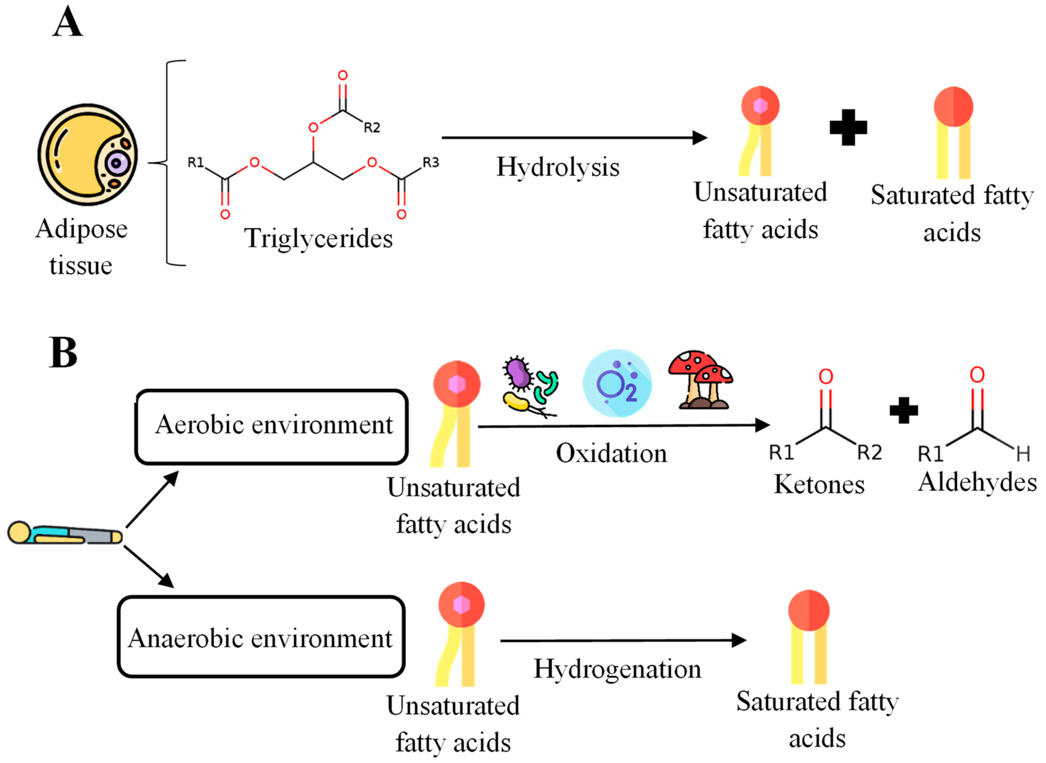
Figure 1. General scheme for the degradation process of postmortem adipose tissue. (A) Initial stages of the degradation process of adipose tissue. (B) Processes in aerobic and anaerobic environments after death.
In an aerobic environment, unsaturated fatty acids (UFAs) can be oxidized by bacteria, fungi, or atmospheric oxygen. The final products obtained in this reaction are ketones and aldehydes [2]. On the other hand, in an anaerobic environment, the mixture of saturated fatty acids (SFAs) and UFAs generated postmortem can undergo additional hydrolysis and hydrogenation. This hydrogenation process introduces a hydrogen bond that causes unsaturated bonds to become saturated.
Additionally, when cadaveric decomposition occurs, adipocere formation will occur if the body is in the appropriate temperature, humidity, and soil conditions. Subsequently, it has been proven that body humidity is sufficient for adipocere and can form in almost any environment [3]. Adipocere is a compound that has an essential use in forensic sciences since its appearance makes the corpse’s decomposition slower or, in some cases, allows the preservation of the remains [4]. Adipocire [3] is formed from the degradation of the body’s adipose tissue, and its composition is a mixture of SFAs and UFAs from the degradation of adipose tissue, fatty oxoacids, hydroxy fatty acids, and fatty acid salts. The composition of the adipocere is shown below [5] (Figure 2).
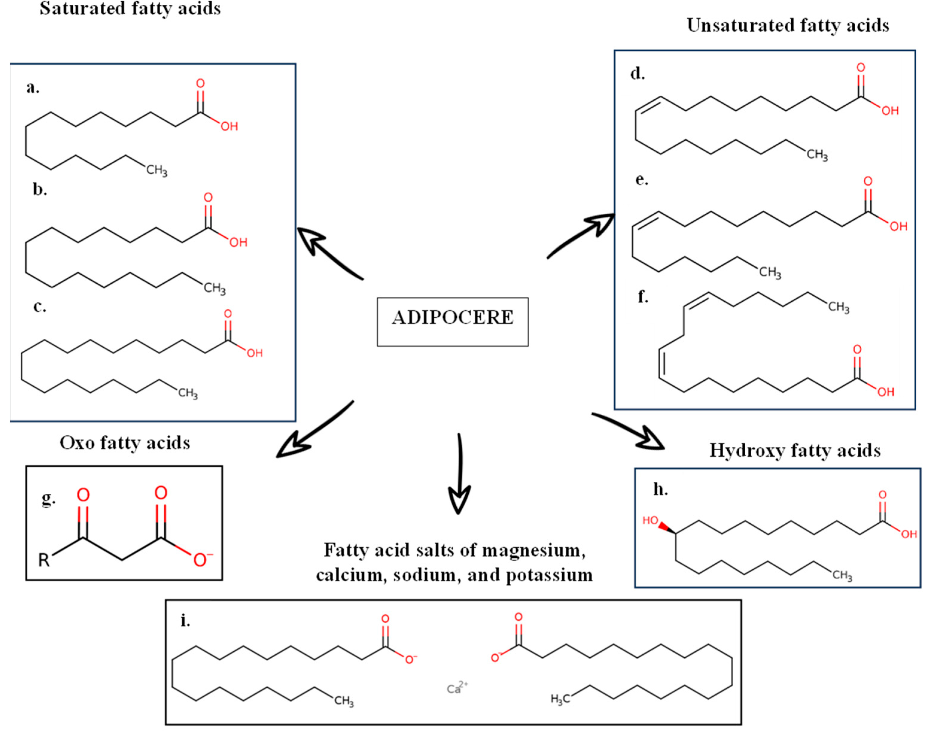
Figure 2. Composition of adipocere. (a) Myristic acid; (b) palmitic acid; (c) stearic acid; (d) oleic acid; (e) palmitoleic acid; (f) linoleic acid; (g) 3-oxo-fatty acid anion; (h) 10-hydroxystearic acid; (i) calcium stearate. Described by [5].
When a corpse is in contact with the ground, compounds generated during decomposition, including lipids, FA, and adipocere, could pass into it via leaching [6]. The soil easily adheres to clothing, the person, or can even remain on certain surfaces or materials, such as vehicles [1][6], making it an accessible sample. It should be noted that lipids and FAs are very stable and can remain for years in the corpse’s remains and its surroundings. Lipids and FAs have been detected in tissue samples taken from mummies [7]. Studies were also found where FAs can be found in the remains of a child from the late Roman period, such as myristic acid (C14:0), palmitic acid (C16:0), and stearic (C18:0) [1][8]. Therefore, due to the degradation process that postmortem lipids undergo and their good preservation over time, they are potential biomarkers for determining the postmortem interval (PMI) and burial places.
2. Postmortem Interval
The PMI is the time elapsed between death and the moment the body is found [9], and its determination is one of the great limitations in Forensic Sciences. Specific mechanisms are used to determine the PMI, the benefits of which vary depending on the stage of decomposition. Determining the short-term PMI has been the subject of some research studies [10]. This has led to the development of some methods for its determination, for example, biochemical studies, such as the analysis of amino acids and tissue organic acids or the determination of potassium in the aqueous humor of the eye, where it is possible to identify a relationship between the potassium concentration in the vitreous humor and PMI. Potassium concentration increases in the vitreous humor after death [10]. However, this method can only be used before the putrefaction process begins [10][11]. After the decomposition stage, the body undergo morphological changes. Some of these changes are the cooling of the body (algor mortis), increased rigidity (rigor mortis), the settling of blood in the sloping areas of the body (livor mortis), and the appearance of a purplish-pink color.
While short-term PMI determination has been widely studied, long-term PMI determination has not proven very interesting [12]. The significant problem in carrying out the determination of the PMI in the long term lies mainly in the inability to observe the morphological changes that are generated once death has occurred, especially if there is a loss of soft tissues of the individual due to scavengers or if the body has been affected by other environmental factors, such as fire, chemical agents, or affected by high temperatures [11].
Recently, there has been an increasing interest in the study of lipids for estimating long-term PMI since lipids undergo a degradation process as cadaveric decomposition occurs. This implies the possibility of finding a pattern of constant change in these compounds that allows researchers to relate it to the PMI.
3. Lipid Markers for Postmortem Interval Estimation
Since tissue lipids are degraded during the breakdown process, leading to FA, and considering the most commonly studied lipids, this section will be divided into studies based on glycerophospholipids (GPL) and FA variation profiles.
3.1. Glycerophospholipids as Markers for Postmortem Interval Estimation
Five studies were found that estimated the PMI by studying GPLs. The main results that concern this part of the review are collected in Table 1. The studies are presented in different study matrices and various organisms, encompassing both animals and humans, and involve different experimental conditions: sample treatment, decomposition time, different ages, and sex.
Table 1. Review of studies on lipid variations and their relationship with PMI. In the case of sex in human samples.
| Ref. | Sample | Human/Animal | Cadaver Sample Data | Postmortem Sampling (Days) | Category | Lipid Type | Results | |||||
|---|---|---|---|---|---|---|---|---|---|---|---|---|
| Sex | PMI of the Samples | Decomposition Environment | Age | Weight (kg) | Cause of Death | |||||||
| [13] | The medial calcaneus, proximal tibia, and vertebral body (fourth lumbar) | Human | - | Between less than 1 year and 30 years | External environment | - | - | - | Sampling every 6 months for 24 months | GPL | PC (34:1) PC (34:2) PC (36:1) PC (36:2) PC (36:4) | Decrease with PMI between the first and sixth month since the beginning of this study. |
| [9] | Anterior midshaft tibia | Human | 4 W | 2, 3, and 10 days | Two subjects in shallow open pits | Between 61 and 91 years old | - | - | Days 0, 219, 790, and 872 | GPL | LysoPC PI PC |
Drastic reduction in intensity of markers between the fresh state and the state after decomposition. |
| Two subjects buried in pits | ||||||||||||
| [14] | Biceps femoris muscle tissue. | Human | 1 W | - | Refrigerated | 69 | - | Suicide | Days 11, 12, 13, 14, and 15 | GPL | Choline phosphate | Shows an increasing pattern with PMI from day 7 to 19 postmortem. |
| 5 M | - | Refrigerated | 60 | - | Hemorrhagic stroke | Days 7, 8, 9, and 10 | ||||||
| Refrigerated | 62 | - | Pulmonary embolism | Days 11, 12, 13, 14, and 15 | ||||||||
| Refrigerated | 69 | - | Metastatic nonsmall cell lung cancer | Days 12, 13, 14, 15, and 16 | ||||||||
| External environment | 59 | - | Acute respiratory distress syndrome | Days 3, 4, and 5 | ||||||||
| External environment | 60 | - | Cardiovascular disease | Days 18 and 19 | ||||||||
| [11] | Vastus lateralis muscle | Human | - | - | External environment | - | - | - | Daily samples up to 2000 ADD or until the muscle is no longer available | GPL | PG (34:0) PC (36:2) PtdE (36:4) |
Decrease with PMI. Regression models were developed with PG 34:0 and PtdE 36:4. |
| Plasmalogen | PlsCh (34:2) PlsEtn (36:4) |
Decrease with PMI. | ||||||||||
| VLCFA | - | Increase with PMI. | ||||||||||
| [12] | Skeletal muscle | Human | - | - | - | - | - | - | Days 1, 9, and 24 | Sterol sulfate | Cholesterol sulfate and DHEA sulfate | Increase with PMI. |
| Plasmalogen | PlsE (36:1) and (40:6) PlsCh (34:1) and (36:4) |
Decrease with PMI. | ||||||||||
| GPL | PG | Decrease with PMI. | ||||||||||
| 5FA | VLCFA | Decrease with PMI. | ||||||||||
| PUFA | Increase with PMI. | |||||||||||
| [14] | Biceps femoris muscle tissue | Animal (rat) | 8 M | 0 days | Two of them immediately dissected | Adult | - | Euthanasia | Day 0, 1, 2, and 3 | GPL | Choline phosphate | There is an increase during the investigation period (3 days). |
| Six remaining were dissected for 3 days. | ||||||||||||
W: women and M: men, in animal samples. (-) Not indicated. ADD, Accumulated degree days; DHEA sulfate, dehydroepiandrosterone sulfate; FA, fatty acid; GPL, glycerophospholipid; LysoPC, Lysophosphatidylcholine; PC, phosphatidylcholine; PG, phosphatidylglycerol; PI, phosphatidylinositol; PlsCh, choline plasmalogen; PlsE, ethanolamine plasmalogen; PlsEtn, ethanolamine plasmalogen; PMI, postmortem interval; PtdE, phosphatidylethanolamine; PUFA, Polyunsaturated fat; VLCFA, very long-chain fatty acid. “Lipid (N:n)” indicates the total number of carbons that the molecule is composed of, and n is the number of double bonds it has.
It should be noted that the study times of human decomposition considered in the articles are highly variable, so the articles on human bone and those that analyze skeletal tissue samples were separated.
3.2. Human Bone Samples
On the other hand, Table 1 shows studies based on human bone samples. When the degradation process is too advanced, obtaining tissue samples from the corpse is impossible since it can be found in a skeletonized state. Bone is one of the matrices studied with the most interest to determine the PMI.
A recent study [13] analyzed fresh samples of human bone that were left deposited outside for 24 months and determined the presence of GPLs, in this case, PC. It was concluded that degradation of these PCs occurs within the first six months, but it indicates that the critical degradation period occurs between the first three months postmortem, and they are one of the GPLs that generate one of the signals more robust. Furthermore, these compounds can be maintained in bone for decades at low concentrations [13]. This means that PC can be a promising biomarker for determining long-term PMI. Nevertheless, although a clear relationship is observed between the degradation of PC and the PMI, the available data are insufficient to formulate regression equations for predicting the PMI. Therefore, the authors emphasize the need to continue investigating to enable the prediction of PMIs longer than three months [13].
Subsequently, Bonicelli et al. [9] obtained fresh human samples (3–10 days postmortem) from the middle anterior tibia of donors between 61 and 91 years old and deposited them in buried pits or pits open to the outside for 219, 790 and 872 days. This study focused on analyzing the concentration of GPLs (LysoPC, phosphatidylinositol (PI), and PC) in bone. After the analysis, they observed how these compounds’ concentrations decreased [9]. Since this study did not carry out continuous sampling over time, the exact moment of the decrease in the concentration of GPL could not be determined [9]. Differences between samples buried in pits and those deposited outside could also not be determined.
Therefore, only two studies have been conducted on human bone samples in which GPL has been analyzed for PMI. This indicates that these compounds decrease as the decomposition progresses (Figure 3), and both studies [9][13] highlight the great potential of GPL as biomarkers for estimating PMI due to their characteristic variation profile with decomposition and their ability to last over time in this sample type.
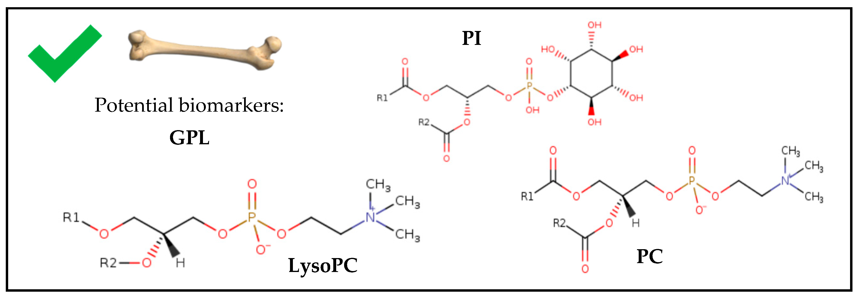
Figure 3. Chemical structures of potential biomarkers for estimating PMI in forensic human bone samples. Chemical structures obtained from ChEBI (Chemical Entities of Biological Interest) [15]. GPL, glycerophospholipid; LysoPC, Lysophosphatidylcholine; PC, phosphatidylcholine; PI, phosphatidylinositol.
Additionally, a timeline shows the variation profile of lipids in human bone samples (Figure 4).
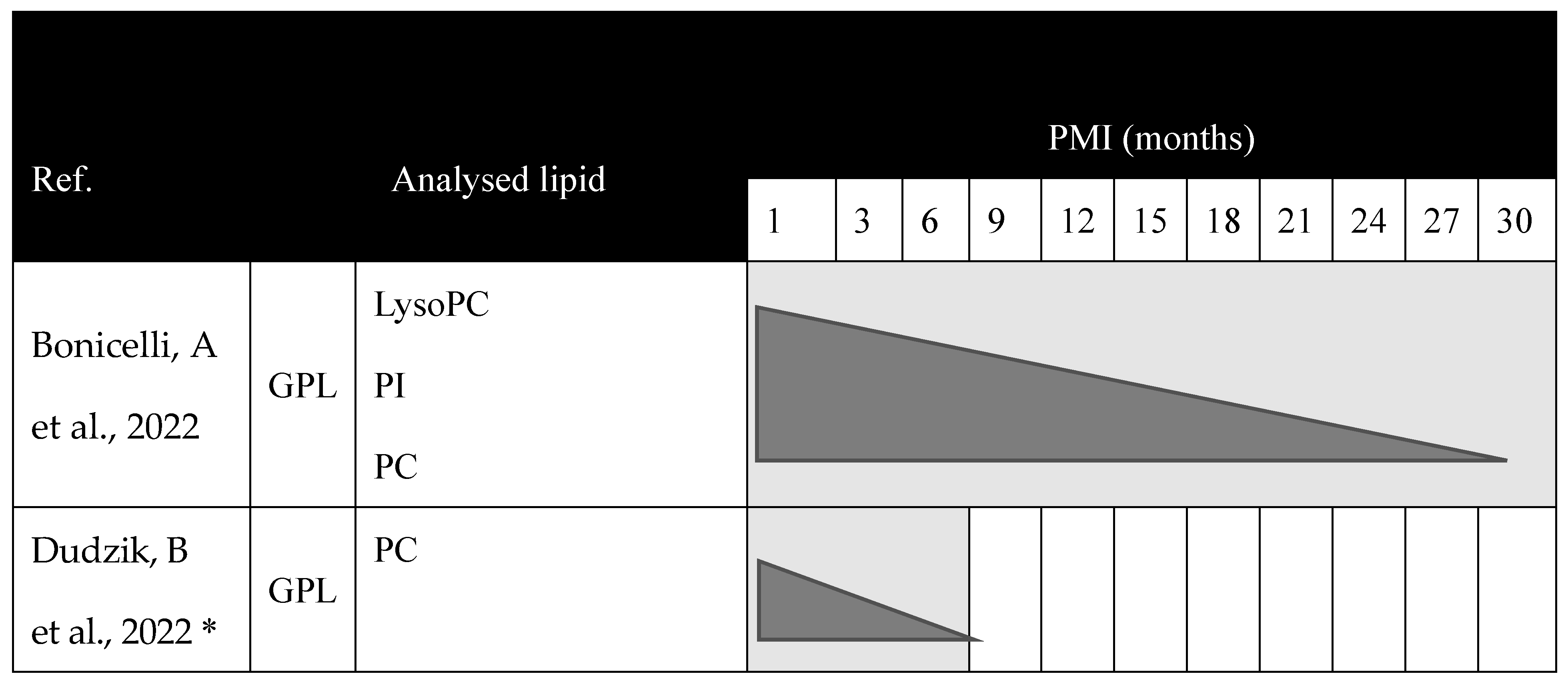
Figure 4. Chronogram of the variation of lipids collected in the systematic review during the process of cadaveric decomposition in human bone samples. * In this case, Dudzik, B et al., 2022 [13] indicates that the critical period for degradation of PC occurs from 0 to 3 months, but degradation occurs during the first six months. References [9][13] are cited in this figure. GPL, glycerophospholipid; LysoPC, Lysophosphatidylcholine; PC, phosphatidylcholine; PI, phosphatidylinositol; PMI, postmortem interval.
References
- Sousa Queirós, S.; von der Lühe, B.; Silva-Bessa, A.; Machado Brito-da-Costa, A.; Caldas, I.M.; Dawson, L.; Madureira-Carvalho, Á. Lipidic Compounds Found in Soils Surrounding Human Decomposing Bodies and Its Use in Forensic Investigations—A Narrative Review. Sci. Justice 2023, 63, 303–312.
- Stuart, B. Decomposition Chemistry: Overview, Analysis, and Interpretation. In Encyclopedia of Forensic Sciences; Elsevier: Amsterdam, The Netherlands, 2013; pp. 11–15. ISBN 978-0-12-382166-9.
- Forbes, S.L.; Keegan, J.; Stuart, B.H.; Dent, B.B. A Gas Chromatography-mass Spectrometry Method for the Detection of Adipocere in Grave Soils. Eur. J. Lipid Sci. Technol. 2003, 105, 761–768.
- Notter, S.J.; Stuart, B.H.; Rowe, R.; Langlois, N. The Initial Changes of Fat Deposits During the Decomposition of Human and Pig Remains. J. Forensic Sci. 2009, 54, 195–201.
- Magni, P.A.; Lawn, J.; Guareschi, E.E. A Practical Review of Adipocere: Key Findings, Case Studies and Operational Considerations from Crime Scene to Autopsy. J. Forensic Leg. Med. 2021, 78, 102109.
- Johari, I.S. Lipidomic Characterisation and Profiling of Soil for Forensic Investigations. Ph.D. Dissertation, University of Bristol, Bristol, UK, 2019.
- Comstock, J. Elucidation of the Lipid Degradation Process in Soft Tissue and Fluid during Decomposition, in the Presence and Absence of Insects. Ph.D. Dissertation, Ontario Tech University, Oshawa, ON, Canada, 2014.
- Fiedler, S.; Buegger, F.; Klaubert, B.; Zipp, K.; Dohrmann, R.; Witteyer, M.; Zarei, M.; Graw, M. Adipocere Withstands 1600 Years of Fluctuating Groundwater Levels in Soil. J. Archaeol. Sci. 2009, 36, 1328–1333.
- Bonicelli, A.; Mickleburgh, H.L.; Chighine, A.; Locci, E.; Wescott, D.J.; Procopio, N. The ‘ForensOMICS’ Approach for Postmortem Interval Estimation from Human Bone by Integrating Metabolomics, Lipidomics, and Proteomics. eLife 2022, 11, e83658.
- Wq, S. The Vitreous Humour: Postmortem Potassium Changes. Lancet 1963, 1, 807–808.
- Langley, N.; Wood, P.; Herring, P.; Steadman, D. Forensic Postmortem Interval Estimation from Skeletal Muscle Tissue: A Lipidomics Approach. Forensic Anthropol. 2019, 2, 152–157.
- Wood, P.L. Lipidomics Analysis of Postmortem Interval: Preliminary Evaluation of Human Skeletal Muscle. Metabolomics 2012, 3, 127.
- Dudzik, B.; Jantz, L.M.; Langley, N.R.; Wood, P. Postmortem Interval Determination from Bone: A Metabolomics and Lipidomics Approach; Office of Justice Programs: Washington, DC, USA, 2022.
- Pesko, B.K.; Weidt, S.; McLaughlin, M.; Wescott, D.J.; Torrance, H.; Burgess, K.; Burchmore, R. Postmortomics: The Potential of Untargeted Metabolomics to Highlight Markers for Time Since Death. OMICS 2020, 24, 649–659.
- Chemical Entities of Biological Interest (ChEBI). Available online: https://www.ebi.ac.uk/chebi/init.do (accessed on 5 July 2023).
More
Information
Subjects:
Medicine, Legal
Contributors
MDPI registered users' name will be linked to their SciProfiles pages. To register with us, please refer to https://encyclopedia.pub/register
:
View Times:
565
Revisions:
2 times
(View History)
Update Date:
25 Jan 2024
Table of Contents


Notice
You are not a member of the advisory board for this topic. If you want to update advisory board member profile, please contact office@encyclopedia.pub.
OK
Confirm
Only members of the Encyclopedia advisory board for this topic are allowed to note entries. Would you like to become an advisory board member of the Encyclopedia?
Yes
No
${ textCharacter }/${ maxCharacter }
Submit
Cancel
Back
Comments
${ item }
|
${ item.createdUser.fullName }
${ item.createdAt }
${ item.vote }
${ item.reply }
Delete
${ reply.createdUser.fullName }
${ reply.createdAt }
${ reply.vote }
Delete
There is no reply to this comment~
${ item.replyTextCharacter }/${ item.replyMaxCharacter }
Submit
Cancel
More
No more~
There is no comment~
${ textCharacter }/${ maxCharacter }
Submit
Cancel
${ selectedItem.replyTextCharacter }/${ selectedItem.replyMaxCharacter }
Submit
Cancel
Confirm
Are you sure to Delete?
Yes
No