Submitted Successfully!
To reward your contribution, here is a gift for you: A free trial for our video production service.
Thank you for your contribution! You can also upload a video entry or images related to this topic.
Version Summary Created by Modification Content Size Created at Operation
1 -- 2631 2024-01-16 12:52:16 |
2 Reference format revised. Meta information modification 2631 2024-01-17 02:16:39 |

Video Upload Options

Do you have a full video?

Confirm

Are you sure to Delete?
Cite
If you have any further questions, please contact Encyclopedia Editorial Office.
Bačenková, D.; Trebuňová, M.; Demeterová, J.; Živčák, J. Articular Cartilage. Encyclopedia. Available online: https://encyclopedia.pub/entry/53895 (accessed on 22 May 2024).
Bačenková D, Trebuňová M, Demeterová J, Živčák J. Articular Cartilage. Encyclopedia. Available at: https://encyclopedia.pub/entry/53895. Accessed May 22, 2024.
Bačenková, Darina, Marianna Trebuňová, Jana Demeterová, Jozef Živčák. "Articular Cartilage" Encyclopedia, https://encyclopedia.pub/entry/53895 (accessed May 22, 2024).
Bačenková, D., Trebuňová, M., Demeterová, J., & Živčák, J. (2024, January 16). Articular Cartilage. In Encyclopedia. https://encyclopedia.pub/entry/53895
Bačenková, Darina, et al. "Articular Cartilage." Encyclopedia. Web. 16 January, 2024.
Articular Cartilage
Edit

Articular cartilage is a load-bearing connective tissue that has a low self-repair potential. There are high demands placed on articular hyaline cartilage in the organism, mainly mechanical flexibility, load-bearing capacity, and the ability to reduce friction. The function of the cartilage in joints is to ensure low friction and the ability to distribute the weight load acting in the joint. An articular cartilage defect can persist without healing, or if it extends into the blood-filled subchondrium, then it is replaced by cartilage tissue that does not have suitable strength properties.

Hyaline cartilage extracellular matrix Chondrocytes

1. Introduction

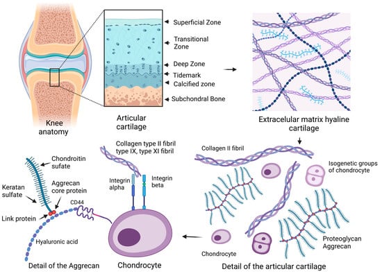
Hyaline cartilage is composed of a complex structured extracellular matrix (ECM) that facilitates friction on the surface of the articular cartilage and is adapted to repeated compression during movement. Synovial fluid has an important function in the nutrition of articular chondrocytes, as this process is ensured by diffusion and improving the lubrication of cartilage joint surfaces [1]. The joint capsule with its cell lining on the surface of the synovial membrane contains cells of the synovial intima called synoviocytes. Fibroblast-like cells are involved in the production of synovial fluid, and the macrophage-like type can be considered resident macrophage cell types [2]. In the organism, articular cartilage is located in the articular surfaces at the end of the epiphyses of long bones and consists of several interconnected zones, namely superficial, transitional, deep, and calcified areas separated from the underlying bone. Parts of the pelvis and the bones of the long limbs are replaced by bone through the process of ossification [3].

2. Articular Cartilage

2.1. Extracellular Matrix

Hyaline cartilage contains 70–80% water from the total volume, and the main components of the ECM are collagen and proteoglycans, comprising up to 50–70% of the dry weight. Specific cartilage is characterized by the presence of collagen type II (COL2) and the proteoglycan aggrecan. It is composed of layers differing in collagen type composition, relative orientation of collagen fibers, and cell density and cytomorphology, which generates a hierarchically organized cartilage structure. Chondrocytes, which represent a low volume percentage of the total tissue share, do not have a direct mechanical role. These cells in the ECM are without detectable mitotic activity in cartilage with a low metabolic turnover. The main functions of chondrocytes are the synthesis and degradation of the ECM [4]. The metabolic activity of chondrocytes is focused on the synthesis and maintenance of the components of the ECM.
Hyaline cartilage has no innervation and no vascularization, and it is characterized by a low pH mainly in deep zones (~6.9), with a minimum pO2 of 2–5% [5][6]. The ECM of hyaline cartilage consists of a highly organized network of interconnected structural proteoglycans and collagens. The ECM of the articular joint creates conditions for the basic mechanical properties of cartilage. Reducing friction and lubrication is made possible by a complex of collagen fibers interacting with hyaluronic acid or hyaluronan (HA) and lubricin on the surface of the joint. Collagens act as a stabilizing element with the ability to resist tension, and proteoglycans, which are negatively charged and connected to the collagen network, attract Na+ and H2O cations. The process of binding water causes an increase in tension in the collagen network, which gives the tissue the power to resist compression [5][7]. In hyaline cartilage, type II collagen fibers are represented the most, and type I, IV, V, VI, IX, and XI collagen are also present but in smaller concentrations, and their role is to mutually strengthen the fibers [8].
In terms of size and content, glycosaminoglycans (GAGs) and aggrecans (ACANs) are significant, containing more than 100 chondroitin sulfate (CS) and keratan sulfate (KS) chains which are bound to the core protein. ACANs are reported to be the main proteoglycans of hyaline cartilage, which interact with HA and form complex proteoglycan aggregates [6]. ACANs are bound to the HA fiber by covalent bonding, while this interaction is stabilized by a termed link protein. An anionic charge is present on the individual molecules of ACANs containing sulfated GAGs chains, while their localization in the matrix ensures the formation of aggregates and is essential for the function of ACANs.
ACAN molecules in the form of proteoglycan aggregates with a high molecular weight mainly ensure the strength properties and increased mechanical load-bearing requirements in cartilage tissue [1]. Proteoglycans characterized by shorter and less structured chains than ACANs have the ability to interact with collagen molecules. Decorin, biglycan, and fibromodulin do have structural properties like proteins, but they differ in their function from GAGs. Both decorin and fibromodulin interact with COL2 fibrils in the matrix and may play a role in fibrillogenesis and interfibril interactions [9]. HA forms aggregate structures with ACANs that are stabilized by a binding protein. The resulting spatial network represents the structures that are anchored within collagen fibrillar networks in articular cartilage [10]. HA-ACAN aggregates affect the water content and influence the hydrostatic pressure in cartilage. In addition to the structural osmotic function, ACANs enable the transport of dissolved substances and essential nutrients in the tissue of hyaline cartilage, while this process is related to the ability of hydration. The structure and content of ACANs in cartilage are not constant during human life. Overall, it can be said that aggregate molecules provide cartilage with specific osmotic properties, thereby optimizing the ability of articular cartilage to withstand high-pressure loads. CS and KS form GAG, in which they act as structural molecules of extracellular matrices [11][12] (Figure 1).
Figure 1. Schematic of an articular joint and details of the extracellular matrix (ECM) and chondrocytes. The arrows between the images mean a more detailed view in the overall scheme. Hyaline cartilage is composed of a complex structured ECM with the presence of collagen type II (COL2) and the proteoglycan aggrecan. Articular cartilage affects collagen fiber orientation, cell density, and cytomorphology, which creates a hierarchically organized cartilage structure. Glycosaminoglycans (GAGs) and aggrecans (ACANs) are present in the ECM, which contain chains of chondroitin sulfate (CS) and keratan sulfate (KS). ACANs, as the main proteoglycans of hyaline cartilage, interact with hyaluronan (HA) fibers and form complex proteoglycan aggregates. ACANs are bound to the HA fiber by link and core proteins. Mature chondrocytes of hyaline cartilage are situated in lacunae in isogenetic groups of several cells in a territorial matrix located in hyaline cartilage in a specific physicochemical environment. The membrane receptor of chondrocytes is the hyaluronic receptor CD44. Articular chondrocytes express alpha and beta integrins of several types, which interact with COL2 and proteoglycans molecules in the ECM (created with BioRender.com, accessed on 1 November 2023).

2.2. Chondrocyte

The cell population of chondrocytes in healthy articular cartilage represents typical resting and differentiated cells, whose task is to maintain a dynamic relationship between anabolism and catabolism of the ECM [13]. Mature chondrocytes of hyaline cartilage are stored in lacunae in isogenetic groups of several cells in the territorial matrix located in hyaline cartilage in a specific physicochemical environment. The mentioned chondrocytes were created by mitotic division from one parent cell [14]. Chondrocytes have a round or oval shape and fill only 5–10% of the cartilage volume in total. The cytoplasm of chondrocytes is characterized by a small spherical nucleus, and it also contains the Golgi apparatus, mitochondria, and less numerous lipid droplets [15].

2.2.1. Progenitor Cells of Cartilage

The process of cartilage formation is initiated in the early period of embryogenesis by a population of prechondrocytic mesenchymal stem cells (MSCs). Chondrogenesis is a process of condensation of MSCs by forming densely packed cell aggregates where, subsequently, differentiation into prechondrocytes occurs [16][17]. The ontogenesis of the organism includes the activity of growth processes in tissues, specifically the complex process of endochondral ossification. The initial differentiation of growth plate chondrocytes during endochondral bone in vivo is affected by signaling activated by homeobox genes and soluble mediators. The SOX gene family is bound by the amino acid sequences and DNA-binding domains of the high-mobility group box (HMGB). SRY-box transcription factor 9 (SOX-9) is present in the process, which has a regulatory role in post-transcriptional and post-translational transcripts. Zhao et al. described cooperation between the high expression of SOX-9 and the high expression of collagen type II alpha-1 gene (Col2A1) with the assumption that it is necessary for the full expression of the chondrocyte phenotype. The Col2A1 gene encodes the COL2 molecule, which is characteristic of hyaline cartilage [18]. The differentiation and proliferation of chondrocytes are influenced by growth factors and, at the same time, interaction through contact with the ECM, which is essential for the maintenance of the cell phenotype. During the differentiation of chondrocytes, the cells and the microenvironment of the ECM cooperate through integrin receptors, which are part of the ECM [19].

2.2.2. Characteristic Phenotype of Chondrocytes

Mature chondrocytes produce structural proteins, COL2, collagen types IX (COL9) and XI (COL11), and ACAN [20]. Articular chondrocytes with the unaffected, natural phenotype also synthesize lubricin, or proteoglycan-4 (PRG4), and glycoprotein. Its lubrication function is significant and has an effect on the reduction of friction between the applied cartilage surfaces. PRG4 has a suppressive effect on the function of inflammatory cytokines and their activation of the proliferation of RA synovial fibroblasts [21].
The integrin family has a role in the process of cell adhesion as well as in cell-to-cell interactions and interactions with the ECM. Integrins as transmembrane receptors recognize specific ECM molecules [14][16][22]. The intracellular part of integrins, extending into the cytoplasm, is connected to the cytoskeleton of the cell. The extracellular part is connected to ligands and triggers cell activation. Signaling through integrins enables transmitting signal molecules in both directions. Articular chondrocytes express α1β1, α3β1, α5β1, α10β1, αVβ1, αVβ3, and αVβ5 integrins [19][23]. The α5β1 and αVβ3 integrins bind to the sequence Arg-Gly-Asp (RGD), which is contained in several ECM proteins [19].
Integrin α10β1 is an important integrin that binds collagen in cartilage tissue. Camper et al. described integrin α10β1 as a type II collagen-binding receptor. It is a unique marker for determining the phenotype of chondrocytes. Integrin α10β1 is part of the cell–matrix interaction which is essential for cartilage development and the chondrogenesis of MSCs [23][24]. The function in the morphogenesis of growth plates was observed by the authors in an animal model with a disorder, namely a deletion for the alpha10 integrin gene. A defect in the ontogenesis and growth of long bones were manifested in mice [25]. Furthermore, chondrocytes express types of integrins binding to ECM molecules [26]. Achorin V belongs to annexins, being a molecule that binds to type II collagen. The most abundant receptors for fibronectin (α5β1) are for COL2 and VI (α1β1, α2β1, and α11β1), vitronectin, osteopontin (αVβ3), and laminin (α6β1) [27][28]. An important membrane receptor of chondrocytes is the hyaluronic receptor CD44 [29]. The binding of chondrocytes and HA affects the homeostasis of the cartilage environment [30]. In the case of blocking the binding of CD44 and HA, damage to and degradation of the ECM occurs [26].

2.2.3. Specific Growth Factors during the Expansion of Articular Chondrocytes

In addition to the binding of integrins to ECM molecules, cell metabolism is also regulated in a paracrine influence (i.e., by the release of soluble factors). Several studies have reported that growth factors have a stimulating effect on the proliferation of mammalian chondrocytes [31][32][33]. The transforming growth factor beta (TGF-β) superfamily has a broad role in physiological and pathological events, affecting the adhesion, growth, and differentiation of a variety of tissue cells [34]. Over 30 proteins belong to this group of activins and bone morphogenetic proteins (BMPs) [35]. It is involved in maintaining homeostasis, embryogenesis, and immune events [36][37]. Regarding biochemical events in connective tissues, TGF-β participates in the formation of cartilage and bone tissue [38]. TGF-β activates signaling cascades, of which the TGF-β/Smad signal pathway is the best known one, for enacting and modulating the gene expression of several proteins [39]. After activation with other factors, TGF-β is able to form a serine/threonine kinase complex [40]. This triggers the signaling pathways that regulate differentiation, proliferation, and immune processes [41][42]. It is an interesting fact that animal models of rodents that have excessive expression of TβRII in cartilage are often affected by joint arthrosis [43]. Furthermore, they have an increased expression of markers for collagen type X (COL10) [44].
The most widely studied growth factor from the TGF-β superfamily is the TGF-β1 factor [45]. TGF-β2 is also a subject of studies on disorders and the treatment of cartilage defects [46]. TGF-β contains the following homologous dimeric isoforms: TGF-β1 and TGF-β3. TGF-β has a downregulating effect on osteogenesis, while its pleiotropic function is dependent on the interaction with the surrounding environment. TGF-β2 is involved in the Smad canonical signaling pathway and is also involved in generic mitogen-activated protein kinase (MAPK) activation in chondrocytes [47]. A possible function of TGF-β2 in chondrocyte redifferentiation through activin receptor kinase 5 (ALK5)/Smad3 under hypoxic conditions was considered [48]. In the experiment, the authors observed the redifferentiation of chondrocytes in culture with the stimulating effect of the growth factors, namely fibroblast growth factor (FGF) and TGFβ [49]. Due to the influence of the mentioned growth factors, the chondrocytes showed an increased proliferation rate compared with the control, as well as differentiation into a chondrocyte phenotype with COL2A1 expression [32].
BMPs belong to the TGF-β family. BMPs have a stimulating effect during chondrogenesis. They preferentially support the condensation step at the beginning of chondrogenesis. The process is related to the stimulation of N-cadherin expression promoting cell-cell contact. BMPs affect the expression of SOX-9 and type II collagen [15].

2.2.4. Biomechanical Principles of Articular Cartilage and Chondrocytes

Movement is an essential part of the life of living organisms. At the cellular level, complex mechanical stress acts on tissues and cells. Cartilage metabolism during ontogeny and throughout the life of organisms is influenced by mechanical factors that directly control the activation and expression of genes for growth, metabolism, and the resulting phenotype of chondrocytes [50]. Articular chondrocytes respond to mechanical signals transmitted through the ECM with metabolic activity. Mechanical influences such as intermittent fluid pressure act to preserve the chondrocyte phenotype. Changes in the shape and volume of chondrocyte nuclei in connection with ECM deformation were discovered. During micromechanical analysis, chondrocytes were observed to have viscoelastic properties but with lower mechanical strength than pericellular ECM [51]. Mild tension and shear action have a stimulating effect on growth and ossification. During ontogenesis, the thickness of the articular cartilage is formed, while it is most massive in the place with the greatest bearing load. In articular joints, the health of the cartilage is proportional to its load [52]. The production of ECM by chondrocytes depends on the zone in which they are located. Superficial zone chondrocytes exposed to fluid flow and mechanical pressure produce higher amounts of type II collagen compared with chondrocytes in the middle and deep radial zones, which synthesize high amounts of GAGs in addition to type II collagen [52][53].
From a mechanical point of view, cartilage has a multiphase nature. Cartilage can be characterized from a physicochemical point of view as a viscoelastic material that contains the following two-phase environment: a solid phase and a fluid phase [54]. The solid phase contains ECM components and collagen fibers linked to ACAN and HA. The liquid phase consists of water with a content of up to 80% wet weight and ions, as well as calcium, sodium, and chloride. Mechanical loading of joints is essential for the nutrition of articular chondrocytes and the synthesis of ECM components. Immobilization and restriction of locomotion causes a decrease in the metabolic activity of chondrocytes and a functional weakening of the cartilage [55]. The experimental results showed that up to 75% of the applied load on the joint surface was compensated by the liquid phase. The viscoelastic properties of the articular cartilage ensure the functionality of the joint under repeated loading. When articular contact forces arise during loading, the pressure of the interstitial fluid increases. When the joint is loaded, the pressure increases. The ECM environment is permeable, and with increased pressure, part of the fluid from the ECM can pass into the liquid phase. After the pressure is removed, the interstitial fluid flows back. The process of multi-phase layers in the articular cartilage ensures low friction and the resulting biomechanical functionality of the articular joints.

References

  1. Roughley, P.J.; Lee, E.R. Cartilage proteoglycans: Structure and potential functions. Microsc. Res. Tech. 1994, 28, 385–397.
  2. Iwanaga, T.; Shikichi, M. Morphology and functional roles of synoviocytes in the joint. Arch. Histol. Cytol. 2000, 63, 17–31.
  3. Hunziker, E.B.; Lippuner, K.; Keel, M.J.B.; Shintani, N. An educational review of cartilage repair: Precepts & practice—Myths &misconceptions—Progress & prospects. Osteoarth Cart. 2015, 23, 334–350.
  4. Kurenkova, A.D.; Romanova, I.A.; Kibirskiy, P.D.; Timashev, P. Strategies to convert cells into hyaline cartilage: Magic spells for adult stem cells. Int. J. Mol. Sci. 2022, 23, 11169.
  5. Hall, A.C.; Horwitz, E.R.; Wilkins, R.J. The cellular physiology of articular cartilage. Exp. Physiol. Transl. Integr. 1996, 81, 535–545.
  6. Dudhia, J. Aggrecan, aging and assembly in articular cartilage. Cell. Mol. Life Sci. 2005, 62, 2241–2256.
  7. Hodgkinson, T.; Kelly, D.C.; Curtin, C.M. Mechanosignalling in cartilage: An emerging target for the treatment of osteoarthritis. Nat. Rev. Rheumatol. 2022, 18, 67–84.
  8. Zhang, L.; Hu, J.; Athanasiou, K.A. The role of tissue engineering in articular cartilage repair and regeneration. Crit. Rev. Biomed. Eng. 2009, 37, 1–57.
  9. Kheir, E.; Shaw, D. Hyaline articular cartilage. Orthop. Trauma 2009, 23, 450–455.
  10. Hayes, A.J.; Melrose, J. Glycosaminoglycan and proteoglycan biotherapeutics in articular cartilage protection and repair strategies: Novel approaches to visco-supplementation in orthobiologics. Adv. Ther. 2019, 2, 1900034.
  11. Pomin, V.H. Keratan sulfate: An up-to-date review. Int. J. Biol. Macromol. 2015, 72, 282–289.
  12. Krishnan, Y.; Grodzinsky, A.J. Cartilage diseases. Matrix Biol. 2018, 71, 51–69.
  13. Smith, L.R.; Trindade, M.C.D.; Ikenoue, T. Effects of shear stress on articular chondrocyte metabolism. Biorheology 2000, 37, 95–107.
  14. Fox, S.; Bedi, A.J.; Rodeo, S.A. The basic science of articular cartilage: Structure, composition, and function. Sport. Health 2009, 1, 461–468.
  15. Lin, Z.; Willers, C.; Xu, J.; Zheng, M.H. The chondrocyte: Biology and clinical application. Tissue Eng. 2006, 12, 1971–1984.
  16. Cancedda, R.; Cancedda, F.D.; Castagnola, P. Chondrocyte differentiation. Int. Rev. Cytol. 1995, 159, 265–358.
  17. Responte, D.J.; Lee, J.K.; Hu, J.C.; Athanasiou, K.A. Biomechanics-driven chondrogenesis: From embryo to adult. FASEB J. 2012, 26, 3614.
  18. Zhao, Q.; Eberspaecher, H.; Lefebvre, V.; de Crombrugghe, B. Parallel expression of Sox9 and Col2a1 in cells undergoing chondrogenesis. Dev. Dyn. Off. Publ. Am. Assoc. Anat. 1997, 209, 377–386.
  19. Loeser, R.F. Integrins and chondrocyte–matrix interactions in articular cartilage. Matrix Biol. 2014, 39, 11–16.
  20. Heinegård, D. Fell-Muir Lecture: Proteoglycans and more–from molecules to biology. Int. J. Exp. Pathol. 2009, 90, 575–586.
  21. Tseng, C.C.; Chen, Y.J.; Chang, W.A.; Tsai, W.C. Dual role of chondrocytes in rheumatoid arthritis: The chicken and the egg. Int. J. Mol. Sci. 2020, 21, 1071.
  22. Ecke, A.; Lutter, A.H.; Scholka, J.; Hansch, A. Tissue specific differentiation of human chondrocytes depends on cell microenvironment and serum selection. Cells 2019, 8, 934.
  23. Camper, L.; Hellman, U.; Lundgren-Åkerlund, E. Isolation, cloning, and sequence analysis of the integrin subunit α10, a β1-associated collagen binding integrin expressed on chondrocytes. J. Biol. Chem. 1998, 273, 20383–20389.
  24. Lundgren-Åkerlund, E.; Aszòdi, A. Integrin α10β1: A collagen receptor critical in skeletal development. Adv. Exp. Med. Biol. 2014, 819, 61–71.
  25. Bengtsson, T.; Aszodi, A.; Nicolae, C.; Hunziker, E.B.; Lundgren-Åkerlund, E. Loss of α10β1 integrin expression leads to moderate dysfunction of growth plate chondrocytes. J. Cell Sci. 2005, 118, 929–936.
  26. Knudson, W.; Casey, B.; Nishida, Y.; Eger, W. Hyaluronan oligosaccharides perturb cartilage matrix homeostasis and induce chondrocytic chondrolysis. Arthritis Rheum. Off. J. Am. Coll. Rheumatol. 2000, 43, 1165–1174.
  27. Salter, D.M.; Hughes, D.E.; Simpson, R. Integrin expression by human articular chondrocytes. Rheumatology 1992, 31, 231–234.
  28. Enomoto, M.; Leboy, P.S.; Menko, A.S. β1 integrins mediate chondrocyte interaction with type I collagen, type II collagen, and fibronectin. Exp. Cell Res. 1993, 205, 276–285.
  29. Aguiar, D.J.; Knudson, W.; Knudson, C.B. Internalization of the hyaluronan receptor CD44 by chondrocytes. Exp. Cell Res. 1999, 252, 292–302.
  30. Ishida, O.; Tanaka, Y.; Morimoto, I.; Takigawa, M. Chondrocytes are regulated by cellular adhesion through CD44 and hyaluronic acid pathway. J. Bone Miner. Res. 1997, 12, 1657–1663.
  31. Hu, X.; Zhang, W.; Li, X.; Zhong, D.; Li, Y.; Li, J. Strategies to modulate the redifferentiation of chondrocytes. Front. Bioeng. Biotechnol. 2021, 9, 764193.
  32. Jakob, M.; Demarteau, O.; Schäfer, D.; Hintermann, B.; Dick, W. Specific growth factors during the expansion and redifferentiation of adult human articular chondrocytes enhance chondrogenesis and cartilaginous tissue formation in vitro. J. Cell. Biochem. 2001, 81, 368–377.
  33. Benya, P.D.; Shaffer, J.D. Dedifferentiated chondrocytes reexpress the differentiated collagen phenotype when cultured in agarose gels. Cell 1982, 30, 215–224.
  34. Burt, D.W.; Law, A.S. Evolution of the transforming growth factor-beta superfamily. Prog. Growth Factor Res. 1994, 5, 99–118.
  35. De Caestecker, M. The transforming growth factor-β superfamily of receptors. Cytokine Growth Factor Rev. 2004, 15, 1–11.
  36. Yang, D.; Dai, F.; Yuan, M.; Zheng, Y. Role of transforming growth factor-β1 in regulating fetal-maternal immune tolerance in normal and pathological pregnancy. Front. Immunol. 2021, 12, 689181.
  37. Mantel, P.Y.; Schmidt-Weber, C.B. Transforming growth factor-beta: Recent advances on its role in immune tolerance. Methods Mol. Biol. 2011, 677, 303–338.
  38. Van Osch, G.J.; Van Der Veen, S.W.; Buma, P.; Verwoerd-Verhoef, H.L. Effect of transforming growth factor-β on proteoglycan synthesis by chondrocytes in relation to differentiation stage and the presence of pericellular matrix. Matrix Biol. 1998, 17, 413–424.
  39. Luo, K. Signaling cross talk between TGF-β/Smad and other signaling pathways. Cold Spring Harb. Perspect. Biol. 2017, 9, a022137.
  40. Duan, D.; Derynck, R. Transforming growth factor–β (TGF-β)–induced up-regulation of TGF-β receptors at the cell surface amplifies the TGF-β response. J. Biol. Chem. 2019, 294, 8490–8504.
  41. Battegay, E.J.; Raines, E.W.; Seifert, R.A.; Bowen-Pope, D.F. TGF-β induces bimodal proliferation of connective tissue cells via complex control of an autocrine PDGF loop. Cell 1990, 63, 515–524.
  42. Zhang, Y.; Alexander, P.B.; Wang, X.F. TGF-β family signaling in the control of cell proliferation and survival. Cold Spring Harb. Perspect. Biol. 2017, 9, a022145.
  43. Bailey, K.N.; Nguyen, J.; Yee, C.S.; Dole, N.S.; Dang, A. Mechanosensitive control of articular cartilage and subchondral bone homeostasis in mice requires osteocytic transforming growth factor β signaling. Arthritis Rheumatol. 2021, 73, 414–425.
  44. Li, T.F.; O’Keefe, R.J.; Chen, D. TGF-β signaling in chondrocytes. Front. Biosci. 2005, 10, 681.
  45. Puolakkainen, P.A.; Twardzik, D.R.; Ranchalis, J.E. The enhancement in wound healing by transforming growth factor-β1 (TGF-β1) depends on the topical delivery system. J. Surg. Res. 1995, 58, 321–329.
  46. Critchlow, M.A.; Bland, Y.S.; Ashhurst, D.E. The effect of exogenous transforming growth factor-β2 on healing fractures in the rabbit. Bone 1995, 16, 521–527.
  47. Wells, R.G. TGF-β signaling pathways. Am. J. Physiol. Gastrointest. Liver Physiol. 2000, 279, 845–850.
  48. Duan, M.; Wang, Q.; Liu, Y.; Xie, J. The role of TGF-β2 in cartilage development and diseases. Bone Jt. Res. 2021, 10, 474–487.
  49. Das, R.; Timur, U.T.; Edip, S.; Haak, E.; Wruck, C.; Weinans, H. TGF-β2 is involved in the preservation of the chondrocyte phenotype under hypoxic conditions. Ann. Anat. Anat. Anz. 2015, 198, 1–10.
  50. Park, J.H.; Ushida, T.; Akimoto, T. Control of cell differentiation by mechanical stress. J. Phys. Fit. Sport. Med. 2013, 2, 49–62.
  51. Guilak, F. The deformation behavior and viscoelastic properties of chondrocytes in articular cartilage. Biorheology 2000, 37, 27–44.
  52. Carter, D.R.; Beaupré, G.S.; Wong, M.; Smith, R.L. The mechanobiology of articular cartilage development and degeneration. Clin. Orthop. Relat. Res. 2004, 427, 69–77.
  53. Franzen, A.; Inerot, S.; Hejderup, S. Variations in the composition of bovine hip articular cartilage with distance from the articular surface. Biochem. J. 1981, 195, 535–543.
  54. Eschweiler, J.; Horn, N.; Rath, B.; Betsch, M.; Baroncini, A. The biomechanics of cartilage—An overview. Life 2021, 11, 302.
  55. Lu, X.L.; Mow, V.C. Biomechanics of articular cartilage and determination of material properties. Med. Sci. Sport. Exerc. 2008, 40, 193–199.
More
Information
Subjects: Orthopedics
Contributors MDPI registered users' name will be linked to their SciProfiles pages. To register with us, please refer to https://encyclopedia.pub/register : , , ,
View Times: 222
Revisions: 2 times (View History)
Update Date: 17 Jan 2024
1000/1000
Video Production Service