Your browser does not fully support modern features. Please upgrade for a smoother experience.
Submitted Successfully!
 +1 credit
Thank you for your contribution! You can also upload a video entry or images related to this topic.
For video creation, please contact our Academic Video Service.
| Version | Summary | Created by | Modification | Content Size | Created at | Operation |
|---|---|---|---|---|---|---|
| 1 | William Tansey | -- | 3583 | 2024-01-11 14:10:41 | | | |
| 2 | Jason Zhu | Meta information modification | 3583 | 2024-01-15 04:06:00 | | |
Video Upload Options
We provide professional Academic Video Service to translate complex research into visually appealing presentations. Would you like to try it?
Cite
If you have any further questions, please contact Encyclopedia Editorial Office.
Weissmiller, A.; Fesik, S.; Tansey, W. WD Repeat Domain 5 Inhibitors for Cancer Therapy. Encyclopedia. Available online: https://encyclopedia.pub/entry/53750 (accessed on 05 July 2026).
Weissmiller A, Fesik S, Tansey W. WD Repeat Domain 5 Inhibitors for Cancer Therapy. Encyclopedia. Available at: https://encyclopedia.pub/entry/53750. Accessed July 05, 2026.
Weissmiller, April, Stephen Fesik, William Tansey. "WD Repeat Domain 5 Inhibitors for Cancer Therapy" Encyclopedia, https://encyclopedia.pub/entry/53750 (accessed July 05, 2026).
Weissmiller, A., Fesik, S., & Tansey, W. (2024, January 11). WD Repeat Domain 5 Inhibitors for Cancer Therapy. In Encyclopedia. https://encyclopedia.pub/entry/53750
Weissmiller, April, et al. "WD Repeat Domain 5 Inhibitors for Cancer Therapy." Encyclopedia. Web. 11 January, 2024.
Copy Citation
WDR5 is a conserved nuclear protein that scaffolds the assembly of epigenetic regulatory complexes and moonlights in functions ranging from recruiting MYC oncoproteins to chromatin to facilitating the integrity of mitosis. It is also a high-value target for anti-cancer therapies, with small molecule WDR5 inhibitors and degraders undergoing extensive preclinical assessment. WDR5 inhibitors were originally conceived as epigenetic modulators, proposed to inhibit cancer cells by reversing oncogenic patterns of histone H3 lysine 4 methylation—a notion that persists to this day.
cancer
cancer therapy
epigenetic
1. Introduction
Since the first small molecule inhibitors of WDR5 (WD Repeat Domain 5) were described a decade ago [1], the field has advanced rapidly. There has been expanding interest in the idea of targeting WDR5 for cancer therapy, a growing list of potential cancer indications, and a profound evolution of chemical matter, with orally bioavailable picomolar inhibitors now in hand for preclinical vetting [2]. Despite this progress, mainstream thinking about how WDR5 inhibitors work has, for the most part, not kept up with the pace of drug discovery. Indeed, tenuous assumptions about targeting WDR5 for cancer therapy are prevalent, and if unchallenged their impact is only likely to be amplified as these agents make their way to the clinic.
2. WDR5
WDR5 is a highly conserved, highly structured, and highly networked protein [3]. As described, its participation in a multitude of tumor-relevant processes has made it a prized target for anti-cancer drug discovery. It is a core component of at least five distinct histone modifying complexes—the SET1/MLL histone methyltransferase (HMT), the NSL and ATAC histone acetyltransferases (HATs), the PRC1.6 histone ubiquitin ligase, and an embryonic cell-specific NuRD nucleosome remodeling and histone deacetylase (HDAC) complex [3]. It is a co-factor for multiple transcription factors, including Oct4, Twist1, the retinoic acid receptor, and MYC (myelocytomatosis) [3]; functions as a “reader” of modified histone H3 tails [3][4]; regulates ribosomal protein gene (RPG) transcription [5]; and associates with the anaphase promoting complex (APC) to bookmark genes for rapid reactivation after mitosis [6]. And if this was not enough, WDR5 also recruits the kinesin motor protein KIF2A to the mitotic spindle [7], promotes ciliogenesis via the stabilization of F-actin [8], and is a hub for interactions with long non-coding RNAs such as HOTTIP [9], NeST [10], ANRIL [11], and NEAT1 [12]. These activities are disparate and not all are established to the same level of rigor. But what unites most of them—apart from epigenetic roles—are two things: they were discovered in the last ten years and they receive little consideration in mainstream thinking about the mechanism of action and potential indications of WDR5 inhibitors as anti-cancer agents.
3. Strategies to Target WDR5
Despite many functions and dozens of binding partners, one curious aspect of WDR5 behavior is that all proteins that directly interact with WDR5 do so through one of two interfaces (Figure 1a) [3]. On one surface of WDR5 is a shallow hydrophobic cleft referred to as the “WBM” (WDR5-binding motif) site. As its name implies, the WBM site docks with proteins carrying a WBM, which includes the SET1/MLL complex member RBBP5, the NSL component KANSL2, and MYC family proteins. Partners that bind the WBM site of WDR5 do so with almost identical modalities. On the opposing surface of WDR5 is the “WIN” (WDR5-interaction) site, a deep arginine-binding cavity that engages an arginine-containing WIN motif present in proteins such as SET1/MLL family members, KANSL1, and histone H3, all of which bind WDR5 in nearly identical fashions. The recurring use of these two sites sets up mutually exclusive interactions that may help WDR5 neatly assemble into its different complexes [3], and it makes devising strategies for pharmacologically targeting WDR5 fairly straightforward: find a drug-like inhibitor of the WBM or WIN sites, or discover a PROTAC (PRoteolysis TArgeting Chimera) (Figure 1b).
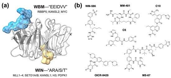
Figure 1. Two sites on WDR5 mediate all known direct protein–protein interactions. (a) Shown is the crystal structure of WDR5 (gray ribbons) in complex with a WBM-containing peptide from RBBP5 (blue surface/sticks) and a WIN-containing peptide from histone H3 (yellow surface/sticks), PDB:2XL [13]. The consensus motifs for the WBM and WIN site binding partners are shown, along with the names of representative examples of proteins that engage WDR5 via each site. (b) Structures of representative WDR5 inhibitors. WM-586—a covalent WBM site inhibitor [14]; MM-401—a peptidomimetic WIN site inhibitor [15]; C6—an early generation WIN site inhibitor [16]; C10—orally bioavailable WIN site inhibitor [2]; OICR-9429—one of the first small molecule WIN site inhibitors [17]; MS-67—a WIN site-directed PROTAC [18].
A number of WBM site inhibitors have been discovered [14][19][20][21], but these have yet to progress sufficiently for thorough interrogation in cells or animal models. The discovery of WIN site inhibitors (WINi), in contrast, has been very successful (e.g., [1][2][15][16][17][22][23][24][25][26][27][28][29][30][31][32][33][34]), due in large part to the tractability of the WIN site and a deep understanding of structure–activity relationships in this class of agents. Given their prominence, advanced state, and the extent to which they have been profiled in vitro and in vivo, it is likely that WINi will dominate the field in the near future.
4. The Premise and the Promise
A set of landmark papers in the early 2000s gave WDR5 its epigenetic credentials by establishing its role in scaffolding the assembly of the so-called “MLL/SET” family of HMTs [35][36][37][38]. In these multiprotein complexes, one of six MLL/SET enzymes (MLL1–MLL4, SETD1A, or SETD1B) [39] associates with a “WRAD” core (WDR5, RBBP5, ASH2L, and DPY30), as well as ancillary proteins, to catalyze histone H3 lysine four di- and tri-methylation (H3K4me2/3)—marks of transcriptionally active chromatin [40]. The enzymatic activity of MLL/SET proteins is stimulated by their association with WRAD, but in different ways depending on the family member [41][42]. MLL1 (Mixed Lineage Leukemia 1) activity as a di- and tri-methylase is dependent on insertion of the WIN motif in its carboxy-terminus into the WIN site of WDR5, and, accordingly, the catalytic activity of MLL1–WRAD complexes is profoundly and uniquely sensitive to WIN site inhibition [15][16]. In broad terms, therefore, the genesis of WDR5 inhibition for cancer therapy—reduced to practice in 2014 [1][15]—is the notion of selective inhibition of MLL1.
At that time, inhibiting MLL1 via WIN site blockade had a clear indication: MLL-rearranged leukemias (MLLr) [15]. In these cancers, one copy of MLL1 is translocated to 1 of up to 100 different loci, leading to the expression of a fusion protein that, despite having no HMT activity, is profoundly oncogenic [39]. Widespread retention of the untranslocated MLL1 allele in MLLr leukemias, together with studies showing that MLL-fusion oncoproteins (e.g., MLL–AF9) require a wild-type copy of MLL1 to drive leukemogenesis [43], fueled the concept that MLL1 activity is a unique vulnerability in these malignancies. And indeed, Dou and colleagues showed that peptidomimetic WINi MM-104 is active against MLLr cancer cells in vitro and works by inhibiting H3K4me2/3 at tumor-critical MLL-fusion target genes, decreasing their expression [15]. The success of this early campaign clearly demonstrated that WDR5 is a viable target for drug discovery, energizing both the quest for drug-like WDR5 inhibitors and the search for additional venues in which they can be used to ameliorate cancer.
Since 2014, potential applications for WDR5 inhibitors have accrued rapidly. Breast cancer [44][45][46][47], C/EBPα-mutant leukemias [17], MYC-driven cancers [48], pancreatic cancer [49][50], p53 gain-of-function mutant cancers [51], colorectal cancer [52][53], neuroblastoma [5][20], hepatocellular carcinoma [54], bladder cancer [55][56], rhabdoid tumors [57], multidrug-resistant cancers [58], and glioblastomas [59] are all cancer settings in which WDR5 inhibition has been proposed as a future therapy. More recently, a wide assortment of non-cancer applications have also been proposed, including improved in vitro fertilization in cattle [60], as well as treating aliments such as chronic kidney disease [61], neuropathic allodynia [62], Alzheimer’s [63], acute kidney injury [64], cardiac fibrosis [65], and atherosclerosis [66]. Clearly, if WDR5 inhibitors can live up to this potential, their impact on human health would be profound.
5. A Mechanistic House of Cards
Regardless of application, most studies of WDR5 inhibitors paint them as a targeted epigenetic therapy, combatting disease by inhibiting pathogenic histone H3K4 methylation. This is the paradigm birthed by the initial work in MLLr leukemia, and it has gained considerable momentum in subsequent years. Mechanism may be trumped by safety and efficacy, but as it also sets expectations for utility and influences clinical implementation, it is worth evaluating how well the notion of WDR5 inhibitors as an epigenetic anti-cancer therapy holds up when viewed a decade from its inception.
5.1. Cracks in the Foundation
In 2014, there was a strong rationale for inhibiting MLL1 activity in MLLr leukemia. The field had compelling in vivo data showing that ablation of wild-type MLL1 suppresses leukemogenesis by MLL-AF9, and there was proof of concept that MLLr cells are exquisitely sensitive to peptidomimetic WIN site blockade. But just months after MM-104 was reported, the Ernst laboratory published that the HMT activity of MLL1 is dispensable for oncogenic transformation by MLL–AF9 [67], and three years later that MLL1 itself is dispensable, with the MLL2 family member playing a major role in sustaining the disease [68]. As the HMT activity of MLL2 complexes is impervious to WIN site inhibition, including by MM-104 [15], the mechanistic premise of using WINi to treat MLLr leukemia—the premise that launched a multitude of other potential applications of these agents—cannot be correct.
5.2. The Importance of Family
As mentioned, genome-wide patterns of H3K4 methylation are governed by the enzymatic activity of six MLL/SET family members, all of which complex with WDR5 but not all of which depend on the availability of the WDR5 WIN site. The division of labor among the six types of MLL/SET complexes is not clearly defined, but where examined they tend to play non-overlapping roles, laying down these epigenetic marks at specific loci or in conjunction with specific co-factors [69]. Recall that the HMT activity of MLL1 complexes is uniquely sensitive to WINi [15][16]—at least under the conditions examined to date—meaning that any invocation of WINi as H3K4me2/3 antagonists has to be tied to MLL1. Unfortunately, as potential applications for WIN site inhibitors have increased over the decade, so too has the tendency to jettison this small but critical distinction. With perhaps as little as 5% of genome-wide H3K4 trimethylation catalyzed by MLL1 [70], odds are that mechanisms tying WIN site inhibitors to response-driving H3K4me2/3 changes at specific genes—without robust supporting data—will be wrong.
5.3. Moonlighting with WDR5
One of the most significant differences between the WDR5 ecosystem in 2014 versus that in 2024 is the burgeoning number of processes in which WDR5 has been found to function. Very few of these have been interrogated for their involvement in the response to WDR5 inhibitors. And it is likely that more processes and pathways await discovery.
5.4. Epigenetic Inertia
In 2019, researchers reported potent small molecule inhibitors that bind the WIN site of WDR5 with picomolar affinity, specifically block the HMT activity of MLL1 complexes, and selectively kill MLLr leukemia cells in culture—recapitulating much of the behavior of MM-104 [16]. Examining chromatin status, however, researchers saw no change in histone H3K4 tri-methylation, either globally [71] or at genes that are rapidly suppressed by WINi [16]. Clearly, these findings do not speak to whether WINi can ever inhibit cancer cells by driving down H3K4 tri-methylation, but they equally clearly demonstrate that their ability to modulate gene expression is not inexorably linked to changes in this epigenetic histone mark.
5.5. H3K4 Methylation—It’s Complicated
Almost all reports evoking an H3K4 methylation-based mechanism of action for WINi rely on the notion that inhibiting H3K4 di- and tri-methylation at a locus will inevitably decrease its transcriptional status. But this is a tenuous assumption. Although H3K4me2/me3 are both marks of active chromatin [40], whether they are activating marks that are instructive for transcription is much less certain. Cause and effect are difficult to separate. Many factors have stymied a clear understanding of the role of H3K4me2/3 in gene regulation—including the complexity of the enzymes involved, the interdependency of epigenetic modifications, the labyrinth of potential H3K4 methyl readers, and the failure of long-term genetic manipulations to expose a clear and consistent relationship between H3K4 methylation and transcriptional output [72][73]. Recent studies employing acute perturbations and focused assays are beginning to disentangle the relationship between H3K4 methylation and gene expression [74]. Until a consensus is reached in this area, however, drawing a straight mechanistic line between the transcriptional consequences of WIN site inhibition and changes in H3K4 methylation status is risky business.
6. Found in Translation: A Unified Mechanism of Action for WINi
If changes in H3K4 methylation cannot adequately explain the action of WINi in cancer cells, is there a mechanism that can? Possibly. Stepping outside the epigenetic framework, there is an alternative explanation for WINi activity that centers on the suppression of a conserved and predictable set of genes with clear ties to malignancy—genes encoding subunits of the ribosome [75].
The studies of WIN site inhibitors in diverse cancer contexts [5][16][48][57][71][76] began to offer a unified three-phase model for the action of these agents (Figure 2). In a nutshell, protein components of the ribosome are encoded by a set of ~80 RPGs, half of which are bound by WDR5 in all cancer cell types profiled to date, including MLLr leukemia cells [16], Burkitt lymphoma cells [48], colorectal cancer cells [5], neuroblastoma cells [5], and rhabdoid tumor cells [57]. WDR5 is tethered to intronic enhancers of these genes via its WIN site [16]. One function of WDR5 at these loci is to recruit MYC, which it does by engaging a WBM found in all extant MYC family members [48]. WIN site inhibitors rapidly displace WDR5—and MYC—from these RPGs, decreasing their transcription two-fold without impacting H3K4 methylation [16][48]. Diminished transcription at target RPGs, in turn, promotes ribosome subunit attrition, choking translation and inducing a nucleolar stress response that activates p53 and its associated cell-inhibitory function [5][16][57][76]. In this scenario, the primary mechanism of action of WIN site inhibitors is the eviction of WDR5 and MYC from chromatin and select RPG suppression, and the ultimate cellular outcome is governed by whether and how cancer cells respond to what is a fairly modest decline in their ribosomal protein inventory.
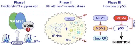
Figure 2. WDR5 WIN site inhibitors as a ribosome-directed anti-cancer strategy. Three-phase model for the mechanism of action of WINi. Phase I, WIN site inhibitoirs (“i”; red circle) rapidly displace WDR5 from chromatin; at RPGs where MYC binding is dependent on interaction with WDR5, MYC is also displaced. The net effect is a two-fold decrease in expression of select RPGs. Phase II, the resulting ribosomal protein (RP) attrition induces a nucleolar stress response in which free ribosomal proteins and nucleophosmin (NPM1) redistribute from the nucleolus into the nucleoplasm. Phase III, free RP and NPM1 inhibit MDM2, which results in induction of p53 and p53-dependent apoptosis. In MLLr cancer cells [76], and potentially other settings, p53 induction is amplified via activation of the splicing factor ZMAT3, which inhibits the MDM family member MDM4 (MDMX), furthering p53 function. p53-independent nucleolar stress responses may also be involved.
Casting WIN site inhibitors as a ribosome-directed anti-cancer therapy has a number of immediate and important implications. First, it predicts that cancer cells with a high MYC burden and pristine p53 signaling will be most sensitive to single agent WINi regimens. Indeed, the involvement of MYC in MLLr leukemia, and the recurring retention of p53 in these malignancies, can explain the sensitivity of these cancer cells to WINi [77][78]. A similar scenario can also explain the sensitivity of neuroblastoma [79] and rhabdoid tumor [80] cells to these inhibitors. This mechanism also predicts additional venues in which single agent WINi activity may be expected, such as Burkitt lymphomas expressing mutant forms of MYC—which typically retain wild-type p53 [81]—as well as the large fraction of “MYC-driven” cancers (e.g., diffuse large B-cell lymphoma) that preserve p53 function on a case-by-case basis [82]. More refined patient selection criteria, including specific patterns of RPG dysregulation [83] and markers of the integrity of the nucleolar stress response [76] can also be developed. Second, it predicts that the action of WINi can be enhanced by mechanistically rationalized drug combination strategies, especially those that conspire to induce p53 or p53-dependent apoptosis in response to nucleolar stress. BET bromodomain inhibitors, for example, which suppress a distinct group of RPGs in MLLr cells, are strongly synergistic with WINi [76], as is the BCL-2 inhibitor venetoclax [76]. Other synergies are possible [57]. Finally, a particularly provocative prediction of a ribosome-directed model for WINi is that the “p53 barrier” to response may not be absolute—a barrier that, if broken, could dramatically expand the utility of these agents. Nucleolar stress is best understood in terms of signaling to p53, but p53-independent nucleolar stress responses do occur [84][85][86][87]. If these responses can be exposed and therapeutically exploited, the reach of WINi could readily extend to solid tumors and other malignant settings bereft of p53.
7. Cancer Runs on Ribosomes
Although the notion of WINi as a ribosome-directed therapy lacks the allure of epigenetic modulation, it is more firmly rooted in cancer etiology than H3K4 methylation, as connections between increased protein synthesis and malignancy are extensive [88]. The relentless rounds of cell division that cancer cells undergo must be supported by the duplication of both the genome and the proteome. MYC itself provides a frank demonstration of the importance of increased translation to cancer mechanisms. It drives the transcription of all three nuclear RNA polymerases—increasing the levels of ribosomal RNAs (rRNAs), ribosomal proteins and ribosome assembly factors, and transfer RNAs (tRNAs)—and reprograms metabolism to meet the high energy demands of ribosome biogenesis [89]. Haploinsufficiency for a single ribosomal protein is sufficient to suppress MYC-driven tumorigenesis in mice [90], and the WBM mutation in MYC, which mimics the impact of WINi on chromatin binding and RPG activation by MYC, renders MYC less able to engraft [91] and sustain [48] tumors in vivo. Add to this the frequent overexpression of RPGs in malignancy [83], the enhanced cancer risk in patients with ribosomopathies, and the prospect of “onco-ribosomes” that support pro-tumorigenic translational programs [92], and the potential of a ribosome-directed anti-cancer therapy comes into sharp focus.
Ribosome- and translation-directed therapies are not new. They are an antibiotic mainstay, and several existing chemotherapies interfere with ribosome production as part of their mechanism of action [93]. But specifically targeting ribosome biogenesis as a way to ameliorate cancer is in its infancy. Perhaps the greatest advances in this area center on the inhibition of ribosomal RNA production, a strategy driven by the discovery of agents that interfere with ribosomal RNA (rDNA) structure or RNA polymerase I activity [84][94]. These agents have undergone extensive preclinical as well as early clinical evaluation [93], and one—CX-5461—has produced significant and durable responses in select patient populations [95], albeit via complex mechanisms [96]. Within this class of agents, WINi occupy a unique place. Because the impact of WDR5 and the MYC–WDR5 interaction on target RPG transcription is around two-fold [16][57][71][76], the self-limiting nature of WINi may provide a greater margin for safety than rRNA inhibitors that have to be dosed to split the difference between ribosome addiction in normal and cancer cells. And most obviously, because WINi suppress ribosomal protein, not rRNA, synthesis, they are likely to distinguish themselves from rRNA poisons by having overlapping but distinct indications, outcomes, and pathways to resistance.
8. The Rise of PROTACs
PROTACs are heterobifunctional molecules that ectopically recruit a ubiquitin-protein ligase to a target protein, thereby triggering its destruction. Although PROTACs are a recent entry into the WDR5 inhibitor arena, their potential for catalytic and comprehensive WDR5 inhibition in cancer is immensely appealing. At first blush, it makes sense that if inhibiting the WIN site of WDR5 offers therapeutic benefit in some malignant settings, then clearing the entirety of WDR5 and its “oncogenic” functions [97] from a cancer cell should amplify and extend those benefits.
Side-by-side modeling of WDR5 degradation versus WIN site inhibition has demonstrated the vastly different consequences of the two approaches [71]. Ribosomal protein genes are suppressed by acute WDR5 degradation—but no more than by WIN site blockade—and MYC is evicted from chromatin at these loci [48], but that is where the similarities end. Acute degradation of WDR5 leads to wholesale loss of H3K4 trimethylation, accompanied by transcriptional changes in thousands of genes, increased anti-sense transcription, and mRNA-level changes from more than half of all active loci. These broad effects are easily rationalized by the highly networked nature of WDR5 and the notion that the destruction of WDR5 eliminates the totality of its actions and may collaterally impact other proteins or complexes that depend on WDR5 for their stability. Such consequences need careful consideration. From a therapeutic perspective, although some activities of WDR5 are neatly classifiable as oncogenic, others may not be, or their indispensability may scupper a therapeutic window for WDR5 PROTACs; this is a sharp point of concern when destroying a pan-essential protein such as WDR5 [98]. WIN site inhibition, in contrast, disrupts only a subset of WDR5 function. Some cells can clearly survive in the presence of these agents [16]. Leaving a portion of WDR5 activities intact may inherently curb the anti-tumor activities of WINi, but it may also underlie the safety of these agents and could contribute to their efficacy, especially if interaction partners that load on to WDR5 in response to WINi contribute to cancer cell inhibition [99].
It is too early to tell if WDR5-directed PROTACs can live up to their promise, or which of the two main approaches, if any, will impact cancer treatment. But as PROTACs and WINi move forward, it behooves the field to remember that these two strategies are fundamentally different and are likely to have very different uses and face very different challenges as they approach the clinic.
References
- Senisterra, G.; Wu, H.; Allali-Hassani, A.; Wasney, G.A.; Barsyte-Lovejoy, D.; Dombrovski, L.; Dong, A.; Nguyen, K.T.; Smil, D.; Bolshan, Y.; et al. Small-molecule inhibition of MLL activity by disruption of its interaction with WDR5. Biochem. J. 2013, 449, 151–159.
- Teuscher, K.B.; Chowdhury, S.; Meyers, K.M.; Tian, J.; Sai, J.; Van Meveren, M.; South, T.M.; Sensintaffar, J.L.; Rietz, T.A.; Goswami, S.; et al. Structure-based discovery of potent WD repeat domain 5 inhibitors that demonstrate efficacy and safety in preclinical animal models. Proc. Natl. Acad. Sci. USA 2023, 120, e2211297120.
- Guarnaccia, A.D.; Tansey, W.P. Moonlighting with WDR5: A Cellular Multitasker. J. Clin. Med. 2018, 7, 21.
- Zhao, J.; Chen, W.; Pan, Y.; Zhang, Y.; Sun, H.; Wang, H.; Yang, F.; Liu, Y.; Shen, N.; Zhang, X.; et al. Structural insights into the recognition of histone H3Q5 serotonylation by WDR5. Sci. Adv. 2021, 7, eabf4291.
- Bryan, A.F.; Wang, J.; Howard, G.C.; Guarnaccia, A.D.; Woodley, C.M.; Aho, E.R.; Rellinger, E.J.; Matlock, B.K.; Flaherty, D.K.; Lorey, S.L.; et al. WDR5 is a conserved regulator of protein synthesis gene expression. Nucleic Acids Res. 2020, 48, 2924–2941.
- Oh, E.; Mark, K.G.; Mocciaro, A.; Watson, E.R.; Prabu, J.R.; Cha, D.D.; Kampmann, M.; Gamarra, N.; Zhou, C.Y.; Rape, M. Gene expression and cell identity controlled by anaphase-promoting complex. Nature 2020, 579, 136–140.
- Ali, A.; Veeranki, S.N.; Chinchole, A.; Tyagi, S. MLL/WDR5 Complex Regulates Kif2A Localization to Ensure Chromosome Congression and Proper Spindle Assembly during Mitosis. Dev. Cell 2017, 41, 605–622.e7.
- Kulkarni, S.S.; Griffin, J.N.; Date, P.P.; Liem, K.F., Jr.; Khokha, M.K. WDR5 Stabilizes Actin Architecture to Promote Multiciliated Cell Formation. Dev. Cell 2018, 46, 595–610.e3.
- Wang, K.C.; Yang, Y.W.; Liu, B.; Sanyal, A.; Corces-Zimmerman, R.; Chen, Y.; Lajoie, B.R.; Protacio, A.; Flynn, R.A.; Gupta, R.A.; et al. A long noncoding RNA maintains active chromatin to coordinate homeotic gene expression. Nature 2011, 472, 120–124.
- Gomez, J.A.; Wapinski, O.L.; Yang, Y.W.; Bureau, J.F.; Gopinath, S.; Monack, D.M.; Chang, H.Y.; Brahic, M.; Kirkegaard, K. The NeST long ncRNA controls microbial susceptibility and epigenetic activation of the interferon-gamma locus. Cell 2013, 152, 743–754.
- Zhang, C.; Ge, S.; Gong, W.; Xu, J.; Guo, Z.; Liu, Z.; Gao, X.; Wei, X.; Ge, S. LncRNA ANRIL acts as a modular scaffold of WDR5 and HDAC3 complexes and promotes alteration of the vascular smooth muscle cell phenotype. Cell Death Dis. 2020, 11, 435.
- Pistoni, M.; Rossi, T.; Donati, B.; Torricelli, F.; Polano, M.; Ciarrocchi, A. Long Noncoding RNA NEAT1 Acts as a Molecular Switch for BRD4 Transcriptional Activity and Mediates Repression of BRD4/WDR5 Target Genes. Mol. Cancer Res. 2021, 19, 799–811.
- Odho, Z.; Southall, S.M.; Wilson, J.R. Characterization of a novel WDR5-binding site that recruits RbBP5 through a conserved motif to enhance methylation of histone H3 lysine 4 by mixed lineage leukemia protein-1. J. Biol. Chem. 2010, 285, 32967–32976.
- Ding, J.; Li, G.; Liu, H.; Liu, L.; Lin, Y.; Gao, J.; Zhou, G.; Shen, L.; Zhao, M.; Yu, Y.; et al. Discovery of Potent Small-Molecule Inhibitors of WDR5-MYC Interaction. ACS Chem. Biol. 2023, 18, 34–40.
- Cao, F.; Townsend, E.C.; Karatas, H.; Xu, J.; Li, L.; Lee, S.; Liu, L.; Chen, Y.; Ouillette, P.; Zhu, J.; et al. Targeting MLL1 H3K4 methyltransferase activity in mixed-lineage leukemia. Mol. Cell 2014, 53, 247–261.
- Aho, E.R.; Wang, J.; Gogliotti, R.D.; Howard, G.C.; Phan, J.; Acharya, P.; Macdonald, J.D.; Cheng, K.; Lorey, S.L.; Lu, B.; et al. Displacement of WDR5 from Chromatin by a WIN Site Inhibitor with Picomolar Affinity. Cell Rep. 2019, 26, 2916–2928.e13.
- Grebien, F.; Vedadi, M.; Getlik, M.; Giambruno, R.; Grover, A.; Avellino, R.; Skucha, A.; Vittori, S.; Kuznetsova, E.; Smil, D.; et al. Pharmacological targeting of the Wdr5-MLL interaction in C/EBPalpha N-terminal leukemia. Nat. Chem. Biol. 2015, 11, 571–578.
- Yu, X.; Li, D.; Kottur, J.; Shen, Y.; Kim, H.S.; Park, K.S.; Tsai, Y.H.; Gong, W.; Wang, J.; Suzuki, K.; et al. A selective WDR5 degrader inhibits acute myeloid leukemia in patient-derived mouse models. Sci. Transl. Med. 2021, 13, eabj1578.
- Ding, J.; Liu, L.; Chiang, Y.L.; Zhao, M.; Liu, H.; Yang, F.; Shen, L.; Lin, Y.; Deng, H.; Gao, J.; et al. Discovery and Structure-Based Design of Inhibitors of the WD Repeat-Containing Protein 5 (WDR5)-MYC Interaction. J. Med. Chem. 2023, 66, 8310–8323.
- Han, Q.L.; Zhang, X.L.; Ren, P.X.; Mei, L.H.; Lin, W.H.; Wang, L.; Cao, Y.; Li, K.; Bai, F. Discovery, evaluation and mechanism study of WDR5-targeted small molecular inhibitors for neuroblastoma. Acta Pharmacol. Sin. 2023, 44, 877–887.
- Song, L.; Cao, J.; Chen, L.; Du, Z.; Zhang, N.; Cao, D.; Xiong, B. Screening and optimization of phage display cyclic peptides against the WDR5 WBM site. RSC Med. Chem. 2023, 14, 2048–2057.
- Karatas, H.; Townsend, E.C.; Cao, F.; Chen, Y.; Bernard, D.; Liu, L.; Lei, M.; Dou, Y.; Wang, S. High-affinity, small-molecule peptidomimetic inhibitors of MLL1/WDR5 protein-protein interaction. J. Am. Chem. Soc. 2013, 135, 669–682.
- Alicea-Velazquez, N.L.; Shinsky, S.A.; Loh, D.M.; Lee, J.H.; Skalnik, D.G.; Cosgrove, M.S. Targeted Disruption of the Interaction between WD-40 Repeat Protein 5 (WDR5) and Mixed Lineage Leukemia (MLL)/SET1 Family Proteins Specifically Inhibits MLL1 and SETd1A Methyltransferase Complexes. J. Biol. Chem. 2016, 291, 22357–22372.
- Getlik, M.; Smil, D.; Zepeda-Velazquez, C.; Bolshan, Y.; Poda, G.; Wu, H.; Dong, A.; Kuznetsova, E.; Marcellus, R.; Senisterra, G.; et al. Structure-Based Optimization of a Small Molecule Antagonist of the Interaction Between WD Repeat-Containing Protein 5 (WDR5) and Mixed-Lineage Leukemia 1 (MLL1). J. Med. Chem. 2016, 59, 2478–2496.
- Li, D.D.; Chen, W.L.; Wang, Z.H.; Xie, Y.Y.; Xu, X.L.; Jiang, Z.Y.; Zhang, X.J.; You, Q.D.; Guo, X.K. High-affinity small molecular blockers of mixed lineage leukemia 1 (MLL1)-WDR5 interaction inhibit MLL1 complex H3K4 methyltransferase activity. Eur. J. Med. Chem. 2016, 124, 480–489.
- Li, D.D.; Chen, W.L.; Xu, X.L.; Jiang, F.; Wang, L.; Xie, Y.Y.; Zhang, X.J.; Guo, X.K.; You, Q.D.; Sun, H.P. Structure-based design and synthesis of small molecular inhibitors disturbing the interaction of MLL1-WDR5. Eur. J. Med. Chem. 2016, 118, 1–8.
- Karatas, H.; Li, Y.; Liu, L.; Ji, J.; Lee, S.; Chen, Y.; Yang, J.; Huang, L.; Bernard, D.; Xu, J.; et al. Discovery of a Highly Potent, Cell-Permeable Macrocyclic Peptidomimetic (MM-589) Targeting the WD Repeat Domain 5 Protein (WDR5)-Mixed Lineage Leukemia (MLL) Protein-Protein Interaction. J. Med. Chem. 2017, 60, 4818–4839.
- Zhang, X.; Zheng, X.; Yang, H.; Yan, J.; Fu, X.; Wei, R.; Xu, X.; Zhang, Z.; Yu, A.; Zhou, K.; et al. Piribedil disrupts the MLL1-WDR5 interaction and sensitizes MLL-rearranged acute myeloid leukemia (AML) to doxorubicin-induced apoptosis. Cancer Lett. 2018, 431, 150–160.
- Dennis, M.L.; Morrow, B.J.; Dolezal, O.; Cuzzupe, A.N.; Stupple, A.E.; Newman, J.; Bentley, J.; Hattarki, M.; Nuttall, S.D.; Foitzik, R.C.; et al. Fragment screening for a protein-protein interaction inhibitor to WDR5. Struct. Dyn. 2019, 6, 064701.
- Ye, X.; Zhang, R.; Lian, F.; Zhang, W.; Lu, W.; Han, J.; Zhang, N.; Jin, J.; Luo, C.; Chen, K.; et al. The identification of novel small-molecule inhibitors targeting WDR5-MLL1 interaction through fluorescence polarization based high-throughput screening. Bioorg. Med. Chem. Lett. 2019, 29, 638–645.
- Chen, W.L.; Li, D.D.; Chen, X.; Wang, Y.Z.; Xu, J.J.; Jiang, Z.Y.; You, Q.D.; Guo, X.K. Proton pump inhibitors selectively suppress MLL rearranged leukemia cells via disrupting MLL1-WDR5 protein-protein interaction. Eur. J. Med. Chem. 2020, 188, 112027.
- Cao, J.; Fan, T.; Li, Y.; Du, Z.; Chen, L.; Wang, Y.; Wang, X.; Shen, J.; Huang, X.; Xiong, B.; et al. Phage-Display Based Discovery and Characterization of Peptide Ligands against WDR5. Molecules 2021, 26, 1225.
- Chen, W.; Chen, X.; Li, D.; Wang, X.; Long, G.; Jiang, Z.; You, Q.; Guo, X. Discovery of a potent MLL1 and WDR5 protein-protein interaction inhibitor with in vivo antitumor activity. Eur. J. Med. Chem. 2021, 223, 113677.
- Chen, W.; Chen, X.; Li, D.; Zhou, J.; Jiang, Z.; You, Q.; Guo, X. Discovery of DDO-2213 as a Potent and Orally Bioavailable Inhibitor of the WDR5-Mixed Lineage Leukemia 1 Protein-Protein Interaction for the Treatment of MLL Fusion Leukemia. J. Med. Chem. 2021, 64, 8221–8245.
- Wysocka, J.; Swigut, T.; Milne, T.A.; Dou, Y.; Zhang, X.; Burlingame, A.L.; Roeder, R.G.; Brivanlou, A.H.; Allis, C.D. WDR5 associates with histone H3 methylated at K4 and is essential for H3 K4 methylation and vertebrate development. Cell 2005, 121, 859–872.
- Dou, Y.; Milne, T.A.; Ruthenburg, A.J.; Lee, S.; Lee, J.W.; Verdine, G.L.; Allis, C.D.; Roeder, R.G. Regulation of MLL1 H3K4 methyltransferase activity by its core components. Nat. Struct. Mol. Biol. 2006, 13, 713–719.
- Ruthenburg, A.J.; Wang, W.; Graybosch, D.M.; Li, H.; Allis, C.D.; Patel, D.J.; Verdine, G.L. Histone H3 recognition and presentation by the WDR5 module of the MLL1 complex. Nat. Struct. Mol. Biol. 2006, 13, 704–712.
- Patel, A.; Vought, V.E.; Dharmarajan, V.; Cosgrove, M.S. A conserved arginine-containing motif crucial for the assembly and enzymatic activity of the mixed lineage leukemia protein-1 core complex. J. Biol. Chem. 2008, 283, 32162–32175.
- Antunes, E.T.B.; Ottersbach, K. The MLL/SET family and haematopoiesis. Biochim. Biophys. Acta Gene Regul. Mech. 2020, 1863, 194579.
- Beacon, T.H.; Delcuve, G.P.; Lopez, C.; Nardocci, G.; Kovalchuk, I.; van Wijnen, A.J.; Davie, J.R. The dynamic broad epigenetic (H3K4me3, H3K27ac) domain as a mark of essential genes. Clin. Epigenetics 2021, 13, 138.
- Li, Y.; Han, J.; Zhang, Y.; Cao, F.; Liu, Z.; Li, S.; Wu, J.; Hu, C.; Wang, Y.; Shuai, J.; et al. Structural basis for activity regulation of MLL family methyltransferases. Nature 2016, 530, 447–452.
- Sha, L.; Ayoub, A.; Cho, U.S.; Dou, Y. Insights on the regulation of the MLL/SET1 family histone methyltransferases. Biochim. Biophys. Acta Gene Regul. Mech. 2020, 1863, 194561.
- Thiel, A.T.; Blessington, P.; Zou, T.; Feather, D.; Wu, X.; Yan, J.; Zhang, H.; Liu, Z.; Ernst, P.; Koretzky, G.A.; et al. MLL-AF9-induced leukemogenesis requires coexpression of the wild-type Mll allele. Cancer Cell 2010, 17, 148–159.
- Mungamuri, S.K.; Murk, W.; Grumolato, L.; Bernstein, E.; Aaronson, S.A. Chromatin modifications sequentially enhance ErbB2 expression in ErbB2-positive breast cancers. Cell Rep. 2013, 5, 302–313.
- Punzi, S.; Balestrieri, C.; D’Alesio, C.; Bossi, D.; Dellino, G.I.; Gatti, E.; Pruneri, G.; Criscitiello, C.; Lovati, G.; Meliksetyan, M.; et al. WDR5 inhibition halts metastasis dissemination by repressing the mesenchymal phenotype of breast cancer cells. Breast Cancer Res. 2019, 21, 123.
- Cai, W.L.; Chen, J.F.; Chen, H.; Wingrove, E.; Kurley, S.J.; Chan, L.H.; Zhang, M.; Arnal-Estape, A.; Zhao, M.; Balabaki, A.; et al. Human WDR5 promotes breast cancer growth and metastasis via KMT2-independent translation regulation. Elife 2022, 11, e78163.
- Li, Z.; Yang, H.Y.; Zhang, X.L.; Zhang, X.; Huang, Y.Z.; Dai, X.Y.; Shi, L.; Zhou, G.R.; Wei, J.F.; Ding, Q. Kinesin family member 23, regulated by FOXM1, promotes triple negative breast cancer progression via activating Wnt/beta-catenin pathway. J. Exp. Clin. Cancer Res. 2022, 41, 168.
- Thomas, L.R.; Adams, C.M.; Wang, J.; Weissmiller, A.M.; Creighton, J.; Lorey, S.L.; Liu, Q.; Fesik, S.W.; Eischen, C.M.; Tansey, W.P. Interaction of the oncoprotein transcription factor MYC with its chromatin cofactor WDR5 is essential for tumor maintenance. Proc. Natl. Acad. Sci. USA 2019, 116, 25260–25268.
- Carugo, A.; Genovese, G.; Seth, S.; Nezi, L.; Rose, J.L.; Bossi, D.; Cicalese, A.; Shah, P.K.; Viale, A.; Pettazzoni, P.F.; et al. In Vivo Functional Platform Targeting Patient-Derived Xenografts Identifies WDR5-Myc Association as a Critical Determinant of Pancreatic Cancer. Cell Rep. 2016, 16, 133–147.
- Mouti, M.A.; Deng, S.; Pook, M.; Malzahn, J.; Rendek, A.; Militi, S.; Nibhani, R.; Soonawalla, Z.; Oppermann, U.; Hwang, C.I.; et al. KMT2A associates with PHF5A-PHF14-HMG20A-RAI1 subcomplex in pancreatic cancer stem cells and epigenetically regulates their characteristics. Nat. Commun. 2023, 14, 5685.
- Zhu, J.; Sammons, M.A.; Donahue, G.; Dou, Z.; Vedadi, M.; Getlik, M.; Barsyte-Lovejoy, D.; Al-awar, R.; Katona, B.W.; Shilatifard, A.; et al. Gain-of-function p53 mutants co-opt chromatin pathways to drive cancer growth. Nature 2015, 525, 206–211.
- Tan, X.; Chen, S.; Wu, J.; Lin, J.; Pan, C.; Ying, X.; Pan, Z.; Qiu, L.; Liu, R.; Geng, R.; et al. PI3K/AKT-mediated upregulation of WDR5 promotes colorectal cancer metastasis by directly targeting ZNF407. Cell Death Dis. 2017, 8, e2686.
- Hao, J.; Huang, J.; Hua, C.; Zuo, Y.; Yu, W.; Wu, X.; Li, L.; Xue, G.; Wan, X.; Ru, L.; et al. A novel TOX3-WDR5-ABCG2 signaling axis regulates the progression of colorectal cancer by accelerating stem-like traits and chemoresistance. PLoS Biol. 2023, 21, e3002256.
- Qu, S.; Jin, L.; Huang, H.; Lin, J.; Gao, W.; Zeng, Z. A positive-feedback loop between HBx and ALKBH5 promotes hepatocellular carcinogenesis. BMC Cancer 2021, 21, 686.
- Lu, C.; Liu, Z.; Klement, J.D.; Yang, D.; Merting, A.D.; Poschel, D.; Albers, T.; Waller, J.L.; Shi, H.; Liu, K. WDR5-H3K4me3 epigenetic axis regulates OPN expression to compensate PD-L1 function to promote pancreatic cancer immune escape. J. Immunother. Cancer 2021, 9, e002624.
- Zhang, J.; Zhou, Q.; Xie, K.; Cheng, L.; Peng, S.; Xie, R.; Liu, L.; Zhang, Y.; Dong, W.; Han, J.; et al. Targeting WD repeat domain 5 enhances chemosensitivity and inhibits proliferation and programmed death-ligand 1 expression in bladder cancer. J. Exp. Clin. Cancer Res. 2021, 40, 203.
- Florian, A.C.; Woodley, C.M.; Wang, J.; Grieb, B.C.; Slota, M.J.; Guerrazzi, K.; Hsu, C.Y.; Matlock, B.K.; Flaherty, D.K.; Lorey, S.L.; et al. Synergistic action of WDR5 and HDM2 inhibitors in SMARCB1-deficient cancer cells. NAR Cancer 2022, 4, zcac007.
- Wu, C.P.; Hsieh, Y.J.; Tseng, H.Y.; Huang, Y.H.; Li, Y.Q.; Hung, T.H.; Wang, S.P.; Wu, Y.S. The WD repeat-containing protein 5 (WDR5) antagonist WDR5-0103 restores the efficacy of cytotoxic drugs in multidrug-resistant cancer cells overexpressing ABCB1 or ABCG2. Biomed. Pharmacother. 2022, 154, 113663.
- Mitchell, K.; Sprowls, S.A.; Arora, S.; Shakya, S.; Silver, D.J.; Goins, C.M.; Wallace, L.; Roversi, G.; Schafer, R.E.; Kay, K.; et al. WDR5 represents a therapeutically exploitable target for cancer stem cells in glioblastoma. Genes Dev. 2023, 37, 86–102.
- Han, X.; Xiang, J.; Li, C.; Wang, J.; Wang, C.; Zhang, Y.; Li, Z.; Lu, Z.; Yue, Y.; Li, X. MLL1 combined with GSK3 and MAP2K inhibition improves the development of in vitro-fertilized embryos. Theriogenology 2020, 146, 58–70.
- Shimoda, H.; Doi, S.; Nakashima, A.; Sasaki, K.; Doi, T.; Masaki, T. Inhibition of the H3K4 methyltransferase MLL1/WDR5 complex attenuates renal senescence in ischemia reperfusion mice by reduction of p16(INK4a). Kidney Int. 2019, 96, 1162–1175.
- Lin, T.B.; Lai, C.Y.; Hsieh, M.C.; Ho, Y.C.; Wang, H.H.; Yang, P.S.; Cheng, J.K.; Chen, G.D.; Ng, S.C.; Peng, H.Y. Inhibiting MLL1-WDR5 interaction ameliorates neuropathic allodynia by attenuating histone H3 lysine 4 trimethylation-dependent spinal mGluR5 transcription. Pain 2020, 161, 1995–2009.
- Cao, Q.; Wang, W.; Williams, J.B.; Yang, F.; Wang, Z.J.; Yan, Z. Targeting histone K4 trimethylation for treatment of cognitive and synaptic deficits in mouse models of Alzheimer’s disease. Sci. Adv. 2020, 6, eabc8096.
- Zhang, C.; Guan, Y.; Zou, J.; Yang, X.; Bayliss, G.; Zhuang, S. Histone methyltransferase MLL1 drives renal tubular cell apoptosis by p53-dependent repression of E-cadherin during cisplatin-induced acute kidney injury. Cell Death Dis. 2022, 13, 770.
- Yuan, J.; Peng, H.; Mo, B.; Yin, C.; Fang, G.; Li, Y.; Wang, Y.; Chen, R.; Wang, Q. Inhibition of Wdr5 Attenuates Ang-II-Induced Fibroblast-to-Myofibroblast Transition in Cardiac Fibrosis by Regulating Mdm2/P53/P21 Pathway. Biomolecules 2022, 12, 1574.
- Yang, K.; Yu, C.; Ruan, L.; Hu, S.; Zhu, W.; Xia, F. MiR-193-3p inhibits the malignant progression of atherosclerosis by targeting WDR5. Clin. Appl. Thromb. Hemost. 2022, 28, 10760296221119458.
- Mishra, B.P.; Zaffuto, K.M.; Artinger, E.L.; Org, T.; Mikkola, H.K.; Cheng, C.; Djabali, M.; Ernst, P. The histone methyltransferase activity of MLL1 is dispensable for hematopoiesis and leukemogenesis. Cell Rep. 2014, 7, 1239–1247.
- Chen, Y.; Anastassiadis, K.; Kranz, A.; Stewart, A.F.; Arndt, K.; Waskow, C.; Yokoyama, A.; Jones, K.; Neff, T.; Lee, Y.; et al. MLL2, Not MLL1, Plays a Major Role in Sustaining MLL-Rearranged Acute Myeloid Leukemia. Cancer Cell 2017, 31, 755–770.e6.
- Yang, W.; Ernst, P. Distinct functions of histone H3, lysine 4 methyltransferases in normal and malignant hematopoiesis. Curr. Opin. Hematol. 2017, 24, 322–328.
- Wang, P.; Lin, C.; Smith, E.R.; Guo, H.; Sanderson, B.W.; Wu, M.; Gogol, M.; Alexander, T.; Seidel, C.; Wiedemann, L.M.; et al. Global analysis of H3K4 methylation defines MLL family member targets and points to a role for MLL1-mediated H3K4 methylation in the regulation of transcriptional initiation by RNA polymerase II. Mol. Cell. Biol. 2009, 29, 6074–6085.
- Siladi, A.J.; Wang, J.; Florian, A.C.; Thomas, L.R.; Creighton, J.H.; Matlock, B.K.; Flaherty, D.K.; Lorey, S.L.; Howard, G.C.; Fesik, S.W.; et al. WIN site inhibition disrupts a subset of WDR5 function. Sci. Rep. 2022, 12, 1848.
- Howe, F.S.; Fischl, H.; Murray, S.C.; Mellor, J. Is H3K4me3 instructive for transcription activation? Bioessays 2017, 39, 1–12.
- Collins, B.E.; Greer, C.B.; Coleman, B.C.; Sweatt, J.D. Histone H3 lysine K4 methylation and its role in learning and memory. Epigenetics Chromatin 2019, 12, 7.
- Wang, H.; Fan, Z.; Shliaha, P.V.; Miele, M.; Hendrickson, R.C.; Jiang, X.; Helin, K. H3K4me3 regulates RNA polymerase II promoter-proximal pause-release. Nature 2023, 615, 339–348.
- Pelletier, J.; Thomas, G.; Volarevic, S. Ribosome biogenesis in cancer: New players and therapeutic avenues. Nat. Rev. Cancer 2018, 18, 51–63.
- Howard, G.C.; Wang, J.; Rose, K.L.; Patel, P.; Tsui, T.; Florian, A.C.; Lorey, S.L.; Grieb, B.C.; Smith, B.N.; Slota, M.J.; et al. Ribosome subunit attrition and activation of the p53-MDM4 axis dominate the response of MLL-rearranged cancer cells to WDR5 WIN site inhibition. bioRxiv 2023.
- Dawson, M.A.; Prinjha, R.K.; Dittmann, A.; Giotopoulos, G.; Bantscheff, M.; Chan, W.I.; Robson, S.C.; Chung, C.W.; Hopf, C.; Savitski, M.M.; et al. Inhibition of BET recruitment to chromatin as an effective treatment for MLL-fusion leukaemia. Nature 2011, 478, 529–533.
- Fitzel, R.; Secker-Grob, K.A.; Keppeler, H.; Korkmaz, F.; Schairer, R.; Erkner, E.; Schneidawind, D.; Lengerke, C.; Hentrich, T.; Schulze-Hentrich, J.M.; et al. Targeting MYC in combination with epigenetic regulators induces synergistic anti-leukemic effects in MLLr leukemia and simultaneously improves immunity. Neoplasia 2023, 41, 100902.
- Bartolucci, D.; Montemurro, L.; Raieli, S.; Lampis, S.; Pession, A.; Hrelia, P.; Tonelli, R. MYCN Impact on High-Risk Neuroblastoma: From Diagnosis and Prognosis to Targeted Treatment. Cancers 2022, 14, 4421.
- Liu, N.Q.; Paassen, I.; Custers, L.; Zeller, P.; Teunissen, H.; Ayyildiz, D.; He, J.; Buhl, J.L.; Hoving, E.W.; van Oudenaarden, A.; et al. SMARCB1 loss activates patient-specific distal oncogenic enhancers in malignant rhabdoid tumors. Nat. Commun. 2023, 14, 7762.
- Berns, A. Cancer: Two in one. Nature 2005, 436, 787–789.
- Xu-Monette, Z.Y.; Wu, L.; Visco, C.; Tai, Y.C.; Tzankov, A.; Liu, W.M.; Montes-Moreno, S.; Dybkaer, K.; Chiu, A.; Orazi, A.; et al. Mutational profile and prognostic significance of TP53 in diffuse large B-cell lymphoma patients treated with R-CHOP: Report from an International DLBCL Rituximab-CHOP Consortium Program Study. Blood 2012, 120, 3986–3996.
- Dolezal, J.M.; Dash, A.P.; Prochownik, E.V. Diagnostic and prognostic implications of ribosomal protein transcript expression patterns in human cancers. BMC Cancer 2018, 18, 275.
- Peltonen, K.; Colis, L.; Liu, H.; Trivedi, R.; Moubarek, M.S.; Moore, H.M.; Bai, B.; Rudek, M.A.; Bieberich, C.J.; Laiho, M. A targeting modality for destruction of RNA polymerase I that possesses anticancer activity. Cancer Cell 2014, 25, 77–90.
- Pfister, A.S.; Keil, M.; Kuhl, M. The Wnt Target Protein Peter Pan Defines a Novel p53-independent Nucleolar Stress-Response Pathway. J. Biol. Chem. 2015, 290, 10905–10918.
- Pfister, A.S. Emerging Role of the Nucleolar Stress Response in Autophagy. Front. Cell Neurosci. 2019, 13, 156.
- Pfister, A.S. An Update on Nucleolar Stress: The Transcriptional Control of Autophagy. Cells 2023, 12, 2071.
- Elhamamsy, A.R.; Metge, B.J.; Alsheikh, H.A.; Shevde, L.A.; Samant, R.S. Ribosome Biogenesis: A Central Player in Cancer Metastasis and Therapeutic Resistance. Cancer Res. 2022, 82, 2344–2353.
- van Riggelen, J.; Yetil, A.; Felsher, D.W. MYC as a regulator of ribosome biogenesis and protein synthesis. Nat. Rev. Cancer 2010, 10, 301–309.
- Barna, M.; Pusic, A.; Zollo, O.; Costa, M.; Kondrashov, N.; Rego, E.; Rao, P.H.; Ruggero, D. Suppression of Myc oncogenic activity by ribosomal protein haploinsufficiency. Nature 2008, 456, 971–975.
- Thomas, L.R.; Wang, Q.; Grieb, B.C.; Phan, J.; Foshage, A.M.; Sun, Q.; Olejniczak, E.T.; Clark, T.; Dey, S.; Lorey, S.; et al. Interaction with WDR5 promotes target gene recognition and tumorigenesis by MYC. Mol. Cell 2015, 58, 440–452.
- Bastide, A.; David, A. The ribosome, (slow) beating heart of cancer (stem) cell. Oncogenesis 2018, 7, 34.
- Temaj, G.; Chichiarelli, S.; Eufemi, M.; Altieri, F.; Hadziselimovic, R.; Farooqi, A.A.; Yaylim, I.; Saso, L. Ribosome-Directed Therapies in Cancer. Biomedicines 2022, 10, 2088.
- Drygin, D.; Lin, A.; Bliesath, J.; Ho, C.B.; O’Brien, S.E.; Proffitt, C.; Omori, M.; Haddach, M.; Schwaebe, M.K.; Siddiqui-Jain, A.; et al. Targeting RNA polymerase I with an oral small molecule CX-5461 inhibits ribosomal RNA synthesis and solid tumor growth. Cancer Res. 2011, 71, 1418–1430.
- Xu, H.; Hurley, L.H. A first-in-class clinical G-quadruplex-targeting drug. The bench-to-bedside translation of the fluoroquinolone QQ58 to CX-5461 (Pidnarulex). Bioorg Med. Chem. Lett. 2022, 77, 129016.
- Hilton, J.; Gelmon, K.; Bedard, P.L.; Tu, D.; Xu, H.; Tinker, A.V.; Goodwin, R.; Laurie, S.A.; Jonker, D.; Hansen, A.R.; et al. Results of the phase I CCTG IND.231 trial of CX-5461 in patients with advanced solid tumors enriched for DNA-repair deficiencies. Nat. Commun. 2022, 13, 3607.
- Schwalm, M.P.; Kramer, A.; Dolle, A.; Weckesser, J.; Yu, X.; Jin, J.; Saxena, K.; Knapp, S. Tracking the PROTAC degradation pathway in living cells highlights the importance of ternary complex measurement for PROTAC optimization. Cell Chem. Biol. 2023, 30, 753–765.e8.
- Tsherniak, A.; Vazquez, F.; Montgomery, P.G.; Weir, B.A.; Kryukov, G.; Cowley, G.S.; Gill, S.; Harrington, W.F.; Pantel, S.; Krill-Burger, J.M.; et al. Defining a Cancer Dependency Map. Cell 2017, 170, 564–576.e16.
- Guarnaccia, A.D.; Rose, K.L.; Wang, J.; Zhao, B.; Popay, T.M.; Wang, C.E.; Guerrazzi, K.; Hill, S.; Woodley, C.M.; Hansen, T.J.; et al. Impact of WIN site inhibitor on the WDR5 interactome. Cell Rep. 2021, 34, 108636.
More
Information
Subjects:
Oncology
Contributors
MDPI registered users' name will be linked to their SciProfiles pages. To register with us, please refer to https://encyclopedia.pub/register
:
View Times:
463
Revisions:
2 times
(View History)
Update Date:
15 Jan 2024
Table of Contents


Notice
You are not a member of the advisory board for this topic. If you want to update advisory board member profile, please contact office@encyclopedia.pub.
OK
Confirm
Only members of the Encyclopedia advisory board for this topic are allowed to note entries. Would you like to become an advisory board member of the Encyclopedia?
Yes
No
${ textCharacter }/${ maxCharacter }
Submit
Cancel
Back
Comments
${ item }
|
${ item.createdUser.fullName }
${ item.createdAt }
${ item.vote }
${ item.reply }
Delete
${ reply.createdUser.fullName }
${ reply.createdAt }
${ reply.vote }
Delete
There is no reply to this comment~
${ item.replyTextCharacter }/${ item.replyMaxCharacter }
Submit
Cancel
More
No more~
There is no comment~
${ textCharacter }/${ maxCharacter }
Submit
Cancel
${ selectedItem.replyTextCharacter }/${ selectedItem.replyMaxCharacter }
Submit
Cancel
Confirm
Are you sure to Delete?
Yes
No