Submitted Successfully!
To reward your contribution, here is a gift for you: A free trial for our video production service.
Thank you for your contribution! You can also upload a video entry or images related to this topic.
Version Summary Created by Modification Content Size Created at Operation
1 -- 5246 2024-01-10 14:02:21

Video Upload Options

Do you have a full video?

Confirm

Are you sure to Delete?
Cite
If you have any further questions, please contact Encyclopedia Editorial Office.
Hassan, N.; Krieg, T.; Zinser, M.; Schröder, K.; Kröger, N. Scaffolds and Biomaterials for Soft Tissue Engineering. Encyclopedia. Available online: https://encyclopedia.pub/entry/53690 (accessed on 04 May 2024).
Hassan N, Krieg T, Zinser M, Schröder K, Kröger N. Scaffolds and Biomaterials for Soft Tissue Engineering. Encyclopedia. Available at: https://encyclopedia.pub/entry/53690. Accessed May 04, 2024.
Hassan, Nourhan, Thomas Krieg, Max Zinser, Kai Schröder, Nadja Kröger. "Scaffolds and Biomaterials for Soft Tissue Engineering" Encyclopedia, https://encyclopedia.pub/entry/53690 (accessed May 04, 2024).
Hassan, N., Krieg, T., Zinser, M., Schröder, K., & Kröger, N. (2024, January 10). Scaffolds and Biomaterials for Soft Tissue Engineering. In Encyclopedia. https://encyclopedia.pub/entry/53690
Hassan, Nourhan, et al. "Scaffolds and Biomaterials for Soft Tissue Engineering." Encyclopedia. Web. 10 January, 2024.
Scaffolds and Biomaterials for Soft Tissue Engineering
Edit

The utilization of materials in medical implants, serving as substitutes for non-functional biological structures, supporting damaged tissues, or reinforcing active organs, holds significant importance in modern healthcare, positively impacting the quality of life for millions of individuals worldwide. Biodegradable metals, including zinc (Zn), magnesium (Mg), iron, and others, present a new paradigm in the realm of implant materials. Research focuses on developing optimized materials that meet medical standards, encompassing controllable corrosion rates, sustained mechanical stability, and favorable biocompatibility. Achieving these objectives involves refining alloy compositions and tailoring processing techniques to carefully control microstructures and mechanical properties. Among the materials under investigation, Mg- and Zn-based biodegradable materials and their alloys demonstrate the ability to provide necessary support during tissue regeneration while gradually degrading over time. Furthermore, as essential elements in the human body, Mg and Zn offer additional benefits, including promoting wound healing, facilitating cell growth, and participating in gene generation while interacting with various vital biological functions. 

skin expansion biomaterials tissue engineering metallic alloys implants

1. Introduction

The human body is made up of various organ systems that are composed of distinct types of cells within an extracellular matrix (ECM), forming different tissues. These cells are highly complex biochemical, molecular, and electrical reaction chambers that are interconnected within the ECM by cellular receptors and cytoskeletal structures [1]. The human body and its constituent cells are subject to diverse mechanical forces on a daily basis, such as tension, compression, shear, gravity, osmotic pressure, and hydrostatic pressure. The impact of microgravity on cellular biology is also acknowledged, particularly in the context of space exploration [2][3][4].
The skin, being the body’s largest organ, acts as a protective barrier separating the internal and external environment and interacts with a variety of forces and deformations caused by the environment [5]. Cutaneous cell populations must sense mechanical cues and respond appropriately to maintain homeostasis and proper mechanical function [6]. Thereby, ECM plays a crucial role in transmitting applied forces to the cells. These forces trigger mechanosignaling pathways in the cells and elicit various biological responses [7][8][9]. The ECM is produced by fibroblasts, which reside in the dermis and are mainly responsible for the tissue’s mechanical properties. The mechanisms of mechanotransduction are similar among many cell types in the body’s various connective tissues [10]. At the molecular level, these responses may involve changes in the configuration of cell membrane channels or receptor sensitivity, enzymatic and protein synthesis in the cytoplasm, and gene expression in the nucleus. In response to these molecular and biochemical reactions, cells can differentiate, proliferate, migrate, or undergo apoptosis [11]. Mechanical forces are increasingly being leveraged to shape cellular and tissue responses in ways that promote tissue regeneration, scar modulation, and wound healing. In the case of the skin, it possesses the remarkable ability for self-regeneration through the presence of stem cells within the epidermis. However, when faced with deep injuries and severe burns, the natural healing process may prove insufficient, resulting in the development of severe scars, wound contraction, and chronic wounds [12]. Therefore, to overcome extensive mechanical forces, sophisticated surgical techniques have to be used to close the wounds with satisfactory scar formation. These also include the application of skin crafts, skin substitutes, and tissue expansion. In the late 1980s, tissue engineering emerged as a distinct field in response to the pressing surgical challenges that needed to be addressed [13][14]. There are several limitations associated with translational applications in soft tissue engineering strategies, including issues related to cell survival, mechanical challenges such as scaffold collapse and availability, considerations of the microenvironment’s composition, potential induction of malignant behavior, cell migration, and cell exhaustion [15]. Tissue expansion is a widely used surgical technique aimed at growing additional skin to address various reconstructive needs such as birth defects, burn injuries, or cancerous breasts [16]. The technique of tissue expansion has been in practice for over three decades and has proven to be a valuable tool in reconstructive surgery across various anatomical regions [17]. One of the major hurdles encountered by reconstructive surgeons is the scarcity of viable soft tissues for such procedures [18]. As a result, there is a growing interest in tissue-engineered skin substitutes as alternative approaches to traditional wound healing, skin expansion strategies, and tissue regeneration [14][19].

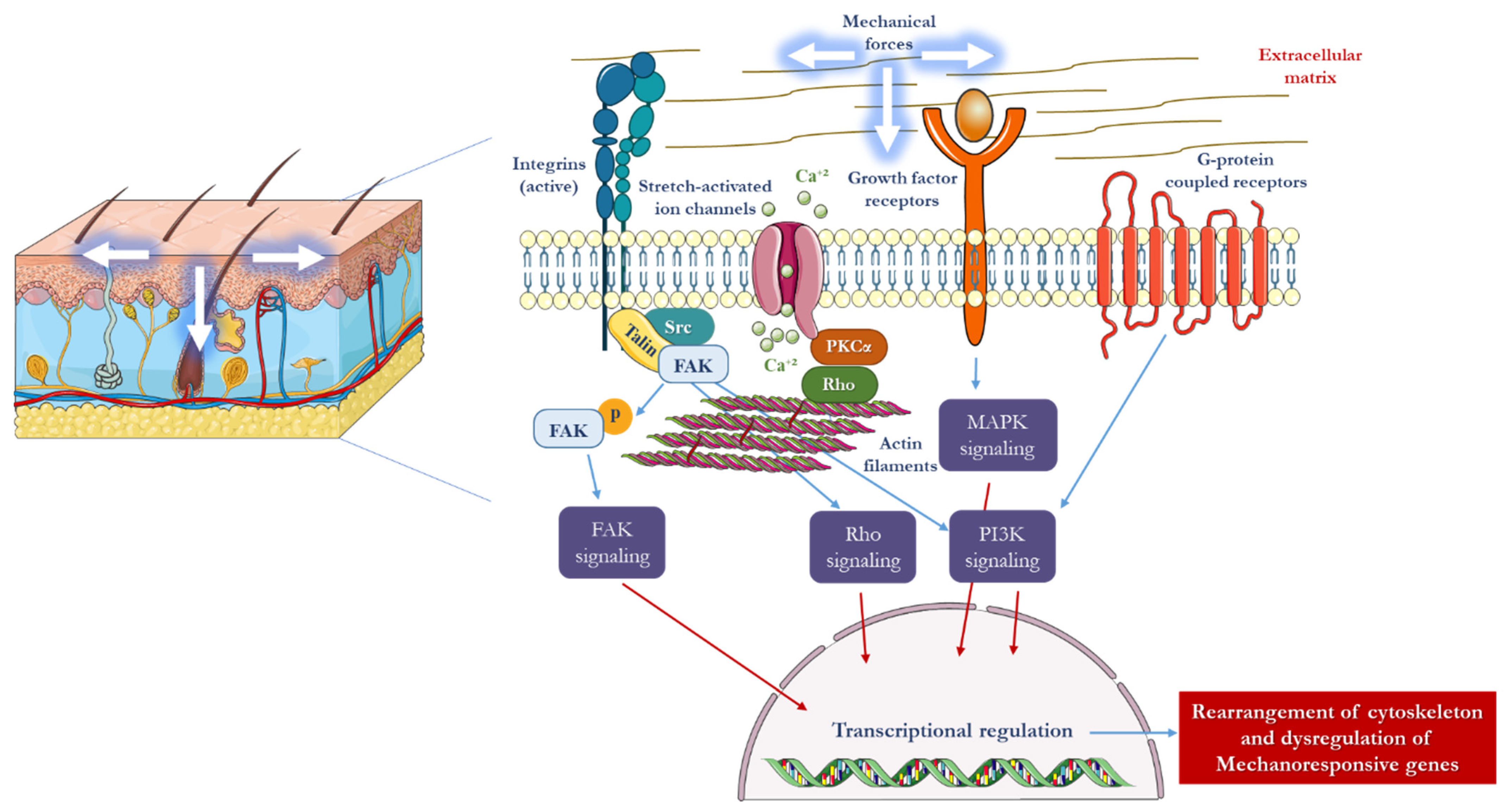
2. Mechanotransduction in Skin

Mechanically sensitive cells, especially fibroblasts, have three types of mechanical sensors at the cell membrane: integrins, G protein-coupled receptors, and stretch-activated ion channels. Furthermore, the cytoskeleton, which provides overall structural support to the cell, can sense deformations through conformational changes, resulting in an additional sensing mechanism [20]. Activation of mechanical sensors directly triggers intracellular signaling pathways, which often activate secondary messengers, such as growth factors [21]. Growth factor receptors located at the cell membrane represent another important sensing mechanism. In response to mechanical stimuli, various cytokines are expressed in connective tissues, including transforming growth factors (TGF-beta), interleukins, fibroblast growth factors (FGF), vascular endothelial growth factors (VEGF), platelet-derived growth factors (PDGF), and tumor necrosis growth factors (TNF-alpha) (Figure 1). These cytokines are particularly important in connective tissues and contribute to various physiological processes [10].
The maintenance of the ECM by dermal fibroblasts involves a constant cycle of collagen and proteoglycan deposition and degradation of the collagen network through matrix metalloproteinases (MMP). Among the various mechanical cues that fibroblasts experience, tension is the most physiologically relevant. Thus, in vitro studies have primarily focused on examining the response of fibroblasts to uniaxial and biaxial strain loading conditions using flexible two-dimensional constructs [22]. In order to better simulate the in vivo environment, mechanical strain has also been applied to fibroblasts embedded within three-dimensional collagen gels [23]. The application of tensile strain to the ECM induces conformational changes in the cytoplasmic tails of the main ECM receptors and the integrins, which activate kinases such as focal adhesion kinase (FAK). FAK activation is then linked to mitogen-activated protein kinase (MAPK) pathways inside the cell [24]. The downstream effects of FAK activation include pro-inflammatory signaling, collagen production, and reduced apoptosis (Figure 1) [25]. Aside from direct mechanical sensing, additional signaling pathways, particularly TGF-beta, play a critical role in controlling how the ECM is remodeled by fibroblasts [26]. TGF-beta exposure results in the up-regulation of collagen genes and the downregulation of the Bax apoptotic gene in fibroblasts [23]. Other mechanical stimuli, including the microstructure and composition of the ECM, also influence fibroblast behavior. For instance, fibroblasts have been found to migrate preferentially along fiber directions, demonstrating the importance of ECM factors in regulating fibroblast behavior [27][28].
Figure 1. In response to mechanical force, a number of intracellular signaling pathways are initiated in mechanotransduction. Membrane-bound mechanosensory complexes such as stretch-activated ion channels, growth factor receptors, integrins, and G-protein-coupled receptors play a crucial role in sensing mechanical strain. In fibroblasts and keratinocytes, where matrix-integrin activation takes place in focal adhesion complexes, FAK is crucial. The mechanical force that is transferred across the cell membrane activates downstream biochemical pathways, such as calcium-dependent targets, nitric oxide (NO) signaling, MAPKs, Rho GTPases, and phosphoinositol-3-kinase (PI3K). When these signals come together, transcription factors are induced to activate mechanoresponsive genes in the nucleus [29]. Parts of the figure were drawn using elements from Servier Medical Art. (https://creativecommons.org/licenses/by/3.0/) (accessed on 16 May 2022).
While the dermis is commonly considered the main load-bearing layer of the skin, it is important to note that keratinocytes in the epidermis also demonstrate mechanosensitivity. Deformations of the dermis are conveyed to the epidermis through hemidesmosome junctions at the basement membrane. Forces are then transmitted to the cytoskeleton inside the keratinocytes via adherens junctions between neighboring cells. Subsequently, intracellular signaling is induced by deformations of the overall cell shape that affect keratinocyte mitosis [30]. Stretch-activated ion channels represent another significant type of mechanoreceptor in the epidermis [31]. When activated downstream of a mechanical stimulus, growth factor receptors in keratinocytes, such as epidermal growth factor (EGF), play a crucial role in controlling cellular proliferation [6]. Experiments on cultured keratinocytes have demonstrated increased mitosis in response to strain [8]. More recently, in vitro studies have explored engineered skin, where both the dermal and epidermal layers respond to strain in a coordinated mechanobiological manner [32].
Mechanotransduction in vivo has mostly been investigated in relation to tissue expansion, which involves implanting a subcutaneous balloon that is persistently inflated over many weeks in order to grow skin [17][33]. By stretching the skin beyond its normal capacity, the expansion process leads to tissue growth (Figure 2) [34]. The ultimate aim of tissue expansion is to avoid donor site morbidity, using the newly created skin as a vascularized flap to reconstruct soft tissue defects during a second operation [35]. However, the procedure does have some limitations, such as potential failures and complications, as well as the need for training and skill development to effectively plan and execute tissue expansion [36]. The technique is also not appropriate for pre-existing open wounds, which represents its biggest drawback [37]. The analysis of expanded tissues has shown an increase in keratinocyte proliferation, activation of the MAPK pathway, and an increase in collagen deposition [38][39][40]. Interestingly, the grown tissue has similar properties to native, unexpanded skin.
Figure 2. Schematic sequence of tissue expander inflation. Initially, the skin is in a state of rest, with no tension present (top). A tissue expander which is inserted underneath the skin, starts to inflate between the epidermis and dermis layers and the hypodermis. Upon inflation of the expander, the skin becomes taut and stretched (middle). The mechanical stretching results in cellular proliferation, leading to either skin growth and proliferation or apoptosis or tumor formation. Eventually, the skin grows enough to return to its original state of rest, with no tension (bottom) [34]. Parts of the figure were drawn using elements from Servier Medical Art. (https://creativecommons.org/licenses/by/3.0/) (accessed on 16 May 2022).

3. The Influence of Mechanical Forces on the Structure and Function of the Skin

Mechanotransduction is the process of converting physical forces into biochemical signals that trigger cellular responses. The mechano-responsiveness of cellular complexes, such as TGFβ/Smad, integrin, and calcium ion pathways, has been demonstrated (Figure 1) [41]. These signals are transmitted into the cell, ultimately reaching the nucleus. In vitro models have shown that mechanical strain can upregulate matrix remodeling genes and downregulate normal cellular apoptosis through an Akt-dependent mechanism, leading to increased production of extracellular matrix [42][43]. The skin is exposed to stretching forces both under normal physiological conditions, such as pregnancy, and through external means, such as tissue expansion using soft tissue expanders, external skin stretching devices, and distraction osteogenesis using external devices in hard tissue.
Skin stretching devices and techniques are useful for treating open wounds in surgery and are referred to as external tissue expanders [44]. Typically, they use hooks, sutures, wires, or loops to engage the skin and apply a mechanical force of tension to promote the approximation of wound edges through mechanical creep over time. The stretched skin edges then heal spontaneously during a consolidation period while the stretching device remains engaged. In most cases, the mechanically stretched skin is surgically freshened at the edges and closed after the device is removed. The impact of mechanical skin stretching devices or techniques on healing wounds is increasingly gaining attention [45].
The process of wound healing engages different types of cells, including inflammatory cells, keratinocytes, fibroblasts, myofibroblasts, and endothelial cells. These cells play a pivotal role in cutaneous healing, and they react to mechanical forces by initiating a cascade of events and pathways at both the cellular and molecular levels. This process takes place in the context of the tensegrity model, which involves the cytoskeletal framework being anchored in ECM [39][46]. The structurally interconnected cells respond to mechanical stimuli. Mechanotherapies are wound healing treatments that use mechanical forces to enhance the healing process. Examples of these therapies include micro-deformational wound therapy (MWT), such as negative pressure wound therapy (NPWT) [47][48], shock wave therapy [49][50], ultrasound [51], and electrotherapy [52]. Each therapy uses a different form of mechanical force to stimulate the cells and tissues involved in wound healing. The effects of NPWT have been extensively studied [53]. When suction is applied, the sponge collapses, causing the wound to shrink. This results in macro- and micro-deformation at the wound-sponge interface, which triggers mechanosignaling in a closed wound-healing environment [54]. This therapy has various biological effects, such as increased gene expression of leucocyte chemoattractants, proliferation and migration of epithelial cells and dermal fibroblasts, decreased activity of matrix metalloproteinases, and increased micro-vessel density [55][56][57][58]. As a result, NPWT promotes moist wound healing, angiogenesis, collagen synthesis, and the breakdown of dead tissue and fibrin [59]. Currently, NPWT is extensively utilized to expedite the healing of different types of wounds across various anatomical locations.
Scar formation is a significant concern after skin injury both functionally and aesthetically [60]. In contrast to the scarring process, which occurs in extra-uterine life, fetal wound healing follows a regenerative process [61][62][63]. This has sparked considerable research interest in the conversion from regenerative healing to scarring, with the ultimate goal of achieving scarless wound healing. Currently, there is growing interest in mechanobiology and scar research, with the aim of utilizing mechanotherapy to prevent and treat abnormal scarring [64]. The relationship between tension and scar growth has been observed clinically, particularly in keloid formation, where different shapes and configurations are seen, often associated with tension [65][66][67][68]. Animal models have demonstrated that mechanosignaling plays a role in the fibroproliferative response to tension [64]. Mechanotherapy, which aims to alter stimuli, processing, or reception, offers strategies to deal with these tension forces, such as skin stabilization by paper and silicone tape, multilayered suturing and plication, flaps, z-plasty, and the addition of radiotherapy [69]. The goal is to reduce skin tension, which is the source of cyclically applied mechanical forces in daily locomotion, and ultimately decrease skin inflammation.
Skin tissue engineering encompasses a multidisciplinary approach, incorporating diverse fields such as biochemistry, polymer chemistry, and stem cell research. The objective of skin tissue engineering is to synergize the expertise from these disciplines in order to develop a substitute that can be efficiently produced and effectively restore the skin’s natural functional, mechanical, and aesthetic characteristics. This involves regenerating ECM to provide support and guidance, improving graft take by establishing a vascular network, and restoring skin appendages for functions such as thermoregulation and sensitivity, as well as the various cell types required for protection. The objective can be achieved through two primary methods. The first method involves creating a biodegradable scaffold that is sophisticated enough to release a specific set of signaling molecules in a controlled manner, which can facilitate the migration, adhesion, and, ultimately, regeneration of skin cells. The second method involves designing a basic, temporary scaffold that can serve as a carrier for stem cells or undifferentiated cells to encourage skin regeneration [70]. Both methods necessitate the creation of a 3D environment that can support cell interactions and foster wound healing.
Even though the researchers have emphasized how important mechanical forces and load-induced events are in skin tissue engineering, it is still critical to provide a quantitative perspective, especially when considering various tissues [71]. Quantitative data can provide a more thorough understanding of how different tissues are impacted by mechanical forces, resulting in more accurate and successful tissue engineering techniques [72]. Depending on the tissue under examination, there are several ways to quantitatively present mechanical forces, including tensile strength and strain, strain distribution, shear forces, compression and stress relaxation, fluid flow and perfusion, frequency and magnitude of mechanical loading, microenvironmental stiffness, and biomechanical properties of biomaterials [73][74][75][76]. Thus, researchers are exploring numerous scaffold materials and techniques to meet the requirements of skin tissue engineering.

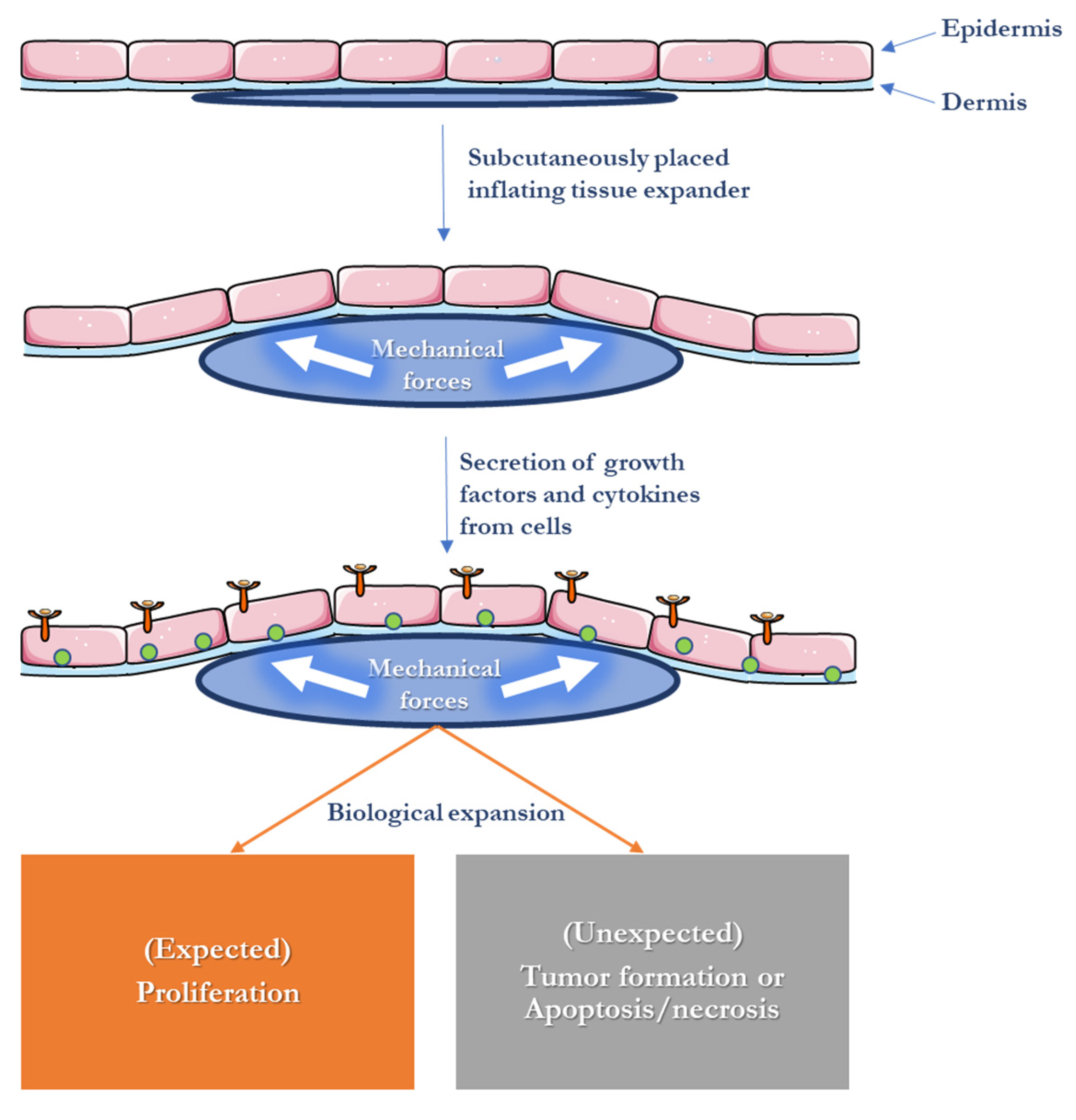
4. Skin Expansion in Reconstructive Surgery

Soft-tissue expanders have emerged as a pre-augmentation technique in implant surgery to circumvent the complications associated with bone grafting procedures [77][78]. The principle of soft tissue expansion is rooted in the biological response of various soft tissues, including skin and mucous membranes, to mechanical forces by producing true tissue growth (cell proliferation) [17]. This phenomenon is evident in various situations, including pregnancy, muscle growth, obesity, and specific cultural practices such as lip and neck expansion in African customs [79]. Tissue expansion offers a remarkable approach to cultivating skin that closely resembles the neighboring healthy skin in terms of texture, color, and hair-bearing characteristics, thereby minimizing scarring and the potential for rejection [80]. The technique of soft tissue expansion is clinically useful in several ways, including preoperative expansion of oral mucosa for large bone augmentations, as well as the intra-oral repair of clefts in the lip and/or palate. Its applications have been popular in plastic surgery since 1976 [81]. Moreover, they are well-established for a variety of indications, such as correcting skin burns after burn wounds, scars, alopecia, congenital nevi, and post-mastectomy breast reconstruction [82][83][84][85]. It has also evolved into one of the principal surgical procedures for creating skin flaps to resurface large congenital defects of the skin, such as giant nevi and vascular anomalies [86][87]. In recent years, this concept has also been introduced in orthopedics, where it was successfully used in a clinical report to achieve skin closure in open fractures using an “external” soft tissue expander. The expansion of soft tissues can reduce the need for periosteal incisions and promote passive flap closure while generating tissues with appropriate color match and texture similar to the original tissues [88].
In 1957, Neumann developed soft tissue expanders using a subcutaneous rubber balloon to repair an ear defect. However, it was not until the early 1980s that soft tissue expanders regained significant interest, particularly in breast reconstruction [89] and the treatment of burns [90]. Early expanders consisted of silicone rubber and featured an external valve that allowed for manual inflation through sequential injections. The extent of soft tissue expansion achieved with conventional expanders has been documented to be influenced by factors such as the specific tissue being expanded and the configuration of the expanders themselves [91][92]. Studies have shown that tissue gain is more pronounced with rectangular and crescent-shaped expanders than with round-based ones [79]. Although conventional soft tissue expanders have shown positive results, they have several disadvantages. The intermittent inflations required for conventional expanders can increase the treatment time by several months and cause pressure peaks, leading to a decrease in tissue vascularity [93] and a higher risk of expander perforation through the soft tissues [88]. The reduction in local oxygen partial pressure increases the risk of expansion failures, and serial injections can result in increased treatment costs, morbidity, and risks for adverse effects [82]. Despite these drawbacks, conventional expanders are still utilized in plastic surgical procedures. However, their use is limited in craniofacial defects due to the aforementioned shortcomings [94].
A self-inflating osmotic soft tissue expander was developed by Austad & Rose (1982) to overcome the drawbacks of conventional soft tissue expanders. It was designed without an external port, and repetitive inflations were not necessary [95]. The new expander was made of a semi-permeable silicone membrane that contained a hypertonic sodium chloride solution. The expansion of the expander and subsequent growth of soft tissue were facilitated by a continuous influx of body fluids driven by an osmotic gradient. This led to an increase in the volume of the expander and the growth of surrounding soft tissues. However, the device had several drawbacks, such as leaks from the expander shell to the surrounding tissues, causing tissue necrosis. Wiese introduced a unique self-inflating osmotic soft tissue expander composed of hydrogel, comprising a polymer network and a variable aqueous component [96][97][98]. This expander, known as Osmed® (Ilmenau, Germany), was developed in 1999 and became the first commercially accessible self-inflatable osmotic expander. It received FDA approval in 2001 and has been available in the market since then. Osmotic expanders eliminate the need for repeated injections and instead inflate continuously by osmotic gradients without requiring any additional interventions. This consistent expansion, in contrast to intermittent inflation, stimulates the generation of new cells, tissue growth [92], and a greater final tissue gain [81][88][99].
The biomaterials used in the hydrogel expanders are the same as those used in contact lenses, providing high biocompatibility and causing no adverse effects such as toxicity, immune reactions, infections, or systemic manifestations [98]. Moreover, they do not provoke any inflammatory responses in the surrounding soft tissues, which is a crucial feature. The presence of methacrylate in ionic hydrogels enhances their osmotic potential, leading to a greater swelling capacity compared to non-ionic hydrogels [96][97][98]. The incorporation of “methyl” methacrylate, specifically in the osmotic hydrogel expanders, results in a higher swelling ratio when compared to “hydroxyethyl” methacrylate [100].
The polymer network of the hydrogel expander is insoluble in water due to the presence of cross-links, making it able to retain large volumes produced by swelling without dissolving [101]. Varga et al. (2009) sought to investigate alternative biomaterials and introduced a hydrogel osmotic soft tissue expander composed of acrylic acid (AAc), acrylamide (AAm), or N-isopropylacrylamide (NIPAAm) [102]. Among these, NIPAAm hydrogels were found to be the most suitable for plastic and reconstructive surgeries in terms of their biological and mechanical properties, although they have only been tested in vivo and require further validation in clinical trials. The next section will delve into the techniques used in scaffold fabrication employed in tissue engineering, shedding light on the advancements, challenges, and future prospects in this exciting field.

5. Scaffold Fabrication Methods Used for Tissue Engineering

Numerous techniques have been devised for constructing and fabricating scaffolds in tissue engineering. The choice of technique depends on the specific properties of the materials employed and the desired characteristics of the scaffold. These methods can be classified into conventional and advanced techniques [103].
Conventional techniques encompass several methods, including solvent-casting and particulate-leaching techniques, which entail the utilization of a polymer solution blended with salt particles of precise dimensions. Subsequent to solvent evaporation and immersion in water, the salt particles dissolve, creating a porous structure [104]. However, gas foaming involves subjecting molded biodegradable polymers to high pressures with gas-foaming agents such as CO2, nitrogen, water, or fluoroform. The polymers become saturated, leading to the nucleation and expansion of gas bubbles within the polymer matrix, typically ranging in size from 100 to 500 μm [105][106]. Moreover, phase separation entails the rapid cooling of a polymer solution, leading to its separation into two separate phases: a polymer-rich phase and a polymer-poor phase. The polymer-rich phase solidifies, while the polymer-poor phase is removed, resulting in the creation of a porous polymer network with high permeability [107]. In melt molding, a combination of polymer powder and porogen components is introduced into a mold, which is then subjected to elevated temperatures beyond the glass-transition temperature of the polymer, accompanied by the application of pressure. This process causes the raw materials to fuse together, forming a scaffold with a predetermined external shape. After removing the mold, the porogen is washed away, leaving behind a porous scaffold that is subsequently dried [108]. Freeze drying, also known as lyophilization, offers a method for the production of polymeric porous scaffolds. The process involves two stages. Initially, the polymer solution is cooled to a specific temperature, causing all components to freeze. During this freezing stage, ice crystals form from the solvent, prompting the polymer molecules to aggregate within the interstitial spaces. In the subsequent phase, the solvent is eliminated by applying a pressure lower than the equilibrium vapor pressure of the frozen solvent. As the solvent undergoes sublimation, a dry polymer scaffold with a well-connected porous microstructure is left behind. The porosity of the scaffolds is contingent upon the concentration of the polymer solution, while the freezing temperatures affect the distribution of pore sizes. In addition to its use in fabricating porous scaffolds, this technique finds application in drying biological samples to safeguard their bioactivities [109][110].
On the other hand, electrospinning and 3D printing technologies are considered advanced techniques in scaffold fabrication. The former methodology is a fabrication technique that utilizes electrical charges to create ultrafine fibers on a nanometer scale. It has found extensive application in the production of porous scaffolds with nanofibrous structures, closely resembling the architecture and biological properties of the native extracellular matrix [111]. This versatile method enables the generation of fibers ranging from 2 nm to several micrometers in diameter, utilizing solutions composed of both natural and synthetic polymers. The resulting scaffolds exhibit small pore sizes and possess a high surface area-to-volume ratio, making them suitable for various biomedical applications [103][112]. Furthermore, 3D printing technologies encompass a range of methods employed for scaffold fabrication, utilizing CAD/CAM technology (computer-aided design/computer-aided manufacturing) [113]. These techniques serve as viable alternatives to address the drawbacks associated with conventional approaches, such as the utilization of cytotoxic solvents and limited control over porosity. By leveraging this technology, it becomes possible to create patient-specific scaffolds with precise shapes guided by computed tomography (CT) images [114]. Multiple 3D printing technologies exist, each distinguished by their unique construction methods and materials employed during the production process [115]. To achieve successful outcomes in the fabrication of skin substitutes, understanding the properties and characteristics of these materials is crucial, which will be highlighted in the next section.

6. Materials Used for Skin Tissue Engineering

Biomaterials are substances that have been specifically designed to assume a particular form, either independently or as part of a more complex system, to influence and guide therapeutic or diagnostic procedures in the field of human or veterinary medicine by controlling their interactions with living systems [49]. Recently, due to the increasing aging of the world’s population, there has been a rise in bone-related diseases and fractures, necessitating treatments that include implants with or without complementary functionality, such as biocompatibility, biodegradability, and antibacterial activity for infection control or growth hormones. To ensure an implant’s success, longevity, and desired function, it is essential to select a suitable biomaterial for the proposed application. These can be classified into natural and synthetic biopolymers, bimetals, bioceramics, and biocomposites.

6.1. Natural Materials

Natural materials, including silk, collagen, elastin, chitosan, and fibronectin, have garnered considerable interest in the development of skin substitutes. The utilization of these biologically-derived components offers a significant advantage, as they enable the creation of scaffolds that are both biocompatible and biodegradable. Moreover, the degradation products resulting from the breakdown of these natural polymers are non-toxic, further enhancing their suitability for biomedical applications [70]. Additionally, natural polymers contain peptides that have evolved over time to provide signals that promote wound healing. However, natural polymers have certain drawbacks, such as batch-to-batch variation, the potential for immune rejection, and the risk of pathogen transfer.

6.2. Synthetic Bioresorbable Polymers

Synthetic polymers, which are manufactured and easily obtainable, have gained attention in skin tissue engineering. Biodegradable, biocompatible, and bioresorbable synthetic polymers are preferred, as they can be naturally degraded and eliminated without surgical intervention. Their predictable mechanical properties, such as tensile strength, offer an advantage in producing reliable treatment outcomes. Nevertheless, synthetic polymers do not possess the inherent biological signals present in natural polymers. Numerous synthetic polymers, including poly(lactic acid) (PLA), poly(glycolic acid) (PGA), and polycaprolactone (PCL), are being researched for use in skin tissue engineering [116].

6.3. Absorbable Metallic Materials

It is possible to significantly enhance the effectiveness of tissue regeneration by introducing certain physiologically active substances, such as metal elements, growth factors, peptides, genes, and stem cells [117][118]. Metal elements play important roles in the structure or expression of several biomacromolecules, including proteins and enzymes, as vital parts of the human body [119][120][121]. Metals are highly desirable for load-bearing implants due to their excellent mechanical properties and biocompatibility. Numerous studies have demonstrated that the regulation of various metal elements, which are essential for cytokine regulation and immunological processes, is closely associated with the tissue regeneration process [122][123][124]. Furthermore, certain metal particles possess inherent antibacterial properties that can effectively combat invading pathogens [125][126]. As a result of advancing research into the mechanisms underlying metal elements in soft tissue regeneration, wound repair techniques incorporating metal elements have gained significant attention [127]. Ideal biomaterials must encompass considerations of biocompatibility, biomechanics, biodegradability, and biofunctionalization (Figure 3) [128]. Currently, stainless steels, titanium, and cobalt-chromium-based alloys are the most commonly used metallic biomaterials [129], and strontium (Sr), iron (Fe), zinc (Zn), and magnesium (Mg) are the most commonly utilized biodegradable metals in clinical practice [130]. Titanium alloys have gained popularity in orthopedic surgeries due to their superior biocompatibility, enhanced corrosion resistance, and lower modulus compared to stainless steels and cobalt-based alloys.
Figure 3. Essential characteristics of an optimal metallic implant [128].
Iron is an indispensable chemical element in the human body and possesses favorable mechanical properties, high biocompatibility, and a slow degradation rate [130]. Its high elastic modulus is associated with high radial strength. However, the degradation rate of Fe is too slow for it to be widely employed in tissue engineering. Further investigations are needed to achieve a desirable corrosion rate, and Fe material properties must be adjusted for it to be suitable for biomedical purposes [131]. Strontium (Sr) is also considered a promising biomaterial with distinct properties that can influence tissue regeneration processes [132], where it was reported that Sr increases osteoblast activity and increases bioactivity when incorporated with HA lattice [133]. Zinc plays a crucial role in various biological functions, including nucleic acid metabolism, DNA synthesis, enzymatic reactions, and apoptosis regulation. It is present in different body parts, such as the skin, liver, bones, and muscles. Mg plays a crucial role in various bodily functions. The Mg ion (Mg2+) acts as a cofactor in over 300 enzymatic reactions, including protein and DNA/RNA synthesis, ion transportation, cell migration and function, and intracellular energy production through the ATP system [134][135]. The interaction between an absorbable metal and human body fluid may lead to the initiation of the anodic reaction, which is accompanied by the generation of electrons, which are subsequently consumed by the cathodic reaction. For mg-based alloys, the cathodic reaction involves water reduction, while for Zn-based alloys and Fe-based alloys, it involves the reduction of dissolved oxygen. In a physiological environment, the presence of high chloride ion concentrations results in the breakdown of the degradation layers and accelerates the degradation process. Depending on the size of the degradation particles, macrophages and/or fibrous tissue may encapsulate these particles until complete degradation of the metal occurs [136].

7. Experimental Studies of Magnesium and Zinc in Soft Tissue Engineering

Magnesium and zinc exhibit distinctive characteristics that make them highly appealing for biomedical applications, including soft tissue engineering and skin expansion. The role of zinc in wound healing has been unequivocally demonstrated in several studies [122][137]. Topical zinc therapy has been shown to effectively reduce wound debris and promote epithelialization in surgical wounds in rat models [138]. Observations of reduced wound debris and necrotic material following topical zinc application in wounds of various origins have led researchers to investigate the action of zinc-dependent MMPs in cultured necrotic tissue from porcine wounds [139]. In vitro experiments using zinc oxide have shown that it enhances the enzymatic breakdown of collagen fragments through the activity of MMPs, which exhibit substrate specificity for various ECM molecules [140][141]. Additionally, locally applied zinc oxide has been found to enhance the repair of ulcerated skin [142]. On the other hand, blocking MMPs has been demonstrated to considerably prolong the wound-healing process [143]. These findings highlight the role of zinc-dependent MMPs in promoting the breakdown of collagen fragments and the repair of damaged skin [144]. In another study, the administration of zinc via intraperitoneal injection immediately after surgery and daily for 4 days (at a dose of 2 mg/kg/day) was found to increase the bursting pressure of colon anastomoses on the seventh day after surgery in both normal rabbits and rabbits treated with a chemotherapeutic agent. Furthermore, the zinc-treated rabbits exhibited increased infiltration of fibroblasts and enhanced epithelialization [145]. However, when these beneficial effects were applied using intraperitoneal zinc sulfate on colon anastomosis repair in a rat model, different results were observed either on the third or seventh day after surgery [146]. In contrast, the research on the role of magnesium in soft tissue engineering and wound healing has been limited. However, new targeted interventions can be investigated by gaining a deeper understanding of the mechanisms underlying the effects of both zinc and magnesium on wound healing and soft tissue regeneration. These interventions have the potential to harness the benefits of these metals, facilitating the healing process and enhancing clinical outcomes.

References

  1. Ingber, D. Integrins as mechanochemical transducers. Curr. Opin. Cell Biol. 1991, 3, 841–848.
  2. Davidson, J.M.; Aquino, A.M.; Woodward, S.C.; Wilfinger, W.W. Sustained microgravity reduces intrinsic wound healing and growth factor responses in the rat. FASEB J. 1999, 13, 325–329.
  3. Jhala, D.V.; Kale, R.K.; Singh, R.P. Microgravity Alters Cancer Growth and Progression. Curr. Cancer Drug Targets 2014, 14, 394–406.
  4. Farahani, R.M.; A DiPietro, L. Microgravity and the implications for wound healing. Int. Wound J. 2008, 5, 552–561.
  5. Evans, N.D.; Oreffo, R.; Healy, E.; Thurner, P.; Man, Y. Epithelial mechanobiology, skin wound healing, and the stem cell niche. J. Mech. Behav. Biomed. 2013, 28, 397–409.
  6. Silver, F.H.; Siperko, L.M.; Seehra, G.P. Mechanobiology of force transduction in dermal tissue. Ski. Res. Technol. 2003, 9, 3–23.
  7. Tranquillo, R.T.; Durrani, M.A.; Moon, A.G. Tissue engineering science: Consequences of cell traction force. Cytotechnology 1992, 10, 225–250.
  8. Takei, T.; Mills, I.; Arai, K.; Sumpio, B.E. Molecular Basis for Tissue Expansion: Clinical Implications for the Surgeon. Plast. Reconstr. Surg. 1998, 102, 247–258.
  9. Huang, S.; Ingber, D.E. The structural and mechanical complexity of cell-growth control. Nature 1999, 1, E131–E138.
  10. Wang, J.H.-C.; Thampatty, B.P. An Introductory Review of Cell Mechanobiology. Biomech. Model. Mechanobiol. 2006, 5, 1–16.
  11. Huang, S.; Ingber, D.E. Shape-Dependent Control of Cell Growth, Differentiation, and Apoptosis: Switching between Attractors in Cell Regulatory Networks. Exp. Cell Res. 2000, 261, 91–103.
  12. Herndon, D.N.; Barrow, R.E.; Rutan, R.L.; Rutan, T.C.; Desai, M.H.; Abston, S. A Comparison of Conservative Versus Early Excision. Therapies in severely burned patients. Ann. Surg. 1989, 209, 547–553; discussion 552–553.
  13. Langer, R.; Vacanti, J. Tissue engineering. Science 1993, 260, 920–926.
  14. Karp, J.M.; Langer, R. Development and therapeutic applications of advanced biomaterials. Curr. Opin. Biotechnol. 2007, 18, 454–459.
  15. Ikada, Y. Challenges in tissue engineering. J. R. Soc. Interface 2006, 3, 589–601.
  16. Tepole, A.B.; Ploch, C.J.; Wong, J.; Gosain, A.K.; Kuhl, E. Growing skin: A computational model for skin expansion in reconstructive surgery. J. Mech. Phys. Solids 2011, 59, 2177–2190.
  17. Neumann, C.G. The expansion of an area of skin by progressive distention of a subcutaneous balloon; use of the method for securing skin for subtotal reconstruction of the ear. Plast. Reconstr. Surg. 1957, 19, 124–130.
  18. Wagh, M.; Dixit, V. Tissue expansion: Concepts, techniques and unfavourable results. Indian J. Plast. Surg. 2013, 46, 333–348.
  19. Langer, R.; Tirrell, D.A. Designing materials for biology and medicine. Nature 2004, 428, 487–492.
  20. Wong, V.W.; Longaker, M.T.; Gurtner, G.C. Soft tissue mechanotransduction in wound healing and fibrosis. Semin. Cell Dev. Biol. 2012, 23, 981–986.
  21. Zöllner, A.M.; Tepole, A.B.; Kuhl, E. On the biomechanics and mechanobiology of growing skin. J. Theor. Biol. 2012, 297, 166–175.
  22. Wang, J.H.-C.; Thampatty, B.P.; Lin, J.-S.; Im, H.-J. Mechanoregulation of gene expression in fibroblasts. Gene 2007, 391, 1–15.
  23. Derderian, C.A.; Bastidas, N.; Lerman, O.Z.; Bhatt, K.A.; Lin, S.-E.; Voss, J.; Holmes, J.W.; Levine, J.P.; Gurtner, G.C. Mechanical Strain Alters Gene Expression in an in Vitro Model of Hypertrophic Scarring. Ann. Plast. Surg. 2005, 55, 69–75; discussion 75.
  24. Silver, F.H.; Siperko, L.M. Mechanosensing and Mechanochemical Transduction: How Is Mechanical Energy Sensed and Converted Into Chemical Energy in an Extracellular Matrix? Crit. Rev. Biomed. Eng. 2003, 31, 78.
  25. Wong, V.; Levi, K.; Akaishi, S.; Schultz, G.; Dauskardt, R. Scar zones: Region-specific differences in skin tension may determine incisional scar formation. Plast. Reconstr. Surg. 2012, 129, 1272–1276.
  26. Montesano, R.; Orci, L. Transforming growth factor beta stimulates collagen-matrix contraction by fibroblasts: Implications for wound healing. Proc. Natl. Acad. Sci. USA 1988, 85, 4894–4897.
  27. Halfter, W.; Liverani, D.; Vigny, M.; Monard, D. Deposition of extracellular matrix along the pathways of migrating fibroblasts. Cell Tissue Res. 1990, 262, 467–481.
  28. Sander, E.A.; Barocas, V.H.; Tranquillo, R.T. Initial Fiber Alignment Pattern Alters Extracellular Matrix Synthesis in Fibroblast-Populated Fibrin Gel Cruciforms and Correlates with Predicted Tension. Ann. Biomed. Eng. 2010, 39, 714–729.
  29. Wong, V.W.; Akaishi, S.; Longaker, M.T.; Gurtner, G.C. Pushing Back: Wound Mechanotransduction in Repair and Regeneration. J. Investig. Dermatol. 2011, 131, 2186–2196.
  30. Ko, K.S.; McCulloch, C.A. Intercellular Mechanotransduction: Cellular Circuits That Coordinate Tissue Responses to Mechanical Loading. Biochem. Biophys. Res. Commun. 2001, 285, 1077–1083.
  31. Geffeney, S.L.; Goodman, M.B. How We Feel: Ion Channel Partnerships that Detect Mechanical Inputs and Give Rise to Touch and Pain Perception. Neuron 2012, 74, 609–619.
  32. Powell, H.M.; McFarland, K.L.; Butler, D.L.; Supp, D.M.; Boyce, S.T. Uniaxial Strain Regulates Morphogenesis, Gene Expression, and Tissue Strength in Engineered Skin. Tissue Eng. Part A 2010, 16, 1083–1092.
  33. De Filippo, R.E.; Atala, A. Stretch and Growth: The Molecular and Physiologic Influences of Tissue Expansion. Plast. Reconstr. Surg. 2002, 109, 2450–2462.
  34. Tepole, A.B.; Gart, M.; Gosain, A.K.; Kuhl, E. Characterization of living skin using multi-view stereo and isogeometric analysis. Acta Biomater. 2014, 10, 4822–4831.
  35. Wilhelmi, B.J.; Blackwell, S.J.; Mancoll, J.S.; Phillips, L.G. Creep vs. Stretch: A Review of the Viscoelastic Properties of Skin. Ann. Plast. Surg. 1998, 41, 215–219.
  36. LoGiudice, J.; Gosain, A.K. Pediatric Tissue Expansion: Indications and Complications. J. Craniofacial Surg. 2003, 14, 866–872.
  37. Filho, P.T.B.; Neves, R.I.; Gemperli, R.; Kaweski, S.; Kahler, S.H.; Banducci, D.R.; Manders, E.K. Soft-Tissue Expansion in Lower Extremity Reconstruction. Clin. Plast. Surg. 1991, 18, 593–599.
  38. Beauchene, J.; Chambers, M.; Peterson, A.; Scott, P. Biochemical, biomechanical, and physical changes in the skin in an experimental animal model of therapeutic tissue expansion. J. Surg. Res. 1989, 47, 507–514.
  39. Pietramaggiori, G.; Liu, P.; Scherer, S.S.; Kaipainen, A.; Prsa, M.J.; Mayer, H.; Newalder, J.; Alperovich, M.; Mentzer, S.J.; Konerding, M.A.; et al. Tensile Forces Stimulate Vascular Remodeling and Epidermal Cell Proliferation in Living Skin. Ann. Surg. 2007, 246, 896–902.
  40. Purnell, C.A.; Gart, M.S.; Buganza-Tepole, A.; Tomaszewski, J.P.; Topczewska, J.M.; Kuhl, E.; Gosain, A.K. Determining the Differential Effects of Stretch and Growth in Tissue-Expanded Skin: Combining Isogeometric Analysis and Continuum Mechanics in a Porcine Model. Dermatol. Surg. 2018, 44, 48–52.
  41. Huang, C.; Akaishi, S.; Ogawa, R. Mechanosignaling pathways in cutaneous scarring. Arch. Dermatol. Res. 2012, 304, 589–597.
  42. Hinz, B.; Gabbiani, G. Mechanisms of force generation and transmission by myofibroblasts. Curr. Opin. Biotechnol. 2003, 14, 538–546.
  43. Aarabi, S.; Bhatt, K.A.; Shi, Y.; Paterno, J.; Chang, E.I.; Loh, S.A.; Holmes, J.W.; Longaker, M.T.; Yee, H.; Gurtner, G.C. Mechanical load initiates hypertrophic scar formation through decreased cellular apoptosis. FASEB J. 2007, 21, 3250–3261.
  44. Ismavel, R.; Samuel, S.; Boopalan, P.R.J.V.C.; Chittaranjan, S.B. A Simple Solution for Wound Coverage by Skin Stretching. J. Orthop. Trauma 2011, 25, 127–132.
  45. Lancerotto, L.; Chin, M.S.; Freniere, B.; Lujan-Hernandez, J.R.; Li, Q.; Vasquez, A.V.; Bassetto, F.; Del Vecchio, D.A.; Lalikos, J.F.; Orgill, D.P. Mechanisms of Action of External Volume Expansion Devices. Plast. Reconstr. Surg. 2013, 132, 569–578.
  46. Ingber, D.E. Tensegrity: The architectural basis of cellular mechanotransduction. Annu. Rev. Physiol. 1997, 59, 575–599.
  47. Orgill, D.P.; Bayer, L. Negative pressure wound therapy: Past, present and future. Int. Wound J. 2013, 10 (Suppl. S1), 15–19.
  48. Lancerotto, L.; Bayer, L.R.; Orgill, D.P. Mechanisms of action of microdeformational wound therapy. Semin. Cell Dev. Biol. 2012, 23, 987–992.
  49. Omar, M.T.; Alghadir, A.; Al-Wahhabi, K.K.; Al-Askar, A.B. Efficacy of shock wave therapy on chronic diabetic foot ulcer: A single-blinded randomized controlled clinical trial. Diabetes Res. Clin. Pract. 2014, 106, 548–554.
  50. Ottomann, C.; Stojadinovic, A.; Lavin, P.T.; Gannon, F.H.; Heggeness, M.H.; Thiele, R.; Schaden, W.; Hartmann, B. Prospective Randomized Phase II Trial of Accelerated Reepithelialization of Superficial Second-Degree Burn Wounds Using Extracorporeal Shock Wave Therapy. Ann. Surg. 2012, 255, 23–29.
  51. Ennis, W.J.; Foremann, P.; Mozen, N.; Massey, J.; Conner-Kerr, T.; Meneses, P. Ultrasound therapy for recalcitrant diabetic foot ulcers: Results of a randomized, double-blind, controlled, multicenter study. Ostomy Wound Manag. 2005, 51, 24–39.
  52. Kloth, L.C. Electrical Stimulation for Wound Healing: A Review of Evidence from In Vitro Studies, Animal Experiments, and Clinical Trials. Int. J. Low. Extrem. Wounds 2005, 4, 23–44.
  53. El-Sabbagh, A.H. Negative pressure wound therapy: An update. Chin. J. Traumatol. 2017, 20, 103–107.
  54. Wiegand, C.; White, R. Microdeformation in wound healing. Wound Repair Regen. 2013, 21, 793–799.
  55. Nuutila, K.; Siltanen, A.; Peura, M.; Harjula, A.; Nieminen, T.; Vuola, J.; Kankuri, E.; Aarnio, P. Gene expression profiling of negative-pressure-treated skin graft donor site wounds. Burns 2013, 39, 687–693.
  56. McNulty, A.K.; Schmidt, M.; Feeley, T.; Kieswetter, K. Effects of negative pressure wound therapy on fibroblast viability, chemotactic signaling, and proliferation in a provisional wound (fibrin) matrix. Wound Repair Regen. 2007, 15, 838–846.
  57. Greene, A.K.; Puder, M.; Roy, R.; Arsenault, D.; Kwei, S.; Moses, M.; Orgill, D. Microdeformational wound therapy: Effects on angiogenesis and matrix metalloproteinases in chronic wounds of 3 debilitated patients. Ann. Plast. Surg. 2006, 56, 418–422.
  58. Lu, F.; Ogawa, R.; Nguyen, D.T.; Chen, B.; Guo, D.; Helm, D.L.; Zhan, Q.; Murphy, G.F.; Orgill, D.P. Microdeformation of Three-Dimensional Cultured Fibroblasts Induces Gene Expression and Morphological Changes. Ann. Plast. Surg. 2011, 66, 296–300.
  59. Junker, J.P.; Kamel, R.A.; Caterson, E.; Eriksson, E.; Nuutila, K.; Patil, P.S.; Fathollahipour, S.; Inmann, A.; Pant, A.; Amini, R.; et al. Clinical Impact Upon Wound Healing and Inflammation in Moist, Wet, and Dry Environments. Adv. Wound Care 2013, 2, 348–356.
  60. Walmsley, G.G.; Maan, Z.N.; Wong, V.W.; Duscher, D.; Hu, M.S.; Zielins, E.R.; Wearda, T.; Muhonen, E.; McArdle, A.; Tevlin, R.; et al. Scarless wound healing: Chasing the holy grail. Plast. Reconstr. Surg. 2015, 135, 907–917.
  61. Ud-Din, S.; Volk, S.W.; Bayat, A. Regenerative healing, scar-free healing and scar formation across the species: Current concepts and future perspectives. Exp. Dermatol. 2014, 23, 615–619.
  62. Nauta, A.; Gurtner, G.; Longaker, M. Wound healing and regenerative strategies. Oral Dis. 2011, 17, 541–549.
  63. Reinke, J.M.; Sorg, H. Wound repair and regeneration. Eur. Surg. Res. 2012, 49, 35–43.
  64. Huang, C.; Holfeld, J.; Schaden, W.; Orgill, D.; Ogawa, R. Mechanotherapy: Revisiting physical therapy and recruiting mechanobiology for a new era in medicine. Trends Mol. Med. 2013, 19, 555–564.
  65. Akaishi, S.; Akimoto, M.; Hyakusoku, H.; Ogawa, R. 142B: The relationship between keloid growth pattern and stretching tension-visual analysis using the finite element method. Ann Plast. Surg. 2008, 60, 445–451.
  66. Akaishi, S.; Ogawa, R.; Hyakusoku, H. Keloid and hypertrophic scar: Neurogenic inflammation hypotheses. Med. Hypotheses 2008, 71, 32–38.
  67. Ogawa, R.; Okai, K.; Tokumura, F.; Mori, K.; Ohmori, Y.; Huang, C.; Hyakusoku, H.; Akaishi, S. The relationship between skin stretching/contraction and pathologic scarring: The important role of mechanical forces in keloid generation. Wound Repair Regen. 2012, 20, 149–157.
  68. Ogawa, R. Mechanobiology of scarring. Wound Repair Regen. 2011, 19 (Suppl. S1), s2–s9.
  69. Ogawa, R.; Akaishi, S.; Kuribayashi, S.; Miyashita, T. Keloids and Hypertrophic Scars Can Now Be Cured Completely: Recent Progress in Our Understanding of the Pathogenesis of Keloids and Hypertrophic Scars and the Most Promising Current Therapeutic Strategy. J. Nippon. Med. Sch. 2016, 83, 46–53.
  70. Metcalfe, A.D.; Ferguson, M.W. Tissue engineering of replacement skin: The crossroads of biomaterials, wound healing, embryonic development, stem cells and regeneration. J. R. Soc. Interface 2006, 4, 413–437.
  71. Rajendran, A.K.; Sankar, D.; Amirthalingam, S.; Kim, H.D.; Rangasamy, J.; Hwang, N.S. Trends in mechanobiology guided tissue engineering and tools to study cell-substrate interactions: A brief review. Biomater. Res. 2023, 27, 1–24.
  72. Butler, D.L.; Goldstein, S.A.; Guldberg, R.E.; Guo, X.E.; Kamm, R.; Laurencin, C.T.; McIntire, L.V.; Mow, V.C.; Nerem, R.M.; Sah, R.L.; et al. The Impact of Biomechanics in Tissue Engineering and Regenerative Medicine. Tissue Eng. Part B Rev. 2009, 15, 477–484.
  73. Huang, C.-Y.; Mow, V.C.; Ateshian, G.A. The Role of Flow-Independent Viscoelasticity in the Biphasic Tensile and Compressive Responses of Articular Cartilage. J. Biomech. Eng. 2001, 123, 410–417.
  74. Walker, M.; Godin, M.; Harden, J.L.; Pelling, A.E. Time dependent stress relaxation and recovery in mechanically strained 3D microtissues. APL Bioeng. 2020, 4, 036107.
  75. Onal, S.; Alkaisi, M.M.; Nock, V. Microdevice-based mechanical compression on living cells. iScience 2022, 25, 105518.
  76. Ning, L.; Gil, C.J.; Hwang, B.; Theus, A.S.; Perez, L.; Tomov, M.L.; Bauser-Heaton, H.; Serpooshan, V. Biomechanical factors in three-dimensional tissue bioprinting. Appl. Phys. Rev. 2020, 7, 041319.
  77. Kaner, D.; Friedmann, A. Soft tissue expansion with self-filling osmotic tissue expanders before vertical ridge augmentation: A proof of principle study. J. Clin. Periodontol. 2010, 38, 95–101.
  78. Mertens, C.; Thiele, O.; Engel, M.; Seeberger, R.; Hoffmann, J.; Freier, K. The Use of Self-Inflating Soft Tissue Expanders Prior to Bone Augmentation of Atrophied Alveolar Ridges. Clin. Implant. Dent. Relat. Res. 2013, 17, 44–51.
  79. Johnson, T.M.; Lowe, L.; Brown, M.D.; Sullivan, M.J.; Nelson, B.R. Histology and Physiology of Tissue Expansion. J. Dermatol. Surg. Oncol. 1993, 19, 1074–1078.
  80. Rivera, R.; LoGiudice, J.; Gosain, A.K. Tissue expansion in pediatric patients. Clin. Plast. Surg. 2005, 32, 35–44.
  81. Uijlenbroek, H.J.J.; Liu, Y.; He, J.F.; Visscher, C.; van Waas, M.A.J.; Wismeyer, D. Expanding soft tissue with Osmed® tissue expanders in the goat maxilla. Clin. Oral Implant. Res. 2010, 22, 121–128.
  82. Berge, S.J.; Wiese, K.G.; von Lindern, J.J.; Niederhagen, B.; Appel, T.; Reich, R.H. Tissue expansion using osmotically active hydrogel systems for direct closure of the donor defect of the radial forearm flap. Plast. Reconstr. Surg. 2001, 108, 1–5, discussion 6–7.
  83. Ronert, M.A.M.; Hofheinz, H.M.; Manassa, E.M.; Asgarouladi, H.; Olbrisch, R.R.M. The Beginning of a New Era in Tissue Expansion: Self-Filling Osmotic Tissue Expander—Four-Year Clinical Experience. Plast. Reconstr. Surg. 2004, 114, 1025–1031.
  84. Obdeijn, M.C.; Nicolai, J.-P.A.; Werker, P.M. The osmotic tissue expander: A three-year clinical experience. J. Plast. Reconstr. Aesthetic Surg. 2009, 62, 1219–1222.
  85. Chummun, S.; Addison, P.; Stewart, K.J. The osmotic tissue expander: A 5-year experience. J. Plast. Reconstr. Aesthet. Surg. 2010, 63, 2128–2132.
  86. Arneja, J.S.; Gosain, A.K. Giant congenital melanocytic nevi. Plast. Reconstr. Surg. 2009, 124 (Suppl. S1), 1e–13e.
  87. Moustafa, D.; Blundell, A.R.; Hawryluk, E.B. Congenital melanocytic nevi. Curr. Opin. Pediatr. 2020, 32, 491–497.
  88. Formby, P.; Flint, J.; Gordon, W.T.; Fleming, M.; Andersen, R.C. Use of a Continuous External Tissue Expander in the Conversion of a Type IIIB Fracture to a Type IIIA Fracture. Orthopedics 2013, 36, e249–e251.
  89. Radovan, C. Breast Reconstruction after Mastectomy Using the Temporary Expander. Plast. Reconstr. Surg. 1982, 69, 207–208.
  90. Argenta, L.C.M.; Marks, M.W.M.; Grabb, W.C.M. Selective Use of Serial Expansion in Breast Reconstruction. Ann. Plast. Surg. 1983, 11, 188–195.
  91. Brobmann, G.F.; Huber, J. Effects of Different-Shaped Tissue Expanders on Transluminal Pressure, Oxygen Tension, Histopathologic Changes, and Skin Expansion in Pigs. Plast. Reconstr. Surg. 1985, 76, 731–745.
  92. Van Rappard, J.H.; Molenaar, J.; Van Doorn, K.; Sonneveld, G.J.; Borghouts, J.M. Surface-area increase in tissue expansion. Plast. Reconstr. Surg. 1988, 82, 833–839.
  93. Pietila, J.P. Tissue expansion and skin circulation. Simultaneous monitoring by laser Doppler flowmetry and transcutaneous oximetry. Scand. J. Plast. Reconstr. Surg. Hand Surg. 1990, 24, 135–140.
  94. Van Damme, P.A.; Heidbüchel, K.L.; Kuijpers-Jagtman, A.-M.; Maltha, J.C.; Freihofer, H.P.M. Cranio-maxillo-facial tissue expansion, experimentally based or clinically empiric? A review of the literature. J. Cranio Maxillofac. Surg. 1992, 20, 61–69.
  95. Austad, E.D.; Rose, G.L. A self-inflating tissue expander. Plast. Reconstr. Surg. 1982, 70, 588–594.
  96. Wiese, K. Osmotically induced tissue expansion with hydrogels: A new dimension in tissue expansion? A preliminary report. J. Cranio-Maxillofac. Surg. 1993, 21, 309–313.
  97. Wiese, K.G.; Vogel, M.; Guthoff, R.; Gundlach, K.K. Treatment of congenital anophthalmos with self-inflating polymer expanders: A new method. J. Cranio-Maxillofac. Surg. 1999, 27, 72–76.
  98. Wiese, K.G.; Heinemann, D.E.H.; Ostermeier, D.; Peters, J.H. Biomaterial properties and biocompatibility in cell culture of a novel self-inflating hydrogel tissue expander. J. Biomed. Mater. Res. 2000, 54, 179–188.
  99. Hoffmann, J.F. Tissue expansion in the head and neck. Facial Plast. Surg. Clin. N. Am. 2005, 13, 315–324, vii.
  100. Downes, R.; Lavin, M.; Collin, R. Hydrophilic expanders for the congenital anophthalmic socket. Adv. Ophthalmic Plast. Reconstr. Surg. 1992, 9, 57–61.
  101. Bell, C.L.; Peppas, N.A. Water, solute and protein diffusion in physiologically responsive hydrogels of poly(methacrylic acid-g-ethylene glycol). Biomaterials 1996, 17, 1203–1218.
  102. Varga, J.; Janovak, L.; Varga, E.; Eros, G.; Dekany, I.; Kemeny, L. Acrylamide, Acrylic Acid and N-Isopropylacrylamide Hydrogels as Osmotic Tissue Expanders. Ski. Pharmacol. Physiol. 2009, 22, 305–312.
  103. Freeman, F.; Browe, D.; Nulty, J.; Von Euw, S.; Grayson, W.; Kelly, D. Biofabrication of multiscale bone extracellular matrix scaffolds for bone tissue engineering. Eur. Cells Mater. 2019, 38, 168–187.
  104. Mikos, A.G.; Thorsen, A.J.; Czerwonka, L.A.; Bao, Y.; Langer, R.; Winslow, D.N.; Vacanti, J.P. Preparation and characterization of poly(l-lactic acid) foams. Polymers 1994, 35, 1068–1077.
  105. Haugen, H.; Ried, V.; Brunner, M.; Will, J.; Wintermantel, E. Water as foaming agent for open cell polyurethane structures. J. Mater. Sci. Mater. Med. 2004, 15, 343–346.
  106. Parks, K.L.; Beckman, E.J. Generation of microcellular polyurethane foams via polymerization in carbon dioxide. II: Foam formation and characterization. Polym. Eng. Sci. 1996, 36, 2417–2431.
  107. Lee, K.-W.D.; Chan, P.K.; Feng, X. Morphology development and characterization of the phase-separated structure resulting from the thermal-induced phase separation phenomenon in polymer solutions under a temperature gradient. Chem. Eng. Sci. 2004, 59, 1491–1504.
  108. Thomson, R.C.; Wake, M.C.; Yaszemski, M.J.; Mikos, A.G. Biodegradable polymer scaffolds to regenerate organs. Adv. Polym. Sci. 1995, 122, 245–274.
  109. Liapis, A.; Bruttini, R. A theory for the primary and secondary drying stages of the freeze-drying of pharmaceutical crystalline and amorphous solutes: Comparison between experimental data and theory. Sep. Technol. 1994, 4, 144–155.
  110. Pikal, M.; Shah, S.; Roy, M.; Putman, R. The secondary drying stage of freeze drying: Drying kinetics as a function of temperature and chamber pressure. Int. J. Pharm. 1990, 60, 203–207.
  111. Vergnol, G.; Ginsac, N.; Rivory, P.; Meille, S.; Chenal, J.M.; Balvay, S.; Chevalier, J.; Hartmann, D.J. In vitro and in vivo evaluation of a polylactic acid-bioactive glass composite for bone fixation devices. J. Biomed. Mater. Res. B 2016, 104, 180–191.
  112. Del Bakhshayesh, A.R.; Annabi, N.; Khalilov, R.; Akbarzadeh, A.; Samiei, M.; Alizadeh, E.; Alizadeh-Ghodsi, M.; Davaran, S.; Montaseri, A. Recent advances on biomedical applications of scaffolds in wound healing and dermal tissue engineering. Artif. Cells Nanomed. Biotechnol. 2018, 46, 691–705.
  113. Bracaglia, L.G.; Smith, B.T.; Watson, E.; Arumugasaamy, N.; Mikos, A.G.; Fisher, J.P. 3D printing for the design and fabrication of polymer-based gradient scaffolds. Acta Biomater. 2017, 56, 3–13.
  114. Nyberg, E.L.; Farris, A.L.; Hung, B.P.; Dias, M.; Garcia, J.R.; Dorafshar, A.H.; Grayson, W.L. 3D-Printing Technologies for Craniofacial Rehabilitation, Reconstruction, and Regeneration. Ann. Biomed. Eng. 2016, 45, 45–57.
  115. Lee, M.J.; Kim, S.E.; Park, J.; Ahn, G.Y.; Yun, T.H.; Choi, I.; Kim, H.; Choi, S. Curcumin-loaded biodegradable polyurethane scaffolds modified with gelatin using 3D printing technology for cartilage tissue engineering. Polym. Adv. Technol. 2019, 30, 3083–3090.
  116. Song, R.; Murphy, M.; Li, C.; Ting, K.; Soo, C.; Zheng, Z. Current development of biodegradable polymeric materials for biomedical applications. Drug Des. Dev. Ther. 2018, 12, 3117–3145.
  117. Dzobo, K.; Thomford, N.E.; Senthebane, D.A.; Shipanga, H.; Rowe, A.; Dandara, C.; Pillay, M.; Motaung, K.S.C.M. Advances in regenerative medicine and tissue engineering: Innovation and transformation of medicine. Stem Cells Int. 2018, 2018, 2495848.
  118. Han, F.; Wang, J.; Ding, L.; Hu, Y.; Li, W.; Yuan, Z.; Guo, Q.; Zhu, C.; Yu, L.; Wang, H.; et al. Tissue Engineering and Regenerative Medicine: Achievements, Future, and Sustainability in Asia. Front. Bioeng. Biotechnol. 2020, 8, 83.
  119. Witkowska, D.; Rowińska-Żyrek, M. Biophysical approaches for the study of metal-protein interactions. J. Inorg. Biochem. 2019, 199, 110783.
  120. Churchfield, L.A.; Tezcan, F.A. Design and Construction of Functional Supramolecular Metalloprotein Assemblies. Acc. Chem. Res. 2019, 52, 345–355.
  121. Nastri, F.; D’alonzo, D.; Leone, L.; Zambrano, G.; Pavone, V.; Lombardi, A. Engineering Metalloprotein Functions in Designed and Native Scaffolds. Trends Biochem. Sci. 2019, 44, 1022–1040.
  122. Lin, P.-H.; Sermersheim, M.; Li, H.; Lee, P.H.U.; Steinberg, S.M.; Ma, J. Zinc in Wound Healing Modulation. Nutrients 2017, 10, 16.
  123. Tenaud, I.; Sainte-Marie, I.; Jumbou, O.; Litoux, P.; Dreno, B. In vitro modulation of keratinocyte wound healing integrins by zinc, copper and manganese. Brit. J. Dermatol. 1999, 140, 26–34.
  124. Huang, J.; Chen, L.; Yuan, Q.; Gu, Z.; Wu, J. Tofu-Based Hybrid Hydrogels with Antioxidant and Low Immunogenicity Activity for Enhanced Wound Healing. J. Biomed. Nanotechnol. 2019, 15, 1371–1383.
  125. Coelho, T.S.; Halicki, P.C.B.; Silva, L.; Vicenti, J.R.d.M.; Gonçalves, B.L.; da Silva, P.E.A.; Ramos, D.F. Metal-based antimicrobial strategies against intramacrophage Mycobacterium tuberculosis. Lett. Appl. Microbiol. 2020, 71, 146–153.
  126. Zhang, E.; Zhao, X.; Hu, J.; Wang, R.; Fu, S.; Qin, G. Antibacterial metals and alloys for potential biomedical implants. Bioact. Mater. 2021, 6, 2569–2612.
  127. Eming, S.A.; Martin, P.; Tomic-Canic, M. Wound repair and regeneration: Mechanisms, signaling, and translation. Sci. Transl. Med. 2014, 6, 265sr6.
  128. Ashtiani, R.E.; Alam, M.; Tavakolizadeh, S.; Abbasi, K. The Role of Biomaterials and Biocompatible Materials in Implant-Supported Dental Prosthesis. Evid. Based Complement. Altern. Med. 2021, 2021, 3349433.
  129. Long, M.; Rack, H.J. Titanium alloys in total joint replacement—A materials science perspective. Biomaterials 1998, 19, 1621–1639.
  130. Li, Y.; Jahr, H.; Lietaert, K.; Pavanram, P.; Yilmaz, A.; Fockaert, L.I.; Leeflang, M.A.; Pouran, B.; Gonzalez-Garcia, Y.; Weinans, H.; et al. Additively manufactured biodegradable porous iron. Acta Biomater. 2018, 77, 380–393.
  131. Wang, X.; Xu, S.; Zhou, S.; Xu, W.; Leary, M.; Choong, P.; Qian, M.; Brandt, M.; Xie, Y.M. Topological design and additive manufacturing of porous metals for bone scaffolds and orthopaedic implants: A review. Biomaterials 2016, 83, 127–141.
  132. You, J.; Zhang, Y.; Zhou, Y. Strontium Functionalized in Biomaterials for Bone Tissue Engineering: A Prominent Role in Osteoimmunomodulation. Front. Bioeng. Biotechnol. 2022, 10, 928799.
  133. Bulina, N.V.; Vinokurova, O.B.; Prosanov, I.Y.; Vorobyev, A.M.; Gerasimov, K.B.; Borodulina, I.A.; Pryadko, A.; Botvin, V.V.; Surmeneva, M.A.; Surmenev, R.A. Mechanochemical synthesis of strontium- and magnesium-substituted and cosubstituted hydroxyapatite powders for a variety of biomedical applications. Ceram. Int. 2022, 48, 35217–35226.
  134. Iseri, L.T.; French, J.H. Magnesium: Nature’s physiologic calcium blocker. Am. Heart J. 1984, 108, 188–193.
  135. Yang, L.; Arora, K.; Beard, W.A.; Wilson, S.H.; Schlick, T. Critical role of magnesium ions in DNA polymerase beta’s closing and active site assembly. J. Am. Chem. Soc. 2004, 126, 8441–8453.
  136. Panaghie, C.; Zegan, G.; Sodor, A.; Cimpoeșu, N.; Lohan, N.-M.; Istrate, B.; Roman, A.-M.; Ioanid, N. Analysis of Degradation Products of Biodegradable ZnMgY Alloy. Materials 2023, 16, 3092.
  137. Sazegar, G.; Reza, A.H.S.; Behravan, E. The effects of supplemental zinc and honey on wound healing in rats. Iran. J. Basic Med. Sci 2011, 14, 391–398.
  138. Lansdown, A. Influence of zinc oxide in the closure of open skin wounds. Int. J. Cosmet. Sci. 1993, 15, 83–85.
  139. Apelqvist, J.; Larsson, J.; Lstenstrom, A. Topical treatment of necrotic foot ulcers in diabetic patients: A comparative trial of DuoDerm and MeZinc. Br. J. Dermatol. 1990, 123, 787–792.
  140. Agren, M. Zinc oxide increases degradation of collagen in necrotic wound tissue. Br. J. Dermatol. 1993, 129, 221–222.
  141. Nagase, H.; Visse, R.; Murphy, G. Structure and function of matrix metalloproteinases and TIMPs. Cardiovasc. Res. 2006, 69, 562–573.
  142. Moreno-Eutimio, M.A.; Espinosa-Monroy, L.; Orozco-Amaro, T.; Torres-Ramos, Y.; Montoya-Estrada, A.; Hicks, J.J.; Rodríguez-Ayala, E.; Del Moral, P.; Moreno, J.; Cueto-García, J. Enhanced healing and anti-inflammatory effects of a carbohydrate polymer with zinc oxide in patients with chronic venous leg ulcers: Preliminary results. Arch. Med. Sci. 2016, 14, 336–344.
  143. Mirastschijski, U.; Haaksma, C.J.; Tomasek, J.J.; Ågren, M.S. Matrix metalloproteinase inhibitor GM 6001 attenuates keratinocyte migration, contraction and myofibroblast formation in skin wounds. Exp. Cell Res. 2004, 299, 465–475.
  144. A Cabral-Pacheco, G.; Garza-Veloz, I.; La Rosa, C.C.-D.; Ramirez-Acuña, J.M.; A Perez-Romero, B.; Guerrero-Rodriguez, J.F.; Martinez-Avila, N.; Martinez-Fierro, M.L. The Roles of Matrix Metalloproteinases and Their Inhibitors in Human Diseases. Int. J. Mol. Sci. 2020, 21, 9739.
  145. Lansdown, A.B.G.; Mirastschijski, U.; Stubbs, N.; Scanlon, E.; Agren, M.S. Zinc in wound healing: Theoretical, experimental, and clinical aspects. Wound Repair Regener. 2007, 15, 2–16.
  146. Ågren, M.S.; Andersen, L.; Heegaard, A.M.; Jorgensen, L.N. Effect of parenteral zinc sulfate on colon anastomosis repair in the rat. Int. J. Color. Dis. 2008, 23, 857–861.
More
Information
Contributors MDPI registered users' name will be linked to their SciProfiles pages. To register with us, please refer to https://encyclopedia.pub/register : , , , ,
View Times: 159
Revision: 1 time (View History)
Update Date: 10 Jan 2024
1000/1000