Submitted Successfully!
To reward your contribution, here is a gift for you: A free trial for our video production service.
Thank you for your contribution! You can also upload a video entry or images related to this topic.
Version Summary Created by Modification Content Size Created at Operation
1 -- 2394 2024-01-10 08:00:15

Video Upload Options

Do you have a full video?

Confirm

Are you sure to Delete?
Cite
If you have any further questions, please contact Encyclopedia Editorial Office.
Taghavi, M.; Jabrane, A.; Jacobs, L.; Mesquita, M.D.C.F.; Demulder, A.; Nortier, J. Antiphospholipid Mediated Arteriovenous Fistula Complications. Encyclopedia. Available online: https://encyclopedia.pub/entry/53643 (accessed on 13 May 2024).
Taghavi M, Jabrane A, Jacobs L, Mesquita MDCF, Demulder A, Nortier J. Antiphospholipid Mediated Arteriovenous Fistula Complications. Encyclopedia. Available at: https://encyclopedia.pub/entry/53643. Accessed May 13, 2024.
Taghavi, Maxime, Abla Jabrane, Lucas Jacobs, Maria Do Carmo Filomena Mesquita, Anne Demulder, Joëlle Nortier. "Antiphospholipid Mediated Arteriovenous Fistula Complications" Encyclopedia, https://encyclopedia.pub/entry/53643 (accessed May 13, 2024).
Taghavi, M., Jabrane, A., Jacobs, L., Mesquita, M.D.C.F., Demulder, A., & Nortier, J. (2024, January 10). Antiphospholipid Mediated Arteriovenous Fistula Complications. In Encyclopedia. https://encyclopedia.pub/entry/53643
Taghavi, Maxime, et al. "Antiphospholipid Mediated Arteriovenous Fistula Complications." Encyclopedia. Web. 10 January, 2024.
Antiphospholipid Mediated Arteriovenous Fistula Complications
Edit

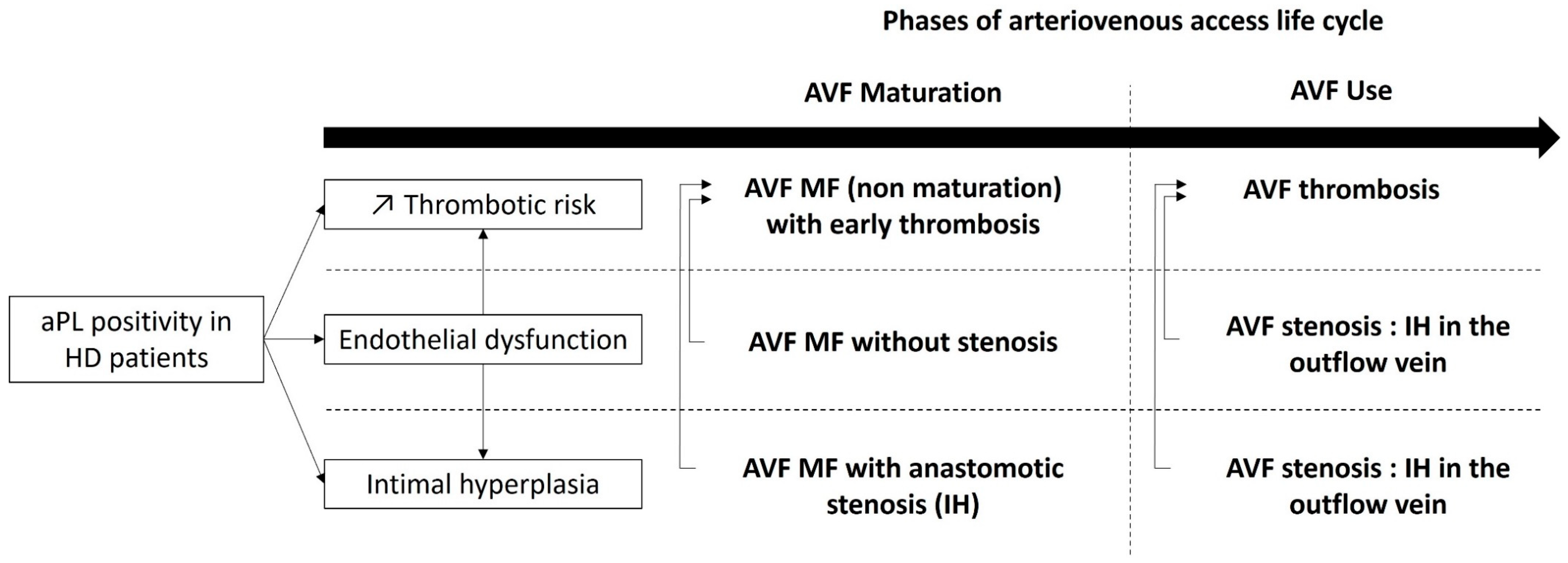
Antiphospholipid antibody (aPL)-persistent positivity is frequent in hemodialysis (HD) patients. Native arteriovenous fistula (AVF) complications such as stenosis and thrombosis are among the most important causes of morbidity and mortality in hemodialysis patients. The association between aPL positivity and AVF thrombosis seems to be well established.

antiphospholipid antibodies antiphospholipid syndrome arteriovenous fistula

1. Introduction

1.1. Hemodialysis and Vascular Access

End-stage kidney disease (ESKD) is typically defined by a glomerular filtration rate of less than 15 mL/min/1.73 m2 and requires the initiation of renal replacement therapy, such as hemodialysis (HD), peritoneal dialysis, or kidney transplantation, for survival [1]. The incidence of ESKD is increasing worldwide and the large majority of the patients remain on chronic HD, a modality requiring an efficient vascular access. The options for vascular access in HD patients include native arteriovenous fistulas (AVF), arteriovenous grafts (AVG), and tunneled central venous catheters (CVC). Native AVF is generally considered as the best option for vascular access in HD patients, because of lower rates of infection and thrombosis compared to AVG and CVC. Additionally, AVF have been associated with improved long-term survival and reduced healthcare costs [2][3]. However, AVF complications are common in HD patients and can lead to significant morbidity and mortality. These complications include thrombosis, stenosis, infection, aneurysm, pseudoaneurysm, and hemorrhage. Thrombosis and stenosis are the most common complications, often requiring intervention with angioplasty or thrombectomy. Regular monitoring and timely intervention can help prevent and manage these complications [4].

1.2. Antiphospholipid Syndrome and Pathophysiology

Antiphospholipid syndrome (APS) is an autoimmune disorder characterized by the persistent positivity of circulating antiphospholipid antibodies (aPL) resulting in arterial, venous, or microvascular thrombosis and obstetrical complications. The pathophysiology of thrombosis in APS is complex and multifactorial. aPL trigger phospholipids and phospholipid binding proteins at different cell surfaces (i.e., endothelial cell, platelets, monocytes, and neutrophils). Endothelial cells are activated by aPL and acquire a phenotype that promotes inflammation, complement activation, leukocyte trafficking, and a procoagulant state. This activation ultimately leads to in situ thrombosis while also promoting other non-thrombotic autoimmune and inflammatory complications [5][6]. APS have also been associated with endothelial cell dysfunction both in vitro and in vivo [7][8]. Distinct from thrombotic events, the chronic occlusive APS vasculopathy is characterized by cell proliferation and infiltration that progressively expands the intima, therefore narrowing the vascular lumen. The latter was first described in aPL nephropathy [9]. The mammalian Target of Rapamycin (mTOR) pathway, implicated in cell proliferation and survival, seems to be an important signaling pathway by which aPL trigger intimal hyperplasia and occlusive vasculopathy [6][10].
A “two-hit” model in the pathogenesis of APS has been proposed, postulating that aPL provide the first hit favoring a procoagulant state but not sufficient to cause thrombosis. Subsequently, a second hit (e.g., an infectious or inflammatory stimuli or a vascular injury) will lead to vascular thrombosis. This second hit is not obvious in many cases [6].

1.3. Classification Criteria of Antiphospholipid Syndrome

The 2006 Revised Sapporo APS classification criteria have been recently revised in 2023 by the American College of Rheumatology (ACR) and the European Alliance of Associations for Rheumatology (EULAR) [11][12]. These new classification criteria are based on a scoring system for both laboratory and clinical criteria. Patients can be classified as APS for research purposes if there are at least 3 points from clinical domains and at least 3 points from laboratory domains. As in the previous criteria, aPL positivity must be confirmed after at least 12 weeks. Three aPL assays are recommended, including Immunoglobulin (Ig) G or IgM anticardiolipin antibody (aCL), IgG or IgM anti-beta2 glycoprotein I antibody (aβ2-GPI), or Lupus Anticoagulant (LA). The 2023 ACR/EULAR classification criteria no longer consider isolated positivity of IgM aCL or IgM aβ2-GPI as sufficient [11][12]. Other non-criteria antibodies potentially predictive of thrombosis in APS such as IgA aCL; IgA aβ2-GPI; IgG, IgA, IgM anti-phosphatidylserine/prothrombin (aPS/PT); IgG anti-phosphatidylserine antibodies (aPS) are not included as well [12][13].
With respect to the clinical manifestations, the new 2023 ACR/EULAR APS classification criteria allow for the stratification of risk for macrovascular events through the assessment of traditional thrombosis risk factors with weighted assessment. The definitions of high-risk venous thromboembolism and cardiovascular disease are presented in the research. These criteria also define microvascular domain items considered mechanistically distinct from moderate-to-large vessel disease. Indeed, features, such as APS Nephropathy, cardiac valve disease, livedo racemose, and thrombocytopenia, have been added to better capture and quantify the diverse manifestations of APS. These new 2023 ACR/EULAR APS classification criteria have a specificity of 99% compared to the 86–91% specificity of the 2006 Revised Sapporo criteria [12]. Table 1 summarizes the main differences between 2006 Revised Sapporo criteria and 2023 ACR/EULAR classification criteria.
Table 1. Main differences between 2006 revised Sapporo and 2023 ACR/EULAR classification criteria for antiphospholipid syndrome.

aCL: anticardiolipin antibody, aβ2GPI: anti-β2 glycoprotein I antibody, LA: lupus anticoagulant.

1.4. Antiphospholipid Antibodies in Hemodialysis Patients

ESKD is rare in APS [14]. On the other hand, aPL-persistent positivity is frequently seen in ESKD. Its prevalence is higher in HD patients when compared to ESKD conservatively treated, to peritoneal dialysis patients and to general population [15]. Indeed, the prevalence of aPL in HD patients varies from 11 to 56% [16][17][18][19][20][21][22][23] and is estimated to range between 40 and 50 cases per 100,000 in the general population [24]. However, aPL positivity is inconsistently associated with AVF complications such as thrombosis and stenosis.

2. AVF Maturation

After native AVF surgical creation, the outflow vein goes through a complex vascular remodeling process called the “maturation process”, usually taking place within 4 to 6 weeks. Actually, this time period can continue during three post-operative months [25]. The outflow vein will experience vasodilatation and wall thickening therefore allowing a two-needle puncture [26]. AVF maturation can be assessed clinically or by using ultrasound imaging mainly based on AVF blood flow and outflow vein diameter [27][28]. AVF maturation failure is a frequent complication that affects more than half of the AVF and requires frequent interventions in order to facilitate maturation (i.e., assisted maturation) [26][29]. Intimal hyperplasia is the main stenosis lesion and the leading cause of AVF non maturation [30]. It has been associated with endothelial dysfunction both in vivo and in vitro [31][32][33]. In the multicenter prospective Hemodialysis Maturation Fistula Study, interventions performed for AVF stenosis were the most frequent interventions aiming to facilitate AVF maturation [29].
To the researchers' knowledge, no study has investigated the association between APS or aPL positivity and native AVF maturation failure. A cross-sectional study by Sunnesh Reddy Anapalli et al., published in 2022, found a statistically significant association between IgG and IgM and aCL and AVF failure, defined as an AVF that never went to the point of successful cannulation, or failed within the first three months. This study focusing on 50 patients with native AVF failure and 50 controls, IgG aCL and IgM aCL were associated with AVF thrombosis. AVF Maturation was not directly assessed in this study. Also, they did not mention if aCL were persistently positive and their cut off for IgG aCL and IgM aCL were > 10 GPL and > 15 MPL, respectively [34]. One clinical case reported AVF maturation failure in a patient with primary APS [35]. One retrospective study on 103 HD patients showed a statistically significant association between AVF maturation failure (defined by the absence or a delay of maturation according to KDOQI guidelines) and aPL or APS. This association was independent of stenosis and intimal hyperplasia in a multivariate analysis. Interestingly, This study reported that patients with a fluctuating aPL profile also have a significant higher prevalence of AVF maturation failure [36]. Therefore, some authors hypothesize that aPL might cause AVF maturation failure, possibly through endothelial dysfunction leading to an impaired vascular remodeling capability without stenosis [7][37][38]. Moreover, because APS is associated with endothelial dysfunction and intimal hyperplasia in some aPL-related manifestations [7][8][39], AVF maturation failure could be related to stenosis and intimal hyperplasia in the setting of aPL positivity. Also, AVF maturation failure could be related to early thrombosis in the setting of aPL positivity. Figure 1 summarizes the putative pathogenesis of aPL-related AVF maturation failure. Further research is needed to better understand the underlying mechanisms and to develop effective strategies for preventing and treating AVF maturation failure in patients with APS or aPL.
Figure 1. Overview of aPL putative pathophysiology in native arteriovenous fistula early and long-term complications. aPL: antiphospholipid antibody, AVF: arteriovenous fistula, HD: hemodialysis, IH intimal hyperplasia, MF maturation failure.

3. AVF Thrombosis

ESKD is associated with more thrombotic manifestations compared to the general population [40][41]. This higher risk is not fully explained by risk factors encountered in HD population. A hypercoagulability state is usually related to uremic toxins accumulation and is mediated by platelet dysfunction as well as an increased level of the procoagulant tissue factor both in vitro and in vivo [42]. Modification of the functional properties of human venous endothelial cells (VEC) and arterial endothelial cells (AEC) have been observed. Indeed, a recent study reported that VEC acquire a prothrombotic phenotype in contact with uremic serum whereas AEC acquire an inflammatory phenotype [43]. Furthermore, HD is associated with elevated levels of procoagulant factors such as prothrombin fragments and thrombin–antithrombin complexes [34]. Native AVF thrombosis remains a major cause of morbidity in HD patients, representing the most common cause of vascular access failure [44]. Such thrombotic event can occur early after AVF creation, usually due to an inflow problem (e.g., juxta anastomotic stenosis, technical errors of construction, and anatomic abnormalities) or later due to outflow vein stenosis or intimal hyperplasia [34].
The role of hereditary and acquired thrombophilia in AVF failure and AVF patency has been reported in few studies, and data suggest that most of the AVF thrombosis in the setting of thrombophilia occurs during the first month after AVF creation [34][45][46]. Because of the higher risk of arterial and venous thrombosis in patients with persistent aPL, AVF thrombosis would be expected to occur more predominantly in these patients. Indeed, several studies have described the association between aPL and AVF thrombosis whereas others did not find such association. A systematic review and meta-analysis confirmed the association between aPL (lupus anticoagulant and IgG aCL) and AVF thrombosis [15]. Non-criteria antibodies that are not included in the 2023 ACR/EULAR classification criteria for APS have been associated with AVF thrombosis such as IgA aβ2GPI [21][22][47]. On the contrary, IgG and IgM aβ2GPI have not been associated with native AVF thrombosis in the literature.
Whether AVF thrombosis in the setting of aPL positivity is favored by stenosis or intimal hyperplasia is not known. Indeed, in the setting of aPL positivity, early thrombosis could be favored by anastomotic intimal hyperplasia [48][49]. Intimal hyperplasia is a well-known non-thrombotic histological lesion of APS, well described in aPL-associated disorders such as aPL-associated nephropathy [39][50]. The latter involves the activation of the mTOR signaling pathway [10]. On the other hand, late AVF stenosis and intimal hyperplasia are caused by tissue remodeling and proliferation which may gradually progress during fistula aging and HD procedure itself (needle injury, change in blood flow, etc.). Indeed, HD duration has been described as a risk factor for AVF thrombosis [47][51].

4. Stenosis and Intimal Hyperplasia

One of the most common complications associated with a native AVF is the stenosis of the outflow vein, resulting in thrombosis, the most common cause of late AVF loss [52]. Both stenosis and thrombosis compromise AVF primary patency and usually coincide, but they cannot always be distinguished [22]. Stenosis is commonly reported in up to 60% of functional AVF. This finding is of interest because AVF thrombosis is usually attributed to the presence of venous stenosis or inflow abnormalities (anastomotic stenosis). The implementation of a surveillance program dedicated to the detection of progressive subclinical stenosis is of importance as data suggest that multiple factors are required for AVF failure [26].
A common cause of stenosis is intimal hyperplasia, which is well described in AVF [48][49]. Intimal hyperplasia is a crucial histopathological injury and forms the basis for vascular stenosis. It implies shear stress, endothelial dysfunction, inflammation, proliferation, and migration of vascular smooth muscle cells and extracellular matrix amalgamation and degradation [30]. It can take place either at anastomotic levels in the outflow vein or playing a role in restenosis after angioplasty. The prevalence of the latter complication has dropped since the use of drug coated balloon or stent mostly using paclitaxel for its anti-proliferative effect [30]. Despite the description of intimal hyperplasia in outflow veins before AVF surgical creation, studies failed to find any association between pre-existing intimal hyperplasia and AVF stenosis [29][48].
There are few studies evaluating AVF stenosis in the setting of aPL positivity in HD patients. In a combined retrospective and prospective cohort study of a single outpatient dialysis unit, the presence of IgM aCL was associated with AVF stenosis. In multivariate analysis, the presence of stenosis was significantly associated with the development of AVF thrombosis [53]. Intrastent restenosis or restenosis after drug-eluted balloon have not been studied in aPL positive HD patients. However, few studies have demonstrated that patients with APS are predisposed to high rates of restenosis of the coronary arteries after percutaneous coronary intervention [54]. As previously said, intimal hyperplasia is a well-known non-thrombotic histological lesion associated with aPL positivity involving the activation of the mTOR signaling pathway [10]. Up to now, no studies comparing AVF restenosis and drug-eluted angioplasty (e.g., sirolimus coated balloon angioplasty) in aPL positive HD patients are currently available.

5. Mortality

Serrano et al. reported in a 2-year prospective study that IgA aβ2GPI positivity was associated with a higher mortality rate, compared to negative patients [21]. However, other authors found no association with mortality [16].

References

  1. Saran, R.; Robinson, B.; Abbott, K.C.; Bragg-Gresham, J.; Chen, X.; Gipson, D.; Gu, H.; Hirth, R.A.; Hutton, D.; Jin, Y.; et al. US Renal Data System 2019 Annual Data Report: Epidemiology of Kidney Disease in the United States. Am. J. Kidney Dis. 2020, 75 (Suppl. S1), A6–A7.
  2. Casserly, L.F.; Dember, L.M. Thrombosis in end-stage renal disease. Semin. Dial. 2003, 16, 245–256.
  3. Roy-Chaudhury, P.; Kelly, B.S.; Melhem, M.; Zhang, J.; Li, J.; Desai, P.; Munda, R.; Heffelfinger, S.C. Vascular access in hemodialysis: Issues, management, and emerging concepts. Cardiol. Clin. 2005, 23, 249–273.
  4. Lawson, J.H.; Niklason, L.E.; Roy-Chaudhury, P. Challenges and novel therapies for vascular access in haemodialysis. Nat. Rev. Nephrol. 2020, 16, 586–602.
  5. Knight, J.S.; Branch, D.W.; Ortel, T.L. Antiphospholipid syndrome: Advances in diagnosis, pathogenesis, and management. BMJ 2023, 380, e069717.
  6. Knight, J.S.; Kanthi, Y. Mechanisms of immunothrombosis and vasculopathy in antiphospholipid syndrome. Semin. Immunopathol. 2022, 44, 347–362.
  7. Velásquez, M.; Rojas, M.; Abrahams, V.M.; Escudero, C.; Cadavid, Á.P. Mechanisms of Endothelial Dysfunction in Antiphospholipid Syndrome: Association with Clinical Manifestations. Front. Physiol. 2018, 9, 1840.
  8. Cugno, M.; Borghi, M.O.; Lonati, L.M.; Ghiadoni, L.; Gerosa, M.; Grossi, C.; De Angelis, V.; Magnaghi, G.; Tincani, A.; Mari, D.; et al. Patients with antiphospholipid syndrome display endothelial perturbation. J. Autoimmun. 2010, 34, 105–110.
  9. Nochy, D.; Daugas, E.; Droz, D.; Beaufils, H.; Grünfeld, J.P.; Piette, J.C.; Bariety, J.; Hill, G. The intrarenal vascular lesions associated with primary antiphospholipid syndrome. J. Am. Soc. Nephrol. 1999, 10, 507–518.
  10. Canaud, G.; Bienaimé, F.; Tabarin, F.; Bataillon, G.; Seilhean, D.; Noël, L.-H.; Dragon-Durey, M.-A.; Snanoudj, R.; Friedlander, G.; Halbwachs-Mecarelli, L.; et al. Inhibition of the mTORC pathway in the antiphospholipid syndrome. N. Engl. J. Med. 2014, 371, 303–312.
  11. Miyakis, S.; Lockshin, M.D.; Atsumi, T.; Branch, D.W.; Brey, R.L.; Cervera, R.; Derksen, R.H.W.M.; DE Groot, P.G.; Koike, T.; Meroni, P.L.; et al. International consensus statement on an update of the classification criteria for definite antiphospholipid syndrome (APS). J. Thromb. Haemost. 2006, 4, 295–306.
  12. Barbhaiya, M.; Zuily, S.; Naden, R.; Hendry, A.; Manneville, F.; Amigo, M.-C.; Amoura, Z.; Andrade, D.; Andreoli, L.; Artim-Esen, B.; et al. 2023 ACR/EULAR antiphospholipid syndrome classification criteria. Ann. Rheum. Dis. 2023, 82, 1258–1270.
  13. Liu, X.; Zhu, L.; Liu, H.; Cai, Q.; Yun, Z.; Sun, F.; Jia, Y.; Guo, J.; Li, C. Non-criteria antiphospholipid antibodies in antiphospholipid syndrome: Diagnostic value added. Front. Immunol. 2022, 13, 972012.
  14. Gracia-Tello, B.; Isenberg, D. Kidney disease in primary anti-phospholipid antibody syndrome. Rheumatology 2017, 56, 1069–1080.
  15. Ames, P.R.J.; Merashli, M.; Bucci, T.; Pastori, D.; Pignatelli, P.; Violi, F.; Bellizzi, V.; Arcaro, A.; Gentile, F. Antiphospholipid antibodies in end-stage renal disease: A systematic review and meta-analysis. Hemodial. Int. 2020, 24, 383–396.
  16. Chew, S.L.; Lins, R.L.; Daelemans, R.; Zachee, P.; De Clerck, L.S.; Vermylen, J. Are antiphospholipid antibodies clinically relevant in dialysis patients? Nephrol. Dial. Transplant. 1992, 7, 1194–1198.
  17. Roozbeh, J.; Serati, A.R.; Malekhoseini, S.A. Arteriovenous fistula thrombosis in patients on regular hemodialysis: A report of 171 patients. Arch. Iran. Med. 2006, 9, 26–32.
  18. Ozmen, S.; Danis, R.; Akin, D.; Batun, S. Anticardiolipin antibodies in hemodialysis patients with hepatitis C and their role in fistula failure. Clin. Nephrol. 2009, 72, 193–198.
  19. Brunet, P.; Aillaud, M.F.; San Marco, M.; Philip-Joet, C.; Dussol, B.; Bernard, D.; Juhan-Vague, I.; Berland, Y. Antiphospholipids in hemodialysis patients: Relationship between lupus anticoagulant and thrombosis. Kidney Int. 1995, 48, 794–800.
  20. Prakash, R.; Miller, C.C.; Suki, W.N. Anticardiolipin antibody in patients on maintenance hemodialysis and its association with recurrent arteriovenous graft thrombosis. Am. J. Kidney Dis. 1995, 26, 347–352.
  21. Serrano, A.; García, F.; Serrano, M.; Ramírez, E.; Alfaro, F.J.; Lora, D.; de la Cámara, A.G.; Paz-Artal, E.; Praga, M.; Morales, J.M. IgA antibodies against β2 glycoprotein I in hemodialysis patients are an independent risk factor for mortality. Kidney Int. 2012, 81, 1239–1244.
  22. Salmela, B.; Hartman, J.; Peltonen, S.; Albäck, A.; Lassila, R. Thrombophilia and arteriovenous fistula survival in ESRD. Clin. J. Am. Soc. Nephrol. 2013, 8, 962–968.
  23. Ghisdal, L.; Broeders, N.; Wissing, K.M.; Mena, J.M.; Lemy, A.; Wijns, W.; Pradier, O.; Donckier, V.; Racapé, J.; Vereerstraeten, P.; et al. Thrombophilic factors in Stage V chronic kidney disease patients are largely corrected by renal transplantation. Nephrol. Dial. Transplant. 2011, 26, 2700–2705.
  24. Dabit, J.Y.; Valenzuela-Almada, M.O.; Vallejo-Ramos, S.; Duarte-García, A. Epidemiology of Antiphospholipid Syndrome in the General Population. Curr. Rheumatol. Rep. 2022, 23, 85.
  25. Zonnebeld, N.; Huberts, W.; van Loon, M.M.; Delhaas, T.; Tordoir, J.H.M. Natural Vascular Remodelling After Arteriovenous Fistula Creation in Dialysis Patients With and Without Previous Ipsilateral Vascular Access. Eur. J. Vasc. Endovasc. Surg. 2020, 59, 277–287.
  26. Gameiro, J.; Ibeas, J. Factors affecting arteriovenous fistula dysfunction: A narrative review. J. Vasc. Access. 2020, 21, 134–147.
  27. Lok, C.E.; Huber, T.S.; Lee, T.; Shenoy, S.; Yevzlin, A.S.; Abreo, K.; Allon, M.; Asif, A.; Astor, B.C.; Glickman, M.H.; et al. KDOQI Clinical Practice Guideline for Vascular Access: 2019 Update. Am. J. Kidney Dis. 2020, 75 (Suppl. S2), S1–S164.
  28. Beathard, G.A.; Lok, C.E.; Glickman, M.H.; Al-Jaishi, A.A.; Bednarski, D.; Cull, D.L.; Lawson, J.H.; Lee, T.C.; Niyyar, V.D.; Syracuse, D.; et al. Definitions and End Points for Interventional Studies for Arteriovenous Dialysis Access. Clin. J. Am. Soc. Nephrol. 2018, 13, 501–512.
  29. Huber, T.S.; Berceli, S.A.; Scali, S.T.; Neal, D.; Anderson, E.M.; Allon, M.; Cheung, A.K.; Dember, L.M.; Himmelfarb, J.; Roy-Chaudhury, P.; et al. Arteriovenous Fistula Maturation, Functional Patency, and Intervention Rates. JAMA Surg. 2021, 156, 1111–1118.
  30. Ma, S.; Duan, S.; Liu, Y.; Wang, H. Intimal Hyperplasia of Arteriovenous Fistula. Ann. Vasc. Surg. 2022, 85, 444–453.
  31. Siddiqui, M.A.; Ashraff, S.; Carline, T. Maturation of arteriovenous fistula: Analysis of key factors. Kidney Res. Clin. Pract. 2017, 36, 318–328.
  32. Somarathna, M.; Hwang, P.T.; Millican, R.C.; Alexander, G.C.; Isayeva-Waldrop, T.; Sherwood, J.A.; Brott, B.C.; Falzon, I.; Northrup, H.; Shiu, Y.-T.; et al. Nitric oxide releasing nanomatrix gel treatment inhibits venous intimal hyperplasia and improves vascular remodeling in a rodent arteriovenous fistula. Biomaterials 2022, 280, 121254.
  33. Singh, M.; Mahapatra, H.S.; Pursnani, L.; Muthukumar, B.; Neeraj Anant, I.; Kumar, A.; Kaur, N.; Singh, A.; Krishnan, C. on prediction of arterio-venous fistula maturation by flow mediated dilatation and AVF blood flow. J. Vasc. Access. 2023, 24, 443–451.
  34. Anapalli, S.R.; N, H.D.; Sarma, P.; Srikanth, L.; V, S.K. Thrombophilic risk factors and ABO blood group profile for arteriovenous access failure in end stage kidney disease patients: A single-center experience. Ren. Fail. 2022, 44, 34–42.
  35. Bolleke, E.; Seferi, S.; Rroji, M.; Idrizi, A.; Barbullushi, M.; Thereska, N. Exhausting multiple hemodialysis access failures. Med. Arch. 2014, 68, 361–363.
  36. Taghavi, M.; Demulder, A.; Mesquita, M.D.C.F.; Dernier, Y.; Nortier, J.; Collart, F.; Pozdzik, A. Underestimated Effect of Antiphospholipid Antibodies on Arteriovenous Fistula Maturation in Hemodialysis Patients, 09 May 2022, Preprint (Version 1). Research Square. Available online: https://www.researchsquare.com/article/rs-1606215/v1 (accessed on 9 May 2022).
  37. Evangelatos, G.; Kravvariti, E.; Konstantonis, G.; Tentolouris, N.; Sfikakis, P.P.; Tektonidou, M.G. Atherosclerosis progression in antiphospholipid syndrome is comparable to diabetes mellitus: A 3 year prospective study. Rheumatology 2022, 61, 3408–3413.
  38. Sorice, M.; Profumo, E.; Capozzi, A.; Recalchi, S.; Riitano, G.; Di Veroli, B.; Saso, L.; Buttari, B. Oxidative Stress as a Regulatory Checkpoint in the Production of Antiphospholipid Autoantibodies: The Protective Role of NRF2 Pathway. Biomolecules 2023, 13, 1221.
  39. Barbhaiya, M.; Taghavi, M.; Zuily, S.; Domingues, V.; Chock, E.Y.; Tektonidou, M.G.; Erkan, D.; Seshan, S.V.; New APS Classification Criteria Steering Committee and APS ACTION Collaborators. Efforts to Better Characterize “Antiphospholipid Antibody Nephropathy” for the 2023 ACR/EULAR Antiphospholipid Syndrome Classification Criteria: Renal Pathology Subcommittee Report. J. Rheumatol. 2023, 50, jrheum.2022-1200.
  40. Wattanakit, K.; Cushman, M.; Stehman-Breen, C.; Heckbert, S.R.; Folsom, A.R. Chronic kidney disease increases risk for venous thromboembolism. J. Am. Soc. Nephrol. 2008, 19, 135–140.
  41. Go, A.S.; Bansal, N.; Chandra, M.; Lathon, P.V.; Fortmann, S.P.; Iribarren, C.; Hsu, C.-Y.; Hlatky, M.A.; ADVANCE Study Investigators. Chronic kidney disease and risk for presenting with acute myocardial infarction versus stable exertional angina in adults with coronary heart disease. J. Am. Coll. Cardiol. 2011, 58, 1600–1607.
  42. Gondouin, B.; Cerini, C.; Dou, L.; Sallée, M.; Duval-Sabatier, A.; Pletinck, A.; Calaf, R.; Lacroix, R.; Jourde-Chiche, N.; Poitevin, S.; et al. Indolic uremic solutes increase tissue factor production in endothelial cells by the aryl hydrocarbon receptor pathway. Kidney Int. 2013, 84, 733–744.
  43. Sosińska-Zawierucha, P.; Bręborowicz, A. Uremic serum induces prothrombotic changes in venous endothelial cells and inflammatory changes in aortic endothelial cells. Ren. Fail. 2021, 43, 401–405.
  44. Montagnana, M.; Meschi, T.; Borghi, L.; Lippi, G. Thrombosis and occlusion of vascular access in hemodialyzed patients. Semin. Thromb. Hemost. 2011, 37, 946–954.
  45. Knoll, G.A.; Wells, P.S.; Young, D.; Perkins, S.L.; Pilkey, R.M.; Clinch, J.J.; Rodger, M.A. Thrombophilia and the risk for hemodialysis vascular access thrombosis. J. Am. Soc. Nephrol. 2005, 16, 1108–1114.
  46. Grupp, C.; Troche-Polzien, I.; Stock, J.; Bramlage, C.; Müller, G.A.; Koziolek, M. Thrombophilic risk factors in hemodialysis: Association with early vascular access occlusion and patient survival in long-term follow-up. PLoS ONE 2019, 14, e0222102.
  47. Hadhri, S.; Rejeb, M.B.; Belarbia, A.; Achour, A.; Skouri, H. Hemodialysis duration, human platelet antigen HPA-3 and IgA isotype of anti-β2glycoprotein I antibodies are associated with native arteriovenous fistula failure in Tunisian hemodialysis patients. Thromb. Res. 2013, 131, e202–e209.
  48. Vazquez-Padron, R.I.; Duque, J.C.; Tabbara, M.; Salman, L.H.; Martinez, L. Intimal Hyperplasia and Arteriovenous Fistula Failure: Looking Beyond Size Differences. Kidney360 2021, 2, 1360–1372.
  49. Hu, H.; Patel, S.; Hanisch, J.J.; Santana, J.M.; Hashimoto, T.; Bai, H.; Kudze, T.; Foster, T.R.; Guo, J.; Yatsula, B.; et al. Future research directions to improve fistula maturation and reduce access failure. Semin. Vasc. Surg. 2016, 29, 153–171.
  50. Martínez-Flores, J.A.; Serrano, M.; Morales, J.M.; Serrano, A. Antiphospholipid Syndrome and Kidney Involvement: New Insights. Antibodies 2016, 5, 17.
  51. Nampoory, M.R.N.; Das, K.C.; Johny, K.V.; Al-Hilali, N.; Abraham, M.; Easow, S.; Saed, T.; Al-Muzeirei, I.A.; Sugathan, T.N.; Al Mousawi, M. Hypercoagulability, a serious problem in patients with ESRD on maintenance hemodialysis, and its correction after kidney transplantation. Am. J. Kidney Dis. 2003, 42, 797–805.
  52. Turmel-Rodrigues, L.; Mouton, A.; Birmelé, B.; Billaux, L.; Ammar, N.; Grézard, O.; Hauss, S.; Pengloan, J. Salvage of immature forearm fistulas for haemodialysis by interventional radiology. Nephrol. Dial. Transplant. 2001, 16, 2365–2371.
  53. Adler, S.; Szczech, L.; Qureshi, A.; Bollu, R.; Thomas-John, R. IgM anticardiolipin antibodies are associated with stenosis of vascular access in hemodialysis patients but do not predict thrombosis. Clin. Nephrol. 2001, 56, 428–434.
  54. Perl, L.; Netzer, A.; Rechavia, E.; Bental, T.; Assali, A.; Codner, P.; Mager, A.; Battler, A.; Kornowski, R.; Lev, E.I. Long-term outcome of patients with antiphospholipid syndrome who undergo percutaneous coronary intervention. Cardiology 2012, 122, 76–82.
More
Information
Contributors MDPI registered users' name will be linked to their SciProfiles pages. To register with us, please refer to https://encyclopedia.pub/register : , , , , ,
View Times: 377
Revision: 1 time (View History)
Update Date: 10 Jan 2024
1000/1000