Submitted Successfully!
To reward your contribution, here is a gift for you: A free trial for our video production service.
Thank you for your contribution! You can also upload a video entry or images related to this topic.
Version Summary Created by Modification Content Size Created at Operation
1 -- 5420 2023-12-18 09:16:16 |
2 format correct + 347 word(s) 5767 2023-12-21 02:01:49 |

Video Upload Options

Do you have a full video?

Confirm

Are you sure to Delete?
Cite
If you have any further questions, please contact Encyclopedia Editorial Office.
Mitaka, T.; Ichinohe, N.; Tanimizu, N. “Small Hepatocytes” in the Liver. Encyclopedia. Available online: https://encyclopedia.pub/entry/52855 (accessed on 28 April 2024).
Mitaka T, Ichinohe N, Tanimizu N. “Small Hepatocytes” in the Liver. Encyclopedia. Available at: https://encyclopedia.pub/entry/52855. Accessed April 28, 2024.
Mitaka, Toshihiro, Norihisa Ichinohe, Naoki Tanimizu. "“Small Hepatocytes” in the Liver" Encyclopedia, https://encyclopedia.pub/entry/52855 (accessed April 28, 2024).
Mitaka, T., Ichinohe, N., & Tanimizu, N. (2023, December 18). “Small Hepatocytes” in the Liver. In Encyclopedia. https://encyclopedia.pub/entry/52855
Mitaka, Toshihiro, et al. "“Small Hepatocytes” in the Liver." Encyclopedia. Web. 18 December, 2023.
“Small Hepatocytes” in the Liver
Edit

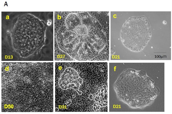
Mature hepatocytes (MHs) in an adult rodent liver are categorized into the following three subpopulations based on their proliferative capability: type I cells (MH-I), which are committed progenitor cells that possess a high growth capability and basal hepatocytic functions; type II cells (MH-II), which possess a limited proliferative capability; and type III cells (MH-III), which lose the ability to divide (replicative senescence) and reach the final differentiated state. These subpopulations may explain the liver’s development and growth after birth. Generally, small-sized hepatocytes emerge in mammal livers. The cells are characterized by being morphologically identical to hepatocytes except for their size, which is substantially smaller than that of ordinary MHs. We initially discovered small hepatocytes (SHs) in the primary culture of rat hepatocytes.

liver stem/progenitor cells regeneration cell transplantation proliferation differentiation self-renewal

1. SHs In Vitro

1.1. Culture Medium

Rat SHs in a healthy adult liver are enriched by centrifugation at 150× g after eliminating MHs by repeated centrifugation at 50× g. The colonies appear at 4–5 days after plating, and SHs continue to expand (Figure 1Aa) [1]. The colonies that consist of SHs were also observed in the primary culture of rat hepatocytes in the serum-free chemically defined medium [2]. The authors demonstrated that the medium that contains diferric transferrin and arginine, as well as nicotinamide, was required for the sustained clonal proliferation of rat hepatocytes, resulting in SH expansion. Hepatocyte growth factor (HGF), transforming growth factor (TGF)-α, and/or EGF were also necessary to stimulate clonal growth. Additionally, the supplementation of nicotinamide in the medium could allow mouse primary hepatocytes to proliferate and survive for >2 months while maintaining their differentiated functions [3]. The proliferating hepatocytes with near-diploid features were immortalized after the long-term cultivation.
Figure 1. Small hepatocytes (SHs) in vitro and in vivo. (A) Photos of SHs isolated from a rat liver (AaAc) and a human liver tissue (AdAf). A photo of an SH colony consisting of small-sized cells showing a flat surface 13 days after plating (Aa). A colony consisting of piled-up cells is surrounded by NPCs cultured for 27 days (Ab). A colony of rat HPPCs of the third passage cultured on thin-Matrigel 21 days after replating (Ac). Primary human hepatocytes isolated from a healthy part of the surgically dissected liver tissue are cultured for 50 days. Many mononuclear SHs are proliferating (Ad). Primary human hepatocytes proliferate to form a large colony and part of the cells pile up on the colony 31 days after plating (Ae). Primary human hepatocytes are cultured in the serum-free medium on a hyaluronan-coated dish for 21 days. A colony consisting of SHs is formed (Af). (B) Cells of an SH fraction isolated from a healthy rat liver are plated on the dishes coated with rat tail collagen (Ba), collagen type IV (Bb), fibronectin (Bc), thin-Matrigel (Bd), and collagen gel (Be,Bf). Cells were cultured in the modified Dulbecco’s modified Eagle medium (DMEM)/F12 medium with 10% fetal bovine serum (FBS) for 20 days. From 4 days after plating, 1% DMSO was added to the medium (BaBd,Bf). Cells were fixed with absolute ethanol at day 20 and immunocytochemically stained with Krt8. Nuclei were stained with hematoxylin. SH colonies show Krt8-positive (Brown). (C) SHs appear in liver tissue (in vivo). (Ca) A cluster of small hepatocyte-like progenitor cells (SHPCs) is observed in the rat liver treated with retrorsine (Ret) and 2/3 PH (HE-staining). (Cb) A photo of the rat liver treated with d-galactosamine at 3 days after administration. (Cc) A focus of SHs observed in the 99-week-old rat liver. (Cd) Magnified image of the area surrounded by white lines in the photo (Cb). (Ce) A photo of the rat liver treated with d-galactosamine at 4 days after administration. (Cb,Cd,Ce) White arrowheads may indicate OC-SHs emerging near elongating bile ductules and yellow arrows may show MH-SHs near resident MHs. Bars show 50 μm.
The emergence of SHs in the primary culture of hepatocytes was observed in the serum-free amino-acid-rich (Leibovitz 15, L-15) medium supplemented with NaHCO3 and EGF in a 5% CO2/95% air incubator, in addition to the condition with high nicotinamide concentrations [4]. SH colonies were often observed in the culture and the SHs continued to proliferate 4–5 days after plating. L-15 is a medium that was originally formulated to make a medium designed for use in a non-bicarbonate buffer system. The buffer with high concentrations of amino acids, especially arginine, was developed to culture cells without the use of a CO2 incubator [5]. Therefore, the researchers added 20 mM NaHCO3 in the medium to culture hepatocytes in a 5% CO2/95% air incubator [4]. The L-15 medium contains approximately three times as many amino acids as Dulbecco’s modified Eagle medium (DMEM). High amino acid and insulin concentrations in hepatocyte cultures are required for protein synthesis and proteolysis inhibition [6]. Additionally, exogenous arginine may be essential for maintaining the proliferative capacity of hepatocytes in culture because proliferating hepatocytes lose their hepatic differentiation function, including arginine production through the urea cycle [2].
The importance of trace elements is well-known in a serum-free hepatocyte culture [7]. The original L-15 medium does not contain trace elements such as CuSO4, FeSO4, MnSO4, ZnSO4, and Na2SeO3. Therefore, these were added into the serum-free L-15 medium [4]. Cable EE and Isom HC [8] also reported that trace metals, such as copper, iron, and zinc, are required for the long-term proliferation of primary hepatocytes cultured in a chemically defined medium. They observed the growth of both smaller hepatocytes and typical MHs in the medium supplemented with 2% dimethylsulfoxide (DMSO) for several months. Isom HC et al. [9] reported that adult rat hepatocytes that were cultured in the medium supplemented with 2% DMSO survived longer and maintained the capability of albumin synthesis longer than when cultured in the commonly used serum-free medium. The addition of 2% DMSO to the culture medium inhibited most hepatocyte proliferation, while 1% DMSO did not inhibit hepatocyte proliferation but did inhibit nonparenchymal cell (NPC) proliferation, especially mesenchymal cells that include fibroblasts (Figure 1Bf) [7]. Fibroblasts (mesenchymal cells) limit the expansion of SH colonies (Figure 1Be), thus 1% DMSO was added to the medium from day 4 after plating [10].
The growth of SHs also depends on an extracellular matrix (ECM) protein that is used to coat the culture devices. SH colonies similarly expanded in dishes coated with collagen (Col) types I and IV and fibronectin (FN), but their expansion was faster in dishes coated with thin-Matrigel or Col-gel. In particular, the expansion of SH colonies was dramatically enhanced (approximately 1.4 times larger than without DMSO) when SHs were cultured on Col-gel in a medium supplemented with 1% DMSO (Figure 1Be,f) [11]. These results indicate that the culture environment can bring out the innate potential of individual hepatocytes. They also indicate that SHs have a very high proliferative capacity and that the extent of their proliferation depends on the composition and three-dimensional ECM structure.

1.2. Purification of SHs

The researchers first used a simple method of low-speed centrifugation with sequential changes in gravity (50 and 150× g) and time (1 and 5 min) to isolate SHs [10][12]. This separation protocol contaminated NPCs, such as stellate cells, liver epithelial cells (LECs), Kupffer cells, and sinusoidal endothelial cells (SECs), as well as MHs, in the cell suspension. Additionally, both nicotinamide and fetal bovine serum (FBS) were required for seeded SHs to proliferate and form colonies [12]. The addition of FBS, as well as growth factors, was also necessary for SHs not only to proliferate but also to secrete plasma proteins such as albumin, transferrin, ceruloplasmin, etc. [7]. However, FBS can be removed from the culture because the proteins secreted by SHs can support their proliferation once SHs have increased in number [7].
SHs start cell division at 2–3 days after plating and proliferate clonally to form colonies when both nicotinamide and growth factors, such as EGF and HGF, are added to the medium [12]. Colony size varies among SHs, with some continuing to expand and others ceasing to grow after a relatively short incubation period. Most SECs disappear within a few days, and contaminated MHs divide two or three times at most. Most SHs can survive for more than a month but never achieve enough confluence to occupy the surface of the dish because many SHs in the colony suddenly die by apoptosis as the culture continues. However, SHs can be cultured for >5 months with repeated death and neogenesis [11]. Many SHs can spontaneously mature into cells with functions equivalent to those of MHs by interacting with NPCs (Figure 1Ab,e) [10][11] or by covering colonies with Engelbreth–Holm–Swam gels (EHS gels, Matrigel®, Growth-factor-reduced, Corning, NY, USA) [13].
Conversely, SHs can continue to proliferate on collagen gel without maturation when NPC proliferation is suppressed by the addition of 1% DMSO (Figure 1Bf), indicating that basement membrane reconstruction through the SH–NPC interaction is important for SHs to differentiate into MHs [10][13]. Additionally, primary hepatocytes have been thought not to proliferate after cryopreservation, but SHs can proliferate after long-term cryopreservation while maintaining their properties [14][15].
The standard method of preparing an SH-rich fraction uses multistep centrifugations and gives a mixture of SHs and NPCs, as described above. The comprehensive analysis identified CD44 as a molecule that is specifically expressed on the surface of SHs but not on MHs [16]. The CD44 gene encodes for a family of alternatively spliced multifunctional molecules, and it plays a role in the adhesion of cells to ECMs such as hyaluronic acid (HA), collagens, and fibronectin [17]. SHs expressed CD44 standard type (CD44s) and variant type 6 (CD44v6) [16]. CD44s becomes positive by immunostaining from day 3 after plating, and CD44v6 expression is delayed compared to CD44s expression, but both CD44s and CD44v6 expression disappear once SHs mature. CD44-positive hepatocytes cannot be identified in normal rat liver, but they transiently appear in the periportal region when the rat liver is severely injured by hepatotoxins such as d-galactosamine (GalN) [16][18][19].
SHs that appeared in GalN-treated rat livers could be isolated using anti-CD44 antibodies. CD44 is not expressed on hepatocytes in the normal adult liver, but it is expressed in cultured SHs forming colonies. Hence, HA, the ligand for CD44, was thought to be able to selectively isolate SH populations. Among NPCs, only SECs express receptors for HA, LYVE-1, and stabilin-1/2 [20][21], but are CD44-negative. Most SECs die within a few days after plating, as mentioned above.
SHs selectively grow and form colonies in the serum-free DMEM/F12 medium when an SH fraction is plated on HA-coated dishes (Figure 1Af) [22]. The effect of HA does not differ among commercially available forms of HA, but the combination of nicotinamide and growth factors, as well as transferrin and selenium, are both required for cell growth in a serum-free culture medium. SHs form colonies that consist of 30–40 cells at 8–10 days after plating. SH colonies gradually detach more easily from the HA-coated dish after approximately 2 weeks of incubation, and SHs can be collected and cryopreserved without trypsin [22]. Long-term cryopreserved SHs could grow without losing their ability to proliferate and mature [15].

1.3. Self-Renewal of Hepatic Progenitor Cells

Most SH colonies uniformly consisted of SHs in early culture. Small mononuclear cells proliferated, while some colonies appeared to have MH-like characteristics with a large cytoplasm and a nucleus, sometimes with binuclei [7]. This finding indicates the existence of parental cells within a population of SHs, a genuine hepatocytic progenitor. Proving that SHs retain the ability to self-renew themselves generation after generation is necessary to identify the parental SH.
SHs were cultured for 9–10 days in HA-coated dishes using a serum-free culture medium. Cells were detached using collagenase and hyaluronidase and then sorted using an anti-CD44 antibody to avoid trypsin injury. The sorted CD44-positive SHs failed to form colonies on either Col-I-coated or HA-coated dishes but proliferated to form colonies on thin-Matrigel-coated dishes. Furthermore, nicotinamide was crucial for colony formation, and the addition of EGF, insulin, and dexamethasone promoted parental SH growth [23]. However, a large number of the sorted cells seeded on thin-Matrigel-coated dishes failed to adhere and proliferate. The colonies demonstrated similar characteristics at the beginning of culture, but two morphologically distinguishable types of colonies emerged with time. One colony was circular and composed of small mononuclear cells, while the other demonstrated an irregular shape and was composed of cells with a large cytoplasm. The size of the colonies also varied. Colonies composed of small mononuclear cells, a typical SH, expanded rapidly to form large colonies, while colonies composed of relatively large cells proliferated slowly. This finding indicates the presence of a highly proliferative cell population. Only approximately 20% of the plated cells could adhere to a new dish that was thinly coated with Matrigel, which was small mononucleate cells when the cells were passaged every 4 weeks after the cultivation (Figure 1Ac). The SHs with high proliferative potential divided >50 times in 17 weeks. 
The HPPCs that grew on dishes coated with thin-Matrigel indicated that a certain component of Matrigel is crucial for HPPCs to maintain their ability to self-renew. Matrigel contains components that make up the basement membrane [24], which consists of laminin (LN), Col-IV, nidogen, and heparan sulfate proteoglycans [25]. Among them, LN is the major adhesion protein and mediates cell adhesion to the basement membrane. LNs are composed of three polypeptide chains, designated as α, β, and γ, and five α(α1–5), three β(β1–3), and three γ(γ1–3) chains are recognized in mammals [26]. Matrigel contains LN111 (α1,β1,γ1) as major constituents [24]. Generally, the LNα1-chain is expressed in fetal and neonatal rat liver lobules but is not found in adults. Instead, the LNα5-chain is present exclusively in the Glisson sheath [27]. Conversely, the transient expression of LNα1 was observed in regenerating liver lobules after 2/3 PH [27]. Integrins play a crucial role in cell adhesion to LNs [26]. They are composed of noncovalently associated α and β subunits. At least 24 separate integrins consisting of distinct combinations of α and β subunits have been identified in mammals to date. The specificity of the LN–integrin interaction is mainly dependent on α chains of LN, and the ligand specificity of the integrin is primarily determined by the α subunits. Integrin β subunits play a crucial role in signal transduction but have an auxiliary role in the ligand specificity [25][26].
Only <20% of SHs could adhere to dishes that were coated with LN111, as observed in thin-Matrigel-coated dishes [28]. The cell adhesion rates were slightly lower on LN111 than on Matrigel, and only <20% of cells adhered to the new dishes when SHs grown on LN111 were passaged. This rate gradually decreased with each successive passage. This indicates that the number of HPPCs decreased with passages. Furthermore, many of the cells that failed to attach to LN111 attached to LN511, indicating that HPPCs do not appear on LN511 and that cells growing on LN511 have lost their self-renewability. Thus, HPPCs generate two distinctive cell populations, which may indicate that HPPCs perform asymmetric cell divisions: LNα1-dependent and LNα5-dependent. HPPCs’ self-renewability is LN111-dependent, whereas LNα5, which is produced by adherent HPPCs, may support the survival and proliferation of LNα5-dependent daughter cells. Among the integrins that are involved in LN-binding, integrins α3 and β1 were expressed more in SHs that proliferate on LN111 than in cells on LN511, while integrin β4 was more strongly expressed in cells growing on LN511 [28]. Integrin α3highα6β1high could form HPPC colonies on LN111, but not α6β1low cells. Neutralizing antibodies against LN111 and integrin β1 could inhibit HPPC colony formation on LN111. These results indicate the importance of signaling from LN111 via integrin β1 for HPPC proliferation.
LNα1 and LNα5 were detected immunohistochemically on the basolateral side of cholangiocytes in fetal mouse liver, whereas only LNα5 was found in the adult liver [29]. Maintaining the self-renewability of LN111 has been reported in hepatoblast-like cells derived from human-induced pluripotent stem cells (hiPSCs) [30]. Conversely, undifferentiated hiPSCs can be maintained on LN511 but not on LN111 [31]. LN111 not only selectively maintains hepatoblast-like cells, but also eliminates the remaining undifferentiated cells. Additionally, CD45TER119c-Kitc-Met+CD29+CD49f+/low cells [32] and CD45TER119Dlk+ cells [33] have been reported to be highly proliferative and bipotential in fetal mouse livers. Furthermore, CD45TER119Dlk+ cells were named hepatic progenitor cells that proliferate on LN (HPPLs) because they can maintain high proliferative potential on LN-coated dishes [34]. HPPLs are DlkKrt19+albumin+, whereas hepatoblasts are Dlk+Krt19albumin+, indicating that HPPLs lose the characteristics of hepatoblasts. However, HPPLs possess not only high proliferative capability but also the potential to differentiate into both hepatocytes and cholangiocytes. LN is a crucial factor in maintaining the proliferation and multilineage differentiation potential of HPPLs. Furthermore, HPPLs strongly expressed integrin α6β1, and signals from LN are transduced through integrin β1 [34]. Conversely, intercellular cell adhesion molecule (ICAM)-1-positive liver progenitor cells separated from late fetal and postnatal mouse livers maintained their self-renewability on LN111 after passaging [35]. Furthermore, RT1A1−OX18lowICAM-1+ cells isolated from E13 rat livers have been reported to express integrin β1, proliferate on LN, and maintain bipotentiality [36]. Additionally, RT1A1+OX18+ICAM-1+ hepatocytes isolated from adult rat livers have also been reported to have a highly proliferative capability, but these cells must be cultured in a serum-free defined medium that contains EGF using STO embryonic cell lines as feeder cells. Thus, the self-renewability of cells with hepatocytic features may be maintained in the LNα1-dependent manner.

1.4. Characteristics of HPPCs

Gene expression patterns are also different between cells on LN111 and LN511. The gene expression levels of Alb, carbamoylphosphate synthetase (Cps) 1, glutamine synthetase (Gs), Keratins (Krts) 8 and 18, hepatocyte nuclear factor (HNF) (Hnf4α), and CCAAT/enhancer binding protein (C/EBP) α (Cebpα), as well as Cd44, a-fetoprotein (Afp), and Dlk-1, were found in both LN111-dependent and LN511-dependent cells [28]. However, absolute values of the genes related to hepatic functions, such as Alb, Cps1, Hnf4α, and Cebpα, were much lower in HPPCs than in MHs. Cholangiocyte-related genes, such as Krts 7 and 19, Sox9, and epithelial cell adhesion molecule (Epcam), were more highly expressed in cells cultured on LN511 than on LN111. Conversely, hepatic stem-cell-related genes, such as Thy1, c-Kit, Ncam1, and Cd24, were more highly expressed in cells cultured on LN111 than on LN511. Additionally, the genes of Cd34, leucine-rich orphan G-protein-coupled receptors (Lgrs) 4 and 5, Axin2, and telomerase reverse transcriptase (Tert), were expressed in cells neither in LN111 nor in LN511 [28]. Recent lineage tracing studies in mouse liver have revealed the presence of cell populations that regenerate the liver under homeostatic or injury conditions. Periportal hepatocytes that express Sox9 [37] or Mfsd2a [38], pericentral Axin2+ [39][40] or Lgr5+ hepatocytes [41], and broadly distributed Terthigh [42] or Lgr4+ hepatocytes [43] have been identified as candidates for generating new hepatocytes in homeostasis although they are strongly debated [44][45][46][47][48][49][50].

2. SH in Liver Lobules

SHs emerge not only in in vitro but also in vivo in rodents and human livers. Isolated hepatocytes are smaller in size than typical MHs, of which morphology is closer to SHs, before weaning. The frequency of the small-sized hepatocytes dramatically decreased with age after weaning [51]. Sigal SH et al. [52] used fluorescence-activated cell sorting (FACS) to reveal that hepatocytes that were isolated from fetal and suckling rat livers were all mononuclear and possessed granularity and autofluorescence comparable to hepatoblasts. The presence of small-sized hepatocytes has also been discovered in adult rat livers. Tateno C et al. [53] revealed that the isolated hepatocytes from male F344 rat livers based on the granularity and the autofluorescence by FACS were categorized into three subpopulations: SH-R3 (17.1 ± 0.2 μm), SH-R2 (22.6 ± 0.5 μm), and PH (24.1 ± 0.5 μm). Subsequently, Asahina K et al. [54] showed that >80% of SH-R3 cells were mononucleate and diploid and possessed high replicative capability.
Conversely, the administration of hepatotoxins, such as [5.1] 2-acetylaminofluorene (2-AAF), [5.2] d-galactosamine (GalN), and [5.3] retrorsine (Ret), induce the appearance of small-sized cells in rodent livers with or without growth stimulation such as PH. [5.4] Intermediate hepatocytes (IHs) have been observed in human livers with acute or chronic liver diseases. 

2.1. The 2-AAF/PH Model

Generally, 2-AAF interacts directly with DNA in hepatocytes to produce DNA adducts. It is used to induce a complete blockage of the proliferative capacity of hepatocytes after PH. This 2-AAF/PH protocol has been used as the most reliable rat OC model [55][56][57][58][59]. However, it cannot be applied to the mouse experiment because the mouse liver lacks the enzyme N-sulfotransferase, which activates 2-AAF [60]. OCs are known to express markers for membrane proteins, such as CD34, c-Kit, and Thy1, which are also regarded as hematopoietic stem cell markers [61]. Therefore, OCs were thought to be derived from BM [62]. Conversely, Thy1 has been reported to be a marker of hepatic myofibroblasts and hepatic stellate cells rather than specific for OCs [63][64]. Whether Thy1 is a specific marker for OCs remains controversial, but the researchers have reported that Thy1+ cells that are isolated from GalN-treated rat liver contain an LSPC (liver stem/progenitor cell) population [17][18].
Two patterns in the differentiation process of OCs into hepatocytes were reported depending on 2-AAF dosage [55][56]. The basic difference between the two doses was recognized a few days after PH. They revealed that OCs were arranged in a straight tubular pattern in rat livers that were treated with a low 2-AAF dosage, and most OCs synchronously differentiated into basophilic SHs (bSHs). The few unchanged OCs were located on the periportal side of the duct, whereas the distal part of the same duct was composed of bSHs. The newly formed hepatocytes maintained their tubular structure during the early stages of the differentiation process. Recovering normal lobular structure takes 10–12 days after PH. In contrast, foci that consist of bSHs appeared in livers treated with high doses of 2-AAF, where the basement membrane is histologically undetectable. Newly formed hepatocytes consisted of foci scattered throughout the livers, not confined in the periportal regions. Reconstructing the liver structure after PH may take approximately a month. The hepatocytic differentiation process of OCs was correlated with HNF4α expression and basement membrane disappearance [55]. Thus, the phenotypes were altered from a hybrid type of cells that express markers of hepatoblasts and BECs (AFP, OV-6, and Cx 43) to hepatocytes (HNF4α, α1-AT, Cx32, and BC formation) during OC differentiation into hepatocytes.

2.2. d-Galactosamine (GalN) Model

GalN is a potent hepatotoxic agent, causing hepatocyte death both by necrosis and apoptosis. GalN is metabolized by hepatocytes mainly in the pericentral region and functions as a transcriptional inhibitor by depleting uridine nucleotides [65][66]. Three unusual epithelial cell types, including OCs, bSHs (<16 μm), and hepatocytes, appear on tubular structures between 3–5 days after the intraperitoneal administration of GalN to rats (Figure 1Cb,d,e) [29][67][68]. The number of bSHs reaches a maximum on day 4 after GalN administration and then decreases. The liver finally regains its normal structure by day 8. The researchers have reported the appearance of SHs to be different from bSHs in GalN-injured livers [16]. Thy1-positive cells emerged in the periportal region on day 2 when rats were treated with GalN (Figure 2A). CD44-positive SHs transiently appeared in the region between Thy1+ cells and viable hepatocytes on days 3–5 after GalN treatment after the appearance of Thy1-positive cells. The number of CD44-positive SHs reached a maximum on day 4, while Thy1-positive cells rapidly declined and almost disappeared from the lobules by day 4. Thereafter, the number of CD44-positive SHs declined rapidly, and finding the cells that expressed CD44 was difficult on day 6.
Figure 2. Origins of small hepatocytes in GalN-treated rat livers. (A) SHs are derived from OCs and MHs. GalN administration activates portal Thy1+ cells and the Canal of Hering to induce OCs, and then OCs differentiate into SHs and BECs within 3 days after the treatment. Conversely, the slightly damaged MHs are delayed to start their proliferation, and MH-SHs are derived from MHs. Some OC-SHs and the proliferated MH-SHs are differentiated into MHs. (B) Typical markers of the specific cell appeared in the process of liver regeneration by GalN administration.
The isolation of the SH fraction, which contains HSCs, SECs, Kupffer cells (KCs), and LECs as well as SHs, yielded approximately 1 × 10⁸ cells from the liver of an adult male F344 rat at day 3 after GalN treatment (GalN-D3). The cells were then divided into Thy1+ and CD44+ fractions using anti-Thy1 and anti-CD44 antibodies, respectively. Thy1-positive cells that were isolated from GalN-D2 mostly demonstrated the characteristics of fibroblasts, while Thy1-positive cells that were isolated from GalN-D3 contained both morphologically polygonal (epithelial-like) and spindle shapes (fibroblast-like) [18][19]. Approximately 45% of fibroblasts predominantly showed Thy1+/desmin+ whereas approximately 55% of epithelial-like cells showed Thy1+/Krt19+, Thy1+/albumin+, or Thy1+/CD44+. In contrast, most of the CD44-positive cells that were isolated from GalN-D3 were polygonal in shape, and their morphology characterized the epithelial cells. Sorted CD44-positive cells expressed Krt19, albumin, and Thy1 at approximately 60%, 62%, and 65%, respectively [19]. Furthermore, approximately 2.0% ± 0.5%, 3.1% ± 0.3%, and 3.0% ± 0.6% of the SH fraction were Thy1+/CD44, Thy1+/CD44+, and Thy1/CD44+ cells, respectively (our unpublished data). The size and the characteristics of CD44+/Krt19+ cells were similar to those of the Thy1+/Krt19+ ones. Conversely, the colony formation efficiency of Thy1/CD44+ cells was five times higher than that of the Thy1+/CD44 and Thy1+/CD44+ ones. Additionally, the average size of colonies composed of CD44+/albumin+ cells was larger than that of the colony composed of the Thy1+/albumin+ ones. Moreover, the average sizes of colonies derived from CD44+ cells (GalN-D4) at day 10 after plating were approximately twice the average size of colonies derived from the CD44+ ones (GalN-D3) [18].
EGF, HGF, and basic fibroblast growth factor (bFGF) must be added to the culture medium alone or in combination to induce CD44+ SH colonies from the Thy1+ cells of GalN-D2 [18]. Conversely, Thy1+ cells that were isolated from GalN-D2 could not differentiate into BECs even in collagen-sandwich culture using the BEC-induction medium, while Thy1+ (GalN-D3) cells could form cord- and/or cyst-like structures. These results indicate that cells, among the Thy1-positive cells isolated from GalN-D3, have acquired the bipotential ability to differentiate into either hepatocytes or BECs. Furthermore, cells that have lost Thy1 expression failed to differentiate into BECs [19]. Comprehensive analysis of gene expressions confirmed that Thy1-positive cells differentiate into hepatocytes, and Thy1-positive epithelial-like cells that appeared in the periportal region on day 2 after GalN treatment may differentiate into Thy1+/CD44, Thy1+/CD44+, Thy1/CD44+ SHs, and finally Thy1/CD44/C/EBPα+ (MHs) cells, in that order (Figure 2B) [18][19]. Transplantation of Thy1+ cells into Ret/PH-treated rat livers verified that the cells possessed bipotentiality [69]. Some donor cells were incorporated into the hepatic cords and differentiated into hepatocytes, with a small number of bile ductules composed of Thy1+ cells derived from donor cells. The relationship between hepatic differentiation and loss of Thy1 expression has also been reported in fetal livers [70][71][72][73]. A small number of AFP+Alb+Krt19+Ecad+ were in the Thy1+ cell fraction isolated from rat E14 fetal liver, while most cells that expressed hepatic markers (AFP+Alb+Krt19+) were in the Thy1-negative fetal liver cell fraction [73].
CD44-positive cells are hepatocytic progenitors, thus CD44-positive cells with SH-colony-forming ability that appear in GalN-injured rat livers are thought to have originated from two distinctly different cells: OCs and MHs. In particular, the newly arising hepatocytes are derived from both the remaining MHs and OCs, but the majority of them are thought to be MH-derived cells since OC proliferation is mild in GalN-injured livers (Figure 3).
Figure 3. The appearance of small hepatocytes in the liver treated with GalN. OC-derived and MH-derived SHs sequentially appear at approximately 3 days after the treatment. MH-SHs majorly contribute to the reconstruction of the injured liver structure. A red arrow indicates that d-galactosamine (GalN) is intraperitoneally administered to a healthy rat. D2, D3, D4, and D6 show the day after GalN administration.

2.3. Retrorsine/PH Model

Ret is a member of the pyrrolizidine alkaloid family of naturally occurring compounds that are toxic to various mammalian tissues [74]. The hepatotoxic effects of Ret are long-lasting, and systemic administration of Ret severely inhibits the proliferative ability of MHs. Ret-treated hepatocytes can synthesize DNA but are unable to complete cell division when the liver is subjected to a strong proliferative stimulus such as PH or massive hepatocellular necrosis. Hence, non-proliferating giant hepatocytes (megalocytes) are formed [75][76][77][78][79]. Hepatocytes were unable to proliferate in the liver of rats treated with Ret/PH, thus small-sized hepatocytes appeared instead, actively proliferating in clusters within the liver lobule [76]. The cluster-forming cells were considered endogenous hepatocyte progenitor cells and were named small hepatocyte-like progenitor cells (SHPCs) [75]. SHPCs are morphologically distinct from the surrounding hepatocytes and are observed from approximately 3 days post-PH, proliferating and forming clusters (Figure 1Ca). SHPC clusters are found in all lobular zones (31%, 43%, and 26% in zones 1, 2, and 3, respectively) [75]. The researchers examined the location of SHPC clusters at 14 days post-PH by measuring their distance from CV and PV and revealed that approximately 55% and 45% of the clusters were localized in zones 1 and 2, respectively. However, no clusters were found in zone 3 (our unpublished data). Conversely, Gordon GJ et al. [80] reported that >90% of cells forming clusters were positive for Ki-67 at 14 days post-PH, while <20% of SHPCs were Ki-67+ in the researchers' experiments [81]. SHPC proliferation was reduced and liver mass was fully restored by 30 days after PH [75]. TUNEL-positive nuclei are often found in the Ret-damaged MHs, typically in megalocytes surrounding SHPC clusters, but rarely in SHPCs. The number of apoptotic cells in Ret-treated livers peaked at 1 day after PH (approximately 6%) and then decreased [80]. OC proliferation was moderate in this model, peaking 7 days after PH [75]. The emergence and expansion of SHPCs may compensate for the lost liver mass by PH instead of MHs receiving the growth suppression by Ret, considering that OC differentiation into bSH was rarely observed. Thus, the Ret/PH-treated livers regained their original mass after approximately 1 month, whereas healthy rat livers recovered in 7–10 days (Figure 4).
Figure 4. The emergence of small hepatocyte-like progenitor cells (SHPCs) in Ret/PH rat livers. Ret is intraperitoneally injected into rats at 2-week intervals, and 2/3 PH is performed 4 weeks after the second injection. SHPCs proliferate to form clusters, and SHPCs gradually differentiate into MHs 30 days later, and the liver mass is recovered. Thereafter, the number of replicative senescent hepatocytes increases. BD, bile duct; CV, central vein.
The cells of origin of SHPCs remained unknown and hotly debated [82][83]. Several possible cells of origin for SHPCs have been indicated, including OCs [84][85], retrorsine-resistant hepatocytes [86][87], and a novel progenitor cell population in the lobules [75][88]. The morphological features of SHPCs observed early after PH are similar to those of MH, except for size [75]. SHPCs share some phenotypes in common with hepatoblasts, OCs, and MHs, making them more like SHs than MHs. A subset of SHPCs express the OC/BEC/hepatoblast markers (OC.2 and OC.5) through 5 days after PH [79][89]. Gene and protein expression analyses in the earliest SHPCs revealed that all of the major liver-enriched transcription factors, Wilms’ tumor 1, AFP, and P-glycoprotein were expressed, whereas the expressions of tyrosine aminotransferase and α1-AT were reduced compared to surrounding MHs [89]. Cytochrome P450 (CYP) 2E1 and CYP3A1 in the rat liver are known to be undetectable until near or at birth [90][91][92]. Ret administration to rat livers has induced Cyp2e1 and Cyp3a1 expression [93], but those expressions were absent [89]. The researchers' comprehensive analysis of SHPC gene expression revealed that many genes related to differentiation functions returned to the MH levels around the SHPC cluster, except for Cyp2b1, in rat liver 14 days after PH [94]. The lack of significant CYP reduction may give SHPCs resistance to the mito-inhibitory effect of Ret, which is required to metabolize Ret to its toxic derivative. SHPCs have reduced the expression of many CYP genes and the differentiation-function-related genes compared to MHs [94]; thus, they are considered hepatocyte-derived and SH-equivalent cells, not OC-derived cells. Additionally, a novel progenitor cell population, as proposed by Gordon GJ et al. [75], is unlikely to exist within the Ret/PH-treated lobules (Figure 4).
Examining whether isolated SHPCs could proliferate in culture is important. Gordon GJ et al. [95] revealed that isolated SHPCs from Ret/PH-treated rat livers at 6–8 days and 13–15 days post-PH did not proliferate or form colonies. The researchers also examined whether isolated SHs from Ret-treated rat livers could proliferate and revealed that no colonies formed when SHPCs were isolated and cultured from Ret-treated livers, but many SH colonies were formed when isolated cells immediately after PH from Ret-treated livers were plated onto thin-Matrigel- or LN-coated dishes (our unpublished data). These results indicate that the cells must be exposed to growth stimuli in vivo for SHPCs to proliferate in vitro.

2.4. Appearance of Intermediate Cells in the Human Liver

Generally, small-sized hepatocytes are present in the human liver. These cells exhibit traits that are intermediate between MHs and BECs. Intermediate hepatobiliary cells (IHBCs) were defined as >6 microns in diameter (the approximate size of normal CoH cells, i.e., the smallest cholangiocytes) but <40 microns (the typical size of a hepatocyte) at a consensus meeting of researchers in liver diseases, with other features indicating dual characteristics of both MHs and BECs [96]. IHBCs are predominantly found in the liver with moderate to severe inflammation. The number of IHBCs gradually increased as inflammation progressed to more severe levels and hepatocyte necrosis expanded in the advanced stages of necrotizing hepatitis and (non)alcoholic steatohepatitis [97][98][99]. The most widely used markers of IHBC are anti-Krt antibodies that target biliary-type keratins (Krt7 and Krt19), and Krt7-positive hepatocytes are recognized in liver cell rosettes and even as single cells or subpopulations distributed heterogeneously throughout the liver lobule [100]. Desmet VJ [101] revealed that the differentiation stage of IHBCs can be recognized by the expression gradient of Krt7 and Krt19, and the differentiation of LSPCs into hepatocytes can be found from Krt19+ LSPCs to Krt19Krt7+ IHBCs, and finally Krt 7 hepatocytes (ductular hepatopoiesis). In contrast, MH dedifferentiation progresses from Krt7 hepatocytes through Krt7+ IHBCs, and finally, to Krt19+Krt7 ductular reaction (DRs). Rodent OCs and human IHBCs share important physiological roles despite the marked morphological and phenotypic differences. They simultaneously express biliary antigens (Krt7, Krt19, and OV-6) and hepatocyte antigens (HepPar1, albumin, α1-AT, and sometimes AFP) [96].
Massive hepatic necrosis (MHN) is associated with fulminant hepatic failure (FHF) and is a rare but very serious complication caused by a variety of etiologies [102]. This condition frequently causes death in patients, but 10–20% are known to recover spontaneously without liver transplantation [103]. MHN in FHF exhibits unique pathophysiologic features, including rapid hepatocyte death and regeneration. The initial regenerative response in FHF is primarily caused by surviving hepatocytes. LSPCs begin to proliferate vigorously to compensate for the massive loss of hepatocytes when the injury persists or most hepatocytes are impaired [104].
The most commonly recognized tissue reaction is the appearance of a tubular response called DR, which corroborates the presence of LSPCs in the human liver. The degree of LSPC activation in acute or subacute liver injury correlates with histopathologic and clinical disease severity [105][106]. LSPC activation and differentiation were more prominent in areas with more severe hepatocyte loss than in areas with less severe hepatocyte loss, and a threshold of 50% loss of hepatocytes (marked reduction in proliferative potential of remaining hepatocytes) was required for extensive LSPC activation. This finding is similar to the dose-dependent differentiation of rat OCs that appeared after 2-AAF treatment [55]. Thus, IHs were already fully differentiated in surviving patients, while this differentiation process (LSPCs-IHs-MHs) was inhibited in deceased patients [106].

References

  1. Mitaka, T.; Mikami, M.; Sattler, G.L.; Pitot, H.C.; Mochizuki, Y. Small cell colonies appear in the primary culture of adult rat hepatocytes in the presence of nicotinamide and epidermal growth factor. Hepatology 1992, 16, 440–447.
  2. Block, G.D.; Locker, J.; Bowen, W.C.; Petersen, B.E.; Katyal, S.; Strom, S.C.; Riley, T.; Howard, T.A.; Michalopoulos, G.K. Population expansion, clonal growth, and specific differentiation patterns in primary cultures of hepatocytes induced by HGF/SF, EGF and TGF alpha in a chemically defined (HGM) medium. J. Cell Biol. 1996, 132, 1133–1149.
  3. Wu, J.C.; Merlino, G.; Cveklova, K.; Mosinger, B., Jr.; Fausto, N. Autonomous growth in serum-free medium and production of hepatocellular carcinomas by differentiated hepatocyte lines that overexpress transforming growth factor alpha 1. Cancer Res. 1994, 54, 5964–5973.
  4. Mitaka, T.; Sattler, G.L.; Pitot, H.C. Amino acid-rich medium (Leibovitz L-15) enhances and prolongs proliferation of primary cultured rat hepatocytes in the absence of serum. J. Cell. Physiol. 1991, 147, 495–504.
  5. Leibovitz, A. THE GROWTH AND MAINTENANCE OF TISSUE–CELL CULTURES IN FREE GAS EXCHANGE WITH THE ATMOSPHERE1. Am. J. Epidemiology 1963, 78, 173–180.
  6. Seglen, P.O.; Gordon, P.B.; Schwarze, P.E. Autophagy and protein degradation in rat hepatocytes. In Isolation, Characterization, and Use of Hepatocytes; Harris, R.A., Cornell, N.W., Eds.; Elsevier: New York, NY, USA, 1983; pp. 153–163.
  7. Mitaka, T. The current status of primary hepatocyte culture. Int. J. Exp. Pathol. 2002, 79, 393–409.
  8. Cable, E.E.; Isom, H.C. Exposure of primary rat hepatocytes in long-term DMSO culture to selected transition metals induces hepatocyte proliferation and formation of duct-like structures. Hepatology 1997, 26, 1444–1457.
  9. Isom, H.C.; Secott, T.; Georgoff, I.; Woodworth, C.; Mummaw, J. Maintenance of differentiated rat hepatocytes in primary culture. Proc. Natl. Acad. Sci. USA 1985, 82, 3252–3256.
  10. Mitaka, T.; Sato, F.; Mizuguchi, T.; Yokono, T.; Mochizuki, Y. Reconstruction of hepatic organoid by rat small hepatocytes and hepatic nonparenchymal cells. Hepatology 1999, 29, 111–125.
  11. Mitaka, T. Reconstruction of hepatic organoid by hepatic stem cells. J. Hepato-Biliary-Pancreatic Surg. 2002, 9, 697–703.
  12. Mitaka, T.; Kojima, T.; Mizuguchi, T.; Mochizuki, Y. Growth and Maturation of Small Hepatocytes Isolated from Adult Rat Liver. Biochem. Biophys. Res. Commun. 1995, 214, 310–317.
  13. Sugimoto, S.; Mitaka, T.; Ikeda, S.; Harada, K.; Ikai, I.; Yamaoka, Y.; Mochizuki, Y. Morphological changes induced by extracellular matrix are correlated with maturation of rat small hepatocytes. J. Cell. Biochem. 2002, 87, 16–28.
  14. Ikeda, S.; Mitaka, T.; Harada, K.; Sugimoto, S.; Hirata, K.; Mochizuki, Y. Proliferation of rat small hepatocytes after long-term cryopreservation. J. Hepatol. 2002, 37, 7–14.
  15. Ooe, H.; Kon, J.; Miyamoto, S.; Ozone, Y.; Ninomiya, S.-I.; Mitaka, T. Cytochrome P450 Expression of Cultured Rat Small Hepatocytes after Long-Term Cryopreservation. Drug Metab. Dispos. 2006, 34, 1667–1671.
  16. Kon, J.; Ooe, H.; Oshima, H.; Kikkawa, Y.; Mitaka, T. Expression of CD44 in rat hepatic progenitor cells. J. Hepatol. 2006, 45, 90–98.
  17. Goodison, S.; Urquidi, V.; Tarin, D. CD44 cell adhesion molecules. Mol. Pathol. 1999, 52, 189–196.
  18. Ichinohe, N.; Tanimizu, N.; Ooe, H.; Nakamura, Y.; Mizuguchi, T.; Kon, J.; Hirata, K.; Mitaka, T. Differentiation capacity of hepatic stem/progenitor cells isolated from D -galactosamine-treated rat livers. Hepatology 2012, 57, 1192–1202.
  19. Kon, J.; Ichinohe, N.; Ooe, H.; Chen, Q.; Sasaki, K.; Mitaka, T. Thy1-Positive Cells Have Bipotential Ability to Differentiate into Hepatocytes and Biliary Epithelial Cells in Galactosamine-Induced Rat Liver Regeneration. Am. J. Pathol. 2009, 175, 2362–2371.
  20. Hansen, B.; Longati, P.; Elvevold, K.; Nedredal, G.-I.; Schledzewski, K.; Olsen, R.; Falkowski, M.; Kzhyshkowska, J.; Carlsson, F.; Johansson, S.; et al. Stabilin-1 and stabilin-2 are both directed into the early endocytic pathway in hepatic sinusoidal endothelium via interactions with clathrin/AP-2, independent of ligand binding. Exp. Cell Res. 2005, 303, 160–173.
  21. Mouta Carreira, C.; Nasser, S.M.; di Tomaso, E.; Padera, T.P.; Boucher, Y.; Tomarev, S.I.; Jain, R.K. LYVE-1 is not restricted to the lymph vessels: Expression in normal liver blood sinusoids and down-regulation in human liver cancer and cirrhosis. Cancer Res. 2001, 61, 8079–8084.
  22. Chen, Q.; Kon, J.; Ooe, H.; Sasaki, K.; Mitaka, T. Selective proliferation of rat hepatocyte progenitor cells in serum-free culture. Nat. Protoc. 2007, 2, 1197–1205.
  23. Ishii, M.; Kino, J.; Ichinohe, N.; Tanimizu, N.; Ninomiya, T.; Suzuki, H.; Mizuguchi, T.; Hirata, K.; Mitaka, T. Hepatocytic parental progenitor cells of rat small hepatocytes maintain self-renewal capability after long-term culture. Sci. Rep. 2017, 7, 46177.
  24. Kleinman, H.K.; McGarvey, M.L.; Liotta, L.A.; Robey, P.G.; Tryggvason, K.; Martin, G.R. Isolation and characterization of type IV procollagen, laminin, and heparan sulfate proteoglycan from the EHS sarcoma. Biochemistry 1982, 21, 6188–6193.
  25. Nishiuchi, R.; Takagi, J.; Hayashi, M.; Ido, H.; Yagi, Y.; Sanzen, N.; Tsuji, T.; Yamada, M.; Sekiguchi, K. Ligand-binding specificities of laminin-binding integrins: A comprehensive survey of laminin–integrin interactions using recombinant α3β1, α6β1, α7β1 and α6β4 integrins. Matrix Biol. 2006, 25, 189–197.
  26. Yamada, M.; Sekiguchi, K. Molecular basis of laminin-integrin interactions. Curr. Top. Membr. 2015, 76, 197–229.
  27. Kikkawa, Y.; Mochizuki, Y.; Miner, J.H.; Mitaka, T. Transient expression of laminin α1 chain in regenerating murine liver: Restricted localization of laminin chains and nidogen-1. Exp. Cell Res. 2005, 305, 99–109.
  28. Kino, J.; Ichinohe, N.; Ishii, M.; Suzuki, H.; Mizuguchi, T.; Tanimizu, N.; Mitaka, T. Self-Renewal Capability of Hepatocytic Parental Progenitor Cells Derived From Adult Rat Liver Is Maintained Long Term When Cultured on Laminin 111 in Serum-Free Medium. Hepatol. Commun. 2019, 4, 21–37.
  29. Tanimizu, N.; Kikkawa, Y.; Mitaka, T.; Miyajima, A. α1- and α5-containing Laminins Regulate the Development of Bile Ducts via β1 Integrin Signals. J. Biol. Chem. 2012, 287, 28586–28597.
  30. Takayama, K.; Nagamoto, Y.; Mimura, N.; Tashiro, K.; Sakurai, F.; Tachibana, M.; Hayakawa, T.; Kawabata, K.; Mizuguchi, H. Long-Term Self-Renewal of Human ES/iPS-Derived Hepatoblast-like Cells on Human Laminin 111-Coated Dishes. Stem Cell Rep. 2013, 1, 322–335.
  31. Rodin, S.; Domogatskaya, A.; Ström, S.; Hansson, E.M.; Chien, K.R.; Inzunza, J.; Hovatta, O.; Tryggvason, K. Long-term self-renewal of human pluripotent stem cells on human recombinant laminin-511. Nat. Biotechnol. 2010, 28, 611–615.
  32. Suzuki, A.; Zheng, Y.-W.; Kaneko, S.; Onodera, M.; Fukao, K.; Nakauchi, H.; Taniguchi, H. Clonal identification and characterization of self-renewing pluripotent stem cells in the developing liver. J. Cell Biol. 2002, 156, 173–184.
  33. Tanimizu, N.; Nishikawa, M.; Saito, H.; Tsujimura, T.; Miyajima, A. Isolation of hepatoblasts based on the expression of Dlk/Pref-1. J. Cell Sci. 2003, 116, 1775–1786.
  34. Tanimizu, N.; Saito, H.; Mostov, K.; Miyajima, A. Long-term culture of hepatic progenitors derived from mouse Dlk+ hepatoblasts. J. Cell Sci. 2004, 117, 6425–6434.
  35. Tanimizu, N.; Ichinohe, N.; Ishii, M.; Kino, J.; Mizuguchi, T.; Hirata, K.; Mitaka, T. Liver Progenitors Isolated from Adult Healthy Mouse Liver Efficiently Differentiate to Functional Hepatocytes In Vitro and Repopulate Liver Tissue. Stem Cells 2016, 34, 2889–2901.
  36. Kubota, H.; Reid, L.M. Clonogenic hepatoblasts, common precursors for hepatocytic and biliary lineages, are lacking classical major histocompatibility complex class I antigen. Proc. Natl. Acad. Sci. USA 2000, 97, 12132–12137.
  37. Font-Burgada, J.; Shalapour, S.; Ramaswamy, S.; Hsueh, B.; Rossell, D.; Umemura, A.; Taniguchi, K.; Nakagawa, H.; Valasek, M.A.; Ye, L.; et al. Hybrid Periportal Hepatocytes Regenerate the Injured Liver without Giving Rise to Cancer. Cell 2015, 162, 766–779.
  38. Pu, W.; Zhang, H.; Huang, X.; Tian, X.; He, L.; Wang, Y.; Zhang, L.; Liu, Q.; Li, Y.; Li, Y.; et al. Mfsd2a+ hepatocytes repopulate the liver during injury and regeneration. Nat. Commun. 2016, 7, 13369.
  39. Wang, B.; Zhao, L.; Fish, M.; Logan, C.Y.; Nusse, R. Self-renewing diploid Axin2+ cells fuel homeostatic renewal of the liver. Nature 2015, 524, 180–185.
  40. Zhao, L.; Jin, Y.; Donahue, K.; Tsui, M.; Fish, M.; Logan, C.Y.; Wang, B.; Nusse, R. Tissue Repair in the Mouse Liver Following Acute Carbon Tetrachloride Depends on Injury-Induced Wnt/β-Catenin Signaling. Hepatology 2019, 69, 2623–2635.
  41. Huch, M.; Dorrell, C.; Boj, S.F.; Van Es, J.H.; Li, V.S.W.; Van De Wetering, M.; Sato, T.; Hamer, K.; Sasaki, N.; Finegold, M.J.; et al. In vitro expansion of single Lgr5+ liver stem cells induced by Wnt-driven regeneration. Nature 2013, 494, 247–250.
  42. Lin, S.; Nascimento, E.M.; Gajera, C.R.; Chen, L.; Neuhöfer, P.; Garbuzov, A.; Wang, S.; Artandi, S.E. Distributed hepatocytes expressing telomerase repopulate the liver in homeostasis and injury. Nature 2018, 556, 244–248.
  43. Planas-Paz, L.; Orsini, V.; Boulter, L.; Calabrese, D.; Pikiolek, M.; Nigsch, F.; Xie, Y.; Roma, G.; Donovan, A.; Marti, P.; et al. The RSPO–LGR4/5–ZNRF3/RNF43 module controls liver zonation and size. Nature 2016, 18, 467–479.
  44. Ang, C.H.; Hsu, S.H.; Guo, F.; Tan, C.T.; Yu, V.C.; Visvader, J.E.; Chow, P.K.H.; Fu, N.Y. Lgr5+ pericentral hepatocytes are self-maintained in normal liver regeneration and susceptible to hepatocarcinogenesis. Proc. Natl. Acad. Sci. USA 2019, 116, 19530–19540.
  45. He, L.; Pu, W.; Liu, X.; Zhang, Z.; Han, M.; Li, Y.; Huang, X.; Han, X.; Li, Y.; Liu, K.; et al. Proliferation tracing reveals regional hepatocyte generation in liver homeostasis and repair. Science 2021, 371, 905.
  46. Matsumoto, T.; Wakefield, L.; Tarlow, B.D.; Grompe, M. In Vivo Lineage Tracing of Polyploid Hepatocytes Reveals Extensive Proliferation during Liver Regeneration. Cell Stem Cell 2019, 26, 34–47.e3.
  47. May, S.; Müller, M.; Livingstone, C.R.; Skalka, G.L.; Walsh, P.J.; Nixon, C.; Hedley, A.; Shaw, R.; Clark, W.; Voorde, J.V.; et al. Absent expansion of AXIN2+ hepatocytes and altered physiology in Axin2CreERT2 mice challenges the role of pericentral hepatocytes in homeostatic liver regeneration. J. Hepatol. 2023, 78, 1028–1036.
  48. Reid, L.M. Paradoxes in studies of liver regeneration: Relevance of the parable of the blind men and the elephant. Hepatology 2015, 62, 330–333.
  49. Sun, T.; Pikiolek, M.; Orsini, V.; Bergling, S.; Holwerda, S.; Morelli, L.; Hoppe, P.S.; Planas-Paz, L.; Yang, Y.; Ruffner, H.; et al. AXIN2+ Pericentral Hepatocytes Have Limited Contributions to Liver Homeostasis and Regeneration. Cell Stem Cell 2019, 26, 97–107.e6.
  50. Wei, Y.; Wang, Y.G.; Jia, Y.; Li, L.; Yoon, J.; Zhang, S.; Wang, Z.; Zhang, Y.; Zhu, M.; Sharma, T.; et al. Liver homeostasis is maintained by midlobular zone 2 hepatocytes. Science 2021, 371, 906.
  51. Mitaka, T.; Norioka, K.; Sattler, G.L.; Pitot, H.C.; Mochizuki, Y. Effect of age on the formation of small-cell colonies in cultures of primary rat hepatocytes. Cancer Res. 1993, 53, 3145–3148.
  52. Sigal, S.H.; Gupta, S.; Gebhard, D.F., Jr.; Holst, P.; Neufeld, D.; Reid, L.M. Evidence for a terminal differentiation process in the liver. Differentiation 1995, 59, 35–42.
  53. Tateno, C.; Takai-Kajihara, K.; Yamasaki, C.; Sato, H.; Yoshizato, K. Heterogeneity of growth potential of adult rat hepatocytesin vitro. Hepatology 2000, 31, 65–74.
  54. Asahina, K.; Shiokawa, M.; Ueki, T.; Yamasaki, C.; Aratani, A.; Tateno, C.; Yoshizato, K. Multiplicative mononuclear small hepatocytes in adult rat liver: Their isolation as a homogeneous population and localization to periportal zone. Biochem. Biophys. Res. Commun. 2006, 342, 1160–1167.
  55. Nagy, P.; Thorgeirsson, S.S.; Paku, S.; Kopper, L. 2-acetylaminofluorene dose-dependent differentiation of rat oval cells into hepatocytes: Confocal and electron microscopic studies. Hepatology 2004, 39, 1353–1361.
  56. Alison, M.R.; Golding, M.; Sarraf, C.; Edwards, R.; Lalani, E. Liver damage in the rat induces hepatocyte stem cells from biliary epithelial cells. Gastroenterology 1996, 110, 1182–1190.
  57. Dezső, K.; Papp, V.; Bugyik, E.; Hegyesi, H.; Sáfrány, G.; Bödör, C.; Nagy, P.; Paku, S. Structural analysis of oval-cell-mediated liver regeneration in rats. Hepatology 2012, 56, 1457–1467.
  58. Evarts, R.P.; Nagy, P.; Marsden, E.; Thorgeirsson, S.S. In situ hybridization studies on expression of albumin and a-fetoprotein during the early stage of neoplastic transformation in rat liver. Cancer Res. 1987, 47, 5469–5475.
  59. Sarraf, C.; Lalani, E.N.; Golding, M.; Anilkumar, T.V.; Poulsom, R.; Alison, M. Cell behavior in the acetylaminofluorene-treated regenerating rat liver. Light. and electron microscopic observations. Am. J. Pathol. 1994, 145, 1114–1126.
  60. DeBaun, J.R.; Rowley, J.Y.; Miller, E.C.; Miller, J.A. Sulfotransferase Activation of N-Hydroxy-2-acetylaminofluorene in Rodent Livers Susceptible and Resistant to this Carcinogen. Exp. Biol. Med. 1968, 129, 268–273.
  61. Petersen, B.E.; Goff, J.P.; Greenberger, J.S.; Michalopoulos, G.K. Hepatic oval cells express the hematopoietic stem cell marker thy-1 in the rat. Hepatology 1998, 27, 433–445.
  62. Petersen, B.E.; Bowen, W.C.; Patrene, K.D.; Mars, W.M.; Sullivan, A.K.; Murase, N.; Boggs, S.S.; Greenberger, J.S.; Goff, J.P. Bone Marrow as a Potential Source of Hepatic Oval Cells. Science 1999, 284, 1168–1170.
  63. Dezső, K.; Jelnes, P.; László, V.; Baghy, K.; Bödör, C.; Paku, S.; Tygstrup, N.; Bisgaard, H.C.; Nagy, P. Thy-1 Is Expressed in Hepatic Myofibroblasts and Not Oval Cells in Stem Cell-Mediated Liver Regeneration. Am. J. Pathol. 2007, 171, 1529–1537.
  64. Dudas, J.; Mansuroglu, T.; Batusic, D.; Saile, B.; Ramadori, G. Thy-1 is an in vivo and in vitro marker of liver myofibroblasts. Cell Tissue Res. 2007, 329, 503–514.
  65. Bachmann, W.; Harms, E.; Hassels, B.; Henninger, H.; Reuitter, W. Studies on rat liver plasma membrane. Altered protein and phospholipid metabolism after injection of d-galactosamine. Biochem. J. 1977, 166, 455–462.
  66. Decker, K.; Keppler, D. Galactosamine hepatitis: Key role of the nucleotide deficiency period in the pathogenesis of cell injury and cell death. Rev. Physiol. Biochem. Pharmacol. 1974, 71, 78–106.
  67. Lesch, R.; Reutter, W.; Keppler, D.; Decker, K. Liver restitution after acute galactosamine hepatitis: Autoradiographic and biochemical studies in rats. Exp. Mol. Pathol. 1970, 12, 58–69.
  68. Medline, A.; Schaffner, F.; Popper, H. Ultrastructural features in galactosamine-induced hepatitis. Exp. Mol. Pathol. 1970, 12, 201–211.
  69. Ichinohe, N.; Kon, J.; Sasaki, K.; Nakamura, Y.; Ooe, H.; Tanimizu, N.; Mitaka, T. Growth Ability and Repopulation Efficiency of Transplanted Hepatic Stem Cells, Progenitor Cells, and Mature Hepatocytes in Retrorsine-Treated Rat Livers. Cell Transplant. 2012, 21, 11–22.
  70. Fiegel, H.C.; Park, J.J.H.; Lioznov, M.V.; Martin, A.; Jaeschke-Melli, S.; Kaufmann, P.M.; Fehse, B.; Zander, A.R.; Kluth, D. Characterization of cell types during rat liver development. Hepatology 2003, 37, 148–154.
  71. Lázaro, C. Establishment, characterization, and long-term maintenance of cultures of human fetal hepatocytes. Hepatology 2003, 38, 1095–1106.
  72. Masson, N.M.; Currie, I.S.; Terrace, J.D.; Garden, O.J.; Parks, R.W.; Ross, J.A. Hepatic progenitor cells in human fetal liver express the oval cell marker Thy-1. Am. J. Physiol. Liver Physiol. 2006, 291, G45–G54.
  73. Oertel, M.; Menthena, A.; Chen, Y.-Q.; Shafritz, D.A. Comparison of hepatic properties and transplantation of Thy-1+ and Thy-1− cells isolated from embryonic day 14 rat fetal liver. Hepatology 2007, 46, 1236–1245.
  74. McLean, E.K. The toxic actions of pyrrolizidine (senecio) alkaloids. Pharmacol. Rev. 1970, 42, 429–483.
  75. Gordon, G.J.; Coleman, W.B.; Hixson, D.C.; Grisham, J.W. Liver Regeneration in Rats with Retrorsine-Induced Hepatocellular Injury Proceeds through a Novel Cellular Response. Am. J. Pathol. 2000, 156, 607–619.
  76. Dabeva, M.D.; Laconi, E.; Oren, R.; Petkov, P.M.; Hurston, E.; Shafritz, D.A. Liver regeneration and a-fetoprotein messenger RNA expression in the retrorsine model for hepatocyte transplantation. Cancer Res. 1998, 58, 5825–5834.
  77. Laconi, E.; Sarma, D.; Pani, P. Transplantation of normal hepatocytes modulates the development of chronic liver lesions induced by a pyrrolizidine alkaloid, lasiocarpinet. Carcinog. 1995, 16, 139–142.
  78. Laconi, E.; Oren, R.; Mukhopadhyay, D.K.; Hurston, E.; Laconi, S.; Pani, P.; Dabeva, M.D.; Shafritz, D.A. Long-Term, Near-Total Liver Replacement by Transplantation of Isolated Hepatocytes in Rats Treated with Retrorsine. Am. J. Pathol. 1998, 153, 319–329.
  79. Oren, R.; Dabeva, M.D.; Karnezis, A.N.; Petkov, P.M.; Rosencrantz, R.; Sandhu, J.P.; Moss, S.F.; Wang, S.; Hurston, E.; Laconi, E.; et al. Role of thyroid hormone in stimulating liver repopulation in the rat by transplanted hepatocytes. Hepatology 1999, 30, 903–913.
  80. Gordon, G.J.; Coleman, W.B.; Grisham, J.W. Bax-mediated apoptosis in the livers of rats after partial hepatectomy in the retrorsine model of hepatocellular injury. Hepatology 2000, 32, 312–320.
  81. Ichinohe, N.; Ishii, M.; Tanimizu, N.; Kon, J.; Yoshioka, Y.; Ochiya, T.; Mizuguchi, T.; Hirata, K.; Mitaka, T. Transplantation of Thy1+ Cells Accelerates Liver Regeneration by Enhancing the Growth of Small Hepatocyte-Like Progenitor Cells via IL17RB Signaling. Stem Cells 2016, 35, 920–931.
  82. Best, D.H.; Coleman, W.B. Cells of origin of small hepatocyte-like progenitor cells in the retrorsine model of rat liver injury and regeneration. J. Hepatol. 2008, 48, 369–371.
  83. Pichard, V.; Ferry, N. Origin of small hepatocyte-like progenitor in retrorsine-treated rats. J. Hepatol. 2008, 48, 368–369.
  84. Chen, Y.-H.; Chang, M.-H.; Chien, C.-S.; Wu, S.-H.; Yu, C.-H.; Chen, H.-L. Contribution of mature hepatocytes to small hepatocyte-like progenitor cells in retrorsine-exposed rats with chimeric livers. Hepatology 2012, 57, 1215–1224.
  85. Vig, P.; Russo, F.P.; Edwards, R.J.; Tadrous, P.J.; Wright, N.A.; Thomas, H.C.; Alison, M.R.; Forbes, S.J. The sources of parenchymal regeneration after chronic hepatocellular liver injury in mice. Hepatology 2006, 43, 316–324.
  86. Avril, A.; Pichard, V.; Bralet, M.-P.; Ferry, N. Mature hepatocytes are the source of small hepatocyte-like progenitor cells in the retrorsine model of liver injury. J. Hepatol. 2004, 41, 737–743.
  87. Pichard, V.; Aubert, D.; Ferry, N. Direct In Vivo Cell Lineage Analysis in the Retrorsine and 2AAF Models of Liver Injury after Genetic Labeling in Adult and Newborn Rats. PLoS ONE 2009, 4, e7267.
  88. Coleman, W.B.; Best, D.H. Cellular responses in experimental liver injury: Possible cellular origins of regenerative stem-like progenitor cells. Hepatology 2005, 41, 1173–1176.
  89. Gordon, G.J.; Coleman, W.B.; Grisham, J.W. Temporal Analysis of Hepatocyte Differentiation by Small Hepatocyte-Like Progenitor Cells during Liver Regeneration in Retrorsine-Exposed Rats. Am. J. Pathol. 2000, 157, 771–786.
  90. Hulla, J.E.; Juchau, M.R. Occurrence and inducibility of cytochrome P 450IIIA in maternal and fetal rats during prenatal development. Biochemistry 1989, 28, 4871–4879.
  91. Omiecinski, C.J.; Hassett, C.; Costa, P. Developmental expression and in situ localization of the phenobarbital-inducible rat hepatic mRNAs for cytochromes CYP2B1, CYP2B2, CYP2C6, and CYP 3A1. Mol. Pharmacol. 1990, 38, 462–470.
  92. Rich, K.J.; Boobis, A.R. Expression and inducibility of P450 enzymes during liver ontogeny. Microsc. Res. Tech. 1997, 39, 424–435.
  93. Gordon, G.J.; Coleman, W.B.; Grisham, J.W. Induction of Cytochrome P450 Enzymes in the Livers of Rats Treated with the Pyrrolizidine Alkaloid Retrorsine. Exp. Mol. Pathol. 2000, 69, 17–26.
  94. Ichinohe, N.; Ishii, M.; Tanimizu, N.; Mizuguchi, T.; Yoshioka, Y.; Ochiya, T.; Suzuki, H.; Mitaka, T. Extracellular vesicles containing miR-146a-5p secreted by bone marrow mesenchymal cells activate hepatocytic progenitors in regenerating rat livers. Stem Cell Res. Ther. 2021, 12, 312.
  95. Gordon, G.J.; Butz, G.M.; Grisham, J.W.; Coleman, W.B. Isolation, short-term culture, and transplantation of small hepatocyte-like progenitor cells from retrorsine-exposed rats1. Transplantation 2002, 73, 1236–1243.
  96. Roskams, T.A.; Theise, N.D.; Balabaud, C.; Bhagat, G.; Bhathal, P.S.; Bioulac-Sage, P.; Brunt, E.M.; Crawford, J.M.; Crosby, H.A.; Desmet, V.; et al. Nomenclature of the finer branches of the biliary tree: Canals, ductules, and ductular reactions in human livers. Hepatology 2004, 39, 1739–1745.
  97. Libbrecht, L.; Desmet, V.; Van Damme, B.; Roskams, T. Deep intralobular extension of human hepatic ‘progenitor cells’ correlates with parenchymal inflammation in chronic viral hepatitis: Can ‘progenitor cells’ migrate? J. Pathol. 2000, 192, 373–378.
  98. Lowes, K.N.; Brennan, B.A.; Yeoh, G.C.; Olynyk, J.K. Oval Cell Numbers in Human Chronic Liver Diseases Are Directly Related to Disease Severity. Am. J. Pathol. 1999, 154, 537–541.
  99. Roskams, T.; Yang, S.Q.; Koteish, A.; Durnez, A.; DeVos, R.; Huang, X.; Achten, R.; Verslype, C.; Diehl, A.M. Oxidative Stress and Oval Cell Accumulation in Mice and Humans with Alcoholic and Nonalcoholic Fatty Liver Disease. Am. J. Pathol. 2003, 163, 1301–1311.
  100. Van Eyken, P.; Sciot, R.; Callea, F.; Van Der Steen, K.; Moerman, P.; Desmet, V.J. The development of the intrahepatic bile ducts in man: A keratin-immunohistochemical study. Hepatology 1988, 8, 1586–1595.
  101. Desmet, V.J. Ductal plates in hepatic ductular reactions. Hypothesis and implications. I. Types of ductular reaction reconsidered. Virchows Arch. 2011, 458, 251–259.
  102. Weng, H.-L.; Cai, X.; Yuan, X.; Liebe, R.; Dooley, S.; Li, H.; Wang, T.-L. Two sides of one coin: Massive hepatic necrosis and progenitor cell-mediated regeneration in acute liver failure. Front. Physiol. 2015, 6, 178.
  103. Dezső, K.; Nagy, P.; Paku, S. Human liver regeneration following massive hepatic necrosis: Two distinct patterns. J. Gastroenterol. Hepatol. 2019, 35, 124–134.
  104. Lefkowitch, J.H. The Pathology of Acute Liver Failure. Adv. Anat. Pathol. 2016, 23, 144–158.
  105. Ko, S.; Russell, J.O.; Molina, L.M.; Monga, S.P. Liver Progenitors and Adult Cell Plasticity in Hepatic Injury and Repair: Knowns and Unknowns. Annu. Rev. Pathol. Mech. Dis. 2020, 15, 23–50.
  106. Katoonizadeh, A.; Nevens, F.; Verslype, C.; Pirenne, J.; Roskams, T. Liver regeneration in acute severe liver impairment: A clinicopathological correlation study. Liver Int. 2006, 26, 1225–1233.
More
Information
Contributors MDPI registered users' name will be linked to their SciProfiles pages. To register with us, please refer to https://encyclopedia.pub/register : , ,
View Times: 110
Revisions: 2 times (View History)
Update Date: 21 Dec 2023
1000/1000