Submitted Successfully!
To reward your contribution, here is a gift for you: A free trial for our video production service.
Thank you for your contribution! You can also upload a video entry or images related to this topic.
Version Summary Created by Modification Content Size Created at Operation
1 -- 2792 2023-12-16 14:28:20 |
2 Reference format revised. Meta information modification 2792 2023-12-18 06:48:03 |

Video Upload Options

Do you have a full video?

Confirm

Are you sure to Delete?
Cite
If you have any further questions, please contact Encyclopedia Editorial Office.
Dzhugashvili, E.; Tamkovich, S. Exosomal Cargo in Ovarian Cancer Dissemination. Encyclopedia. Available online: https://encyclopedia.pub/entry/52832 (accessed on 27 July 2024).
Dzhugashvili E, Tamkovich S. Exosomal Cargo in Ovarian Cancer Dissemination. Encyclopedia. Available at: https://encyclopedia.pub/entry/52832. Accessed July 27, 2024.
Dzhugashvili, Ekaterina, Svetlana Tamkovich. "Exosomal Cargo in Ovarian Cancer Dissemination" Encyclopedia, https://encyclopedia.pub/entry/52832 (accessed July 27, 2024).
Dzhugashvili, E., & Tamkovich, S. (2023, December 16). Exosomal Cargo in Ovarian Cancer Dissemination. In Encyclopedia. https://encyclopedia.pub/entry/52832
Dzhugashvili, Ekaterina and Svetlana Tamkovich. "Exosomal Cargo in Ovarian Cancer Dissemination." Encyclopedia. Web. 16 December, 2023.
Exosomal Cargo in Ovarian Cancer Dissemination
Edit

Ovarian cancer (OC) has the highest mortality rate among all gynecologic cancers and is characterized by early peritoneal spread. The growth and development of OC are associated with the formation of ascitic fluid, creating a unique tumor microenvironment. Understanding the mechanisms of tumor progression is crucial in identifying new diagnostic biomarkers and developing novel therapeutic strategies. Exosomes, lipid bilayer vesicles measuring 30–150 nm in size, are known to establish a crucial link between malignant cells and their microenvironment. Additionally, the confirmed involvement of exosomes in carcinogenesis enables them to mediate the invasion, migration, metastasis, and angiogenesis of tumor cells. Functionally active non-coding RNAs (such as microRNAs, long non-coding RNAs, circRNAs), proteins, and lipid rafts transported within exosomes can activate numerous signaling pathways and modify gene expression.

exosomes exosomal cargo microRNA proteins liquid biopsy ovarian cancer

1. Introduction

Ovarian cancer (OC) ranks among the top ten causes of cancer mortality in women. According to Global Cancer Statistics, there were 313,959 new cases and 207,252 deaths diagnosed worldwide in 2020 [1]. The incidence of OC continues to rise annually, primarily due to the challenges of early diagnosis with only 20% of cases being detectable at stages I–II [2]. The challenge of early OC diagnosis stems not only from its asymptomatic development but also from the prolonged course of nonspecific symptoms, such as asthenic syndrome, weight loss, abdominal pain, urinary symptoms, etc. The primary methods for diagnosing OC and determining the possibility of optimal cytoreduction involve preoperative biopsy, histological analysis, and molecular study of tumor tissues. In cases where obtaining tissue samples is not feasible, clinical manifestations, ascites cytology, measurement of serum tumor marker levels (CA-125, CEA, HE4), and instrumental methods (CT, ultrasound, MRI) are used for diagnosis [3][4]. However, these biochemical markers are not reliable for the early detection of OC due to their limited specificity and sensitivity. Approximately 50–60% of OC cases do not exhibit elevated CA-125 levels [2], and approximately 30% do not show elevated HE4 levels [4][5].
Instrumental methods are commonly employed for OC diagnosis, but their effectiveness in detecting tumors smaller than 10 cm is limited. Transvaginal ultrasonography is the most common diagnostic, offering a sensitivity of 86% and a specificity of 91%. Additionally, it provides accurate visualization of the peritoneum, retroperitoneum, and inguinal lymph nodes, which is crucial for planning cytoreductive surgery [6].
High-field MRI is often used to determine the spread of malignant processes with a sensitivity of 91% and specificity of 88% [7]. One of the most promising instrumental diagnostic methods worth mentioning is positron emission tomography (PET). This method relies on the ability. One of the most promising instrumental diagnostic methods worth mentioning is positron emission tomography (PET). This method relies on the ability of actively dividing tumor cells to accumulate 18F-dGlu, which can be detected by PET [7]. Therefore, PET effectively addresses the issue of visualization of small-sized primary focus, metastases, and lymph nodes [3], and plays a crucial role in planning cytoreductive surgery [8][9]. Its sensitivity and specificity for detecting primary OC tumors are 67% and 79%, respectively. However, for recurrent OC, the sensitivity and specificity increased to 94.5% and 100%, respectively [7][9]. Despite the availability and widespread use of instrumental diagnostic methods, the detection of neoplasms remains a challenging endeavor.
One of the promising methods for early OC diagnosis is the detection of tumor biomarkers in extracellular vesicles (EVs) within biological fluids such as blood plasma and ascites, referred to as liquid biopsy. Liquid biopsy offers a comprehensive view of the molecular profile of the neoplasm since all tumor cells secrete EVs into the extracellular space.
Exosomes, which are lipid bilayer vesicles measuring 30–150 nm, have been shown to carry the tetraspanins CD9, CD63, and CD81 on their surface [10]. Exosomes are produced by all cells in the body, and the content of these vesicles varies depending on the condition of the donor cells. An elevated concentration of exosomes has been observed in tissues and biological fluids of various cancer patients, including those with OC [11][12].

2. Formation and Secretion of Exosomes

The process of exosome formation includes the following steps:
(1)
 Invagination of the plasmalemma and formation of early endosomes.
(2)
 Early endosomes mature into multivesicular bodies (MVBs), which contain intraluminal vesicles (ILVs) filled with various proteins, lipids, and nucleic acids. Notably, the cargo composition of ILVs is specific to the parent cell.
(3)
 Fusion of MVB with the plasmalemma results in the secretion of ILVs into the extracellular space. However, MVBs can also fuse with lysosomes or autophagosomes, leading to the degradation of ILVs [10][13].
Various published sources indicate that the regulation of exosome secretion involves proteins such as T-SNARE, SNAP23, and STX4 [13]. Additionally, the NSF and SNARE complexes play a role in the fusion of MVBs with the plasmalemma [14]. The Rab5 family GTPase is a key regulator of exosome formation and secretion. In particular, the conversion of Rab5 to Rab7, loss of Rab4, Rab11, Rab22, and the attachment of Rab9 are required during the maturation of MVBs [15]. Rab35 and Rab11, in turn, together with the GTPase activator protein TBC1D10A-C regulate the secretion of exosomes into the extracellular space [16].
The ESCRT complexes also play an important role in the formation of exosomes. Several types of these complexes have been identified: ESCRT-0, -I, -II, -III, and Vps4. All complexes closely interact with each other. ESCRT complexes are involved not only in the process of ILVs formation but also in the sorting of biologically active molecules in them. It has been observed that ubiquitinated proteins can be sorted into ILVs, a process that requires ESCRT-0. ESCRT-0, working in conjunction with clathrin-rich sites, captures ubiquitinated proteins and isolates them within the endosomal membrane. The ESCRT-I and -II complexes are involved in the invagination of the MVBs membrane and the formation of vesicles with clusters of ubiquitinated proteins inside. The ESCRT-III complex with its CHMP6 subunit, in turn, promotes the separation from the membrane and the release of vesicles within the MVBs by recruiting the CHMP4 protein. The ESCRT-0 complex requires the HRS protein, which is involved in recognizing monoubiquitinated proteins and sorting them into phosphatidylinositol-3-phosphate-rich endosomal compartments. HRS, together with Tsg101, recruits ESCRT-I [13][17]
An ESCRT-independent pathway of exosome formation has also been shown [18]. According to some studies, ceramides are an important part of the ESCRT-independent pathway due to their involvement in the invagination of the MVBs membrane and the formation of ILVs [15][19]. Ceramides are known to be formed by the action of nSMase from sphingomyelin. During this process, ceramide-rich platforms (CPRs) containing more than 20 ceramides can be formed. Some researchers propose that exosomes might transport CPRs or ‘mobile lipid rafts’ and activate specific signaling pathways in recipient cells that were previously active in donor cells. This mechanism could potentially play a pivotal role in the formation of pre-metastatic niches and carcinogenesis. Inhibition of nSMase has been found to lead to reduced exosome secretion both in vitro and in vivo [19]
Tetraspanins are also involved in exosome biogenesis within the ESCRT-independent pathway. Tetraspanins CD9, CD81, and CD63 are abundantly present in exosomes and are considered key markers of vesicles of exosomal origin [10][11]. CD63 is known to be involved in exosome biogenesis. A recent study showed a decrease in exosome secretion in HEK293 cell culture after CRISPR/Cas9 knockout of CD63. The interaction of CD9 and CD82 with ceramides leads to the enrichment of exosomes with β-catenin, which plays an important role in intercellular adhesion [13][20]
Exosomes have the ability to influence both nearby and distant cells as they spread through tissues via the blood and lymphatic system. As a result of exosomes spreading throughout the body, these vesicles can be found in saliva [21], blood plasma [22][23], ascites [24], urine [25], cerebrospinal fluid [26], breast milk [27], and tears [28]. It is known that 1 mL of blood from healthy donors contains 5–30 × 107 exosomes [12][23][29]. Each exosome contains metabolites, lipids, proteins, and nucleic acids (Figure 1) [25][30].
Figure 1. Scheme of the molecular content of the exosomes. Created with BioRender.com, accessed on 1 April 2023.

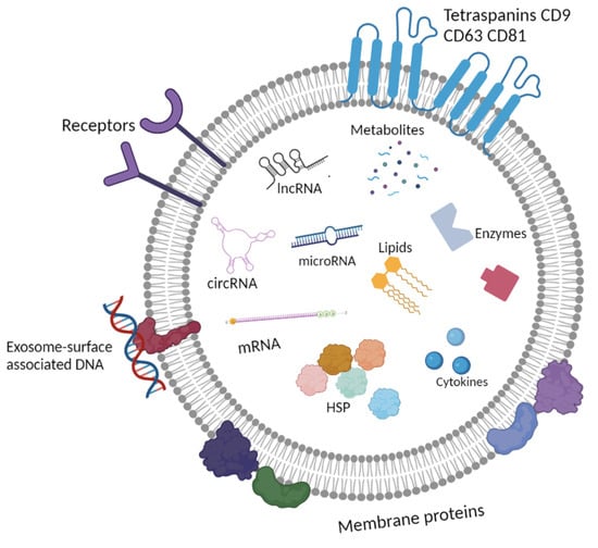
3. Morphology and Content of Exosomes

The extensive use of transmission electron microscopy (TEM) has confirmed that exosomes are extracellular vesicles (EVs) characterized by low electron density and a cup-shaped morphology [31][32] in both the plasma and ascites of the OC patients (Figure 2). Additionally, cryo-electron microscopy has identified subpopulations of exosomes with single-, double-, and multi-layer membranes [31][33][34]. Although fundamental differences in the structure of exosomes in the blood of healthy women and cancer patients have been identified, data specific to OC are currently unavailable.
Figure 2. Total view of exosome preparation obtained from blood plasma of HFs (a), blood plasma of OCPs (b), ascitic fluid of OCPs (c). Inserts and arrows show exosomes. Scale bars correspond to 100 nm. Electron microscopy, negative staining using phosphotungstate acid [35].

3.1. Lipidome of Exosomes

The lipid composition of exosomes is very similar to those in the plasma membrane of the parent cell. The curvature of exosomal membranes, which defines their size and biological functions, depends on their lipid content. The most commonly found lipids in exosomes are phosphoglycerolipids, sphingolipids, and sterols [10]. Exosomes exhibit a lipid composition that is notably enriched with phosphatidylserine, disaturated phosphatidylethanolamine, disaturated phosphatidylcholine, sphingomyelin, ganglioside GM3, and cholesterol when compared to their parent cells [36]. It’s important to note that the lipid subset of exosomes can vary among different subpopulations. For instance, the lipid composition of the CD61-positive exosomes significantly differs from that of other subtypes [37]. Mass spectrometry analysis of exosomes from 12 healthy donors and blood plasma revealed a higher lipid content in exosomes in comparison to plasma (244 and 191, respectively).
The role of exosomal lipids in carcinogenesis is an area of active research. Lipidomic analysis of exosomes derived from the SKOV3 and HOSEPiC cell culture identified 1227 lipid species and 30 lipid classes. Notably, a significant difference in the lipid content of exosomes from the SKOV3 and HOSEPiC cell cultures was observed. The SKOV3-derived exosomes contained higher levels of GM3, zymosterol ester, lysophosphatidylinositol, lysophosphatidylserine, and cholesterol ester [38]. Exosomal lipids are also considered prospective biomarkers for tumorigenesis.

3.2. Proteome of Exosomes

The tetraspanins CD9, CD81, and CD63 are the most surface proteins on exosomes, making them markers for the exosomal nature of EVs. It is also known that the high heterogeneity of the exosomal proteins is due to the protein composition of parental cells as well as the conditions of their cultivation (hypoxia, acidic environment, etc.). According to the ExoCarta database (www.exocarta.org, accessed on 1 April 2023), which contains data from independent studies on the composition of exosomal proteins, lipids, microRNAs, and mRNAs, 9769 exosomal proteins have been identified as of 1 April 2023. The most common exosomal proteins and their functions are summarized in Table 1.
Table 1. Most frequently identified exosomal proteins due to ExoCarta data (as of April 2023).
No Gene Symbol Uniprot ID Function
1 CD9 P21926 Membrane protein. Identified on membranes of oocytes and extracellular exosomes
2 HSPA8 P11142 Chaperone protein
3 PDCD6IP Q8WUM4 Involved in sorting of cargo proteins of the MVBs for incorporation into ILVs
4 GAPDH P04406 Modulates the organization and assembly of the cytoskeleton
5 ACTB P60709 Protein that polymerizes to produce filaments
6 ANXA2 P07355 Calcium-regulated membrane-binding protein
7 CD63 P08962 Cell surface receptor for TIMP1 and plays a role in the activation of cellular signaling cascades AKT, FAK/PTK2 and MAPK
8 SDCBP O00560 Involved in the trafficking of transmembrane proteins, exosome biogenesis, and tumorigenesis
9 ENO1 P06733 Involved in glycolysis, growth control, hypoxia tolerance, and allergic responses
10 HSP90AA1 P07900 Chaperone protein
11 TSG101 Q99816 The component of the ESCRT-I complex mediates the association between the ESCRT-0 and ESCRT-I complex
12 PKM P14618 Catalyzes the final rate-limiting step of glycolysis generating ATP
13 LDHA P00338 Interconverts simultaneously and stereospecifically pyruvate and lactate with concomitant interconversion of NADH and NAD+.
14 EEF1A1 P68104 Translation elongation factor that catalyzes the GTP-dependent binding of aminoacyl-tRNA (aa-tRNA) to the A-site of ribosomes
15 YWHAZ P63104 Adapter protein implicated in the regulation of a large spectrum of signaling pathways
16 PGK1 P00558 Catalyzes one of the two ATP-producing reactions; acts as a polymerase alpha cofactor protein
17 EEF2 P13639 Catalyzes the GTP-dependent ribosomal translocation step during translation elongation
18 ALDOA P04075 Plays a key role in glycolysis and gluconeogenesis; scaffolding protein
19 HSP90AB1 P08238 Chaperone protein
20 ANXA5 P08758 Acts as an indirect inhibitor of the thromboplastin-specific complex

Proteomic analysis of exosomes obtained from the SKOV3 and HOSEPiC cell cultures revealed 659 universal proteins out of all 1433 identified exosomal proteins [38]. COX2 is one of the most abundant exosomal proteins whose increased expression is associated with hypoxia. Probably, the formation of tumor spheroids and metastasis process are caused by overexpression of this protein [39].

Proteins implicated in carcinogenesis have been identified in the cargo of exosomes obtained from the blood plasma and ascitic fluid of OC patients. Specifically, proteins like ATF2, MTA1, ROCK1/2, and CD147 are involved in tumor angiogenesis, while GNA12, EPHA2, and COIA1 promote migration and metastasis. The Nanog protein plays a role in mediating the proliferation and invasion of tumor cells. Exosomes also contain Hsp90 and Hsc70, MHCI, and MHCII. Additionally, various enzymes have been detected in exosomes, such as phosphate isomerase, peroxiredoxins, aldehyde reductase, fatty acid synthase, and Dicer, which is involved in microRNA maturation, among others [40][41].

The important role of the PKR1 protein in the initiation of angiogenesis in vitro was demonstrated by the study of tumor-associated exosomes derived from A2780 and HO-8910 cell cultures. The PKR1 signaling pathway can be mediated by phosphorylation of STAT3. When the HUVECs cells were treated with the PKR1-positive exosomes, migration and tube formation were increased compared to controls [42].
It is known that ascitic fluid can be detected in small amounts even in the early stages of OC. As the tumor grows, ascites become a key component of the tumor microenvironment. Thus, proteomic analysis of exosomes derived from ascites may improve our understanding of OC dissemination as exosomes provide communication between cells and the tumor microenvironment. It has also been shown that up to 40% of the unique proteins of ascitic fluid are part of exosomes [43].
The formation of premetastatic niches is one of the key mechanisms of OC progression. Several studies have shown that exosomes secreted by the tumor play an important role in this process. For example, exosomes carry bioactive molecules such as TGFβ, TNFα, interleukins, MMPs, etc. that mediate EMT. Exosomes of cancer-associated fibroblasts are enriched with TGFβ and thus can activate the SMAD pathway in OC cells. This signaling pathway leads to enhanced cell migration and invasion. It’s well-documented that OC, like many other solid tumors, often occurs under hypoxic conditions. Hypoxia impacts various stages of carcinogenesis, including the formation and secretion of tumor-associated exosomes. For instance, hypoxia enhances exosome secretion by promoting the fusion of MVBs with the cell membrane [44]
Immunosuppression is crucial for tumor proliferation and metastasis. The ability of exosomes to influence this process in tumor cells has also been shown. For example, exosomes derived from ascitic fluid express the ganglioside GD3 on their surface. This molecule can interact with the T-cell receptor, resulting in the arrest of T cells. In addition to immunosuppressive mechanisms, the Fas ligand on the surface of the OC exosomes can suppress the T-cell receptor expression and mediate the T-cell apoptosis [40].

3.3. Nucleic Acids Transported by Exosomes

In addition to proteins and lipids, exosomes carry functionally active nucleic acids. It has been shown that exosomes have DNA in their crown, but the proportion of such DNA does not exceed 0.025% of blood plasma DNA in healthy women [30]. It is also known that exosomes contain various types of RNA: mRNA, microRNA, long noncoding RNA, rRNA, tRNA, circRNA, etc. [45]. According to the Exocarta database (www.exocarta.org, accessed on 1 April 2023), exosomes are involved in the transport of more than 2838 microRNAs and 3408 mRNAs. Some researchers also consider lncRNAs and circRNAs as promising diagnostic biomarkers for liquid biopsy due to their ability to influence the carcinogenesis of tumors, including OCs.

4. Conclusions

Despite the effectiveness of instrumental methods in diagnosing OC, there are several limitations when it comes to detecting stage I disease, identifying cancer in situ, distinguishing between benign and malignant neoplasms, and conducting screening studies of both healthy women and cancer patients following courses of therapy. The high expectations associated with CA-125, CA 19-9, CEA, HE4, and αFP markers for OC detection have also given way to disappointment. To successfully detect neoplasms, there is now a recognized need to discover more sensitive and specific tumor markers for liquid biopsy and explore their combination with instrumental analysis methods. Furthermore, in the realm of modern personalized medicine, there is a pressing need for markers that can effectively predict aggressive tumor behavior and assess the efficacy of anti-cancer therapies. As tumor-secreted exosomes transport biopolymers critical for tumor growth and dissemination, analyzing the cargo of exosomes can provide insights into the molecular mechanisms that stimulate cancer cell proliferation, migration, invasion, angiogenesis, and immunosuppression. Research into the carcinogenic pathways involving proteins and non-coding RNAs (such as lncRNA, circRNA, and microRNA) present in exosomes circulating in the blood of OC patients holds the promise, in the near future, to not only enable the identification of effective markers for disease diagnosis and prognosis but also enhance the assessment of anti-tumor therapy.

References

  1. Sung, H.; Ferlay, J.; Siegel, R.L.; Laversanne, M.; Soerjomataram, I.; Jemal, A.; Bray, F. Global Cancer Statistics 2020: GLOBOCAN Estimates of Incidence and Mortality Worldwide for 36 Cancers in 185 Countries. CA Cancer J. Clin. 2021, 71, 209–249.
  2. Elias, K.M.; Guo, J.; Bast, R.C., Jr. Early Detection of Ovarian Cancer. Hematol. Oncol. Clin. N. Am. 2018, 32, 903–914.
  3. Ledermann, J.A.; Raja, F.A.; Fotopoulou, C.; Gonzalez-Martin, A.; Colombo, N.; Sessa, C.; ESMO Guidelines Working Group. Newly diagnosed and relapsed epithelial ovarian carcinoma: ESMO Clinical Practice Guideline for diagnosis, treatment and follow-up. Ann. Oncol. 2013, 6, 24–32.
  4. Nebgen, D.R.; Lu, K.H.; Bast, R.C., Jr. Novel Approaches to Ovarian Cancer Screening. Curr. Oncol. Rep. 2019, 21, 75.
  5. Dochez, V.; Caillon, H.; Vaucel, E.; Dimet, J.; Winer, N.; Ducarme, G. Biomarkers and algorithms for diagnosis of ovarian cancer: CA125, HE4, RMI and ROMA, a review. J. Ovarian Res. 2019, 12, 28.
  6. Fischerova, D.; Zikan, M.; Semeradova, I.; Slama, J.; Kocian, R.; Dundr, P.; Nemejcova, K.; Burgetova, A.; Dusek, L.; Cibula, D. Ultrasound in preoperative assessment of pelvic and abdominal spread in patients with ovarian cancer: A prospective study. Ultrasound Obstet. Gynecol. 2017, 49, 263–274.
  7. Roett, M.A.; Evans, P. Ovarian cancer: An overview. Am. Fam. Physician 2009, 80, 609–616.
  8. Roze, J.F.; Hoogendam, J.P.; van de Wetering, F.T.; Spijker, R.; Verleye, L.; Vlayen, J.; Veldhuis, W.B.; Scholten, R.J.; Zweemer, R.P. Positron emission tomography (PET) and magnetic resonance imaging (MRI) for assessing tumour resectability in advanced epithelial ovarian/fallopian tube/primary peritoneal cancer. Cochrane Database Syst. Rev. 2018, 10, CD012567.
  9. Gu, P.; Pan, L.L.; Wu, S.Q.; Sun, L.; Huang, G. CA 125, PET alone, PET–CT, CT and MRI in diagnosing recurrent ovarian carcinoma. Eur. J. Radiol. 2009, 71, 164–174.
  10. Tenchov, R.; Sasso, J.M.; Wang, X.; Liaw, W.S.; Chen, C.A.; Zhou, Q.A. Exosomes—Nature’s Lipid Nanoparticles, a Rising Star in Drug Delivery and Diagnostics. ACS Nano 2022, 16, 17802–17846.
  11. Shefer, A.; Yalovaya, A.; Tamkovich, S. Exosomes in Breast Cancer: Involvement in Tumor Dissemination and Prospects for Liquid Biopsy. Int. J. Mol. Sci. 2022, 23, 8845.
  12. Yunusova, N.; Kolegova, E.; Sereda, E.; Kolomiets, L.; Villert, A.; Patysheva, M.; Rekeda, I.; Grigor’eva, A.; Tarabanovskaya, N.; Kondakova, I.; et al. Plasma Exosomes of Patients with Breast and Ovarian Tumors Contain an Inactive 20S Proteasome. Molecules 2021, 26, 6965.
  13. Gurung, S.; Perocheau, D.; Touramanidou, L.; Baruteau, J. The exosome journey: From biogenesis to uptake and intracellular signalling. Cell Commun. Signal. 2021, 19, 47.
  14. Zhang, L.; Yu, D. Exosomes in cancer development, metastasis, and immunity. Biochim. Biophys. Acta Rev. Cancer 2019, 1871, 455–468.
  15. Huotari, J.; Helenius, A. Endosome maturation. EMBO J. 2011, 30, 3481–3500.
  16. Beach, A.; Zhang, H.G.; Ratajczak, M.Z.; Kakar, S.S. Exosomes: An overview of biogenesis, composition and role in ovarian cancer. J. Ovarian Res. 2014, 7, 14.
  17. Colombo, M.; Moita, C.; van Niel, G.; Kowal, J.; Vigneron, J.; Benaroch, P.; Manel, N.; Moita, L.F.; Théry, C.; Raposo, G. Analysis of ESCRT functions in exosome biogenesis, composition and secretion highlights the heterogeneity of extracellular vesicles. J. Cell Sci. 2013, 126, 5553–5565.
  18. Babst, M. MVB vesicle formation: ESCRT-dependent, ESCRT-independent and everything in between. Curr. Opin. Cell Biol. 2011, 23, 452–457.
  19. Elsherbini, A.; Bieberich, E. Ceramide and Exosomes: A Novel Target in Cancer Biology and Therapy. Adv. Cancer Res. 2018, 140, 121–154.
  20. Hurwitz, S.N.; Conlon, M.M.; Rider, M.A.; Brownstein, N.C.; Meckes, D.G., Jr. Nanoparticle analysis sheds budding insights into genetic drivers of extracellular vesicle biogenesis. J. Extracell. Vesicles 2016, 5, 31295.
  21. Nonaka, T.; Wong, D.T.W. Saliva-Exosomics in Cancer: Molecular Characterization of Cancer-Derived Exosomes in Saliva. Enzymes 2017, 42, 125–151.
  22. Cao, J.; Zhang, Y.; Mu, J.; Yang, D.; Gu, X.; Zhang, J. Exosomal miR-21-5p contributes to ovarian cancer progression by regulating CDK6. Hum. Cell 2021, 34, 1185–1196.
  23. Tutanov, O.; Orlova, E.; Proskura, K.; Grigor’eva, A.; Yunusova, N.; Tsentalovich, Y.; Alexandrova, A.; Tamkovich, S. Proteomic Analysis of Blood Exosomes from Healthy Females and Breast Cancer Patients Reveals an Association between Different Exosomal Bioactivity on Non-Tumorigenic Epithelial Cell and Breast Cancer Cell Migration in Vitro. Biomolecules 2020, 10, 495.
  24. Yunusova, N.V.; Patysheva, M.R.; Molchanov, S.V.; Zambalova, E.A.; Grigor’eva, A.E.; Kolomiets, L.A.; Ochirov, M.O.; Tamkovich, S.N.; Kondakova, I.V. Metalloproteinases at the surface of small extrcellular vesicles in advanced ovarian cancer: Relationships with ascites volume and peritoneal canceromatosis index. Clin. Chim. Acta 2019, 494, 116–122.
  25. Li, M.; Zeringer, E.; Barta, T.; Schageman, J.; Cheng, A.; Vlassov, A.V. Analysis of the RNA content of the exosomes derived from blood serum and urine and its potential as biomarkers. Philos. Trans. R. Soc. Lond. B Biol. Sci. 2014, 369, 20130502.
  26. Xu, H.; Li, M.; Pan, Z.; Zhang, Z.; Gao, Z.; Zhao, R.; Li, B.; Qi, Y.; Qiu, W.; Guo, Q.; et al. miR-3184-3p enriched in cerebrospinal fluid exosomes contributes to progression of glioma and promotes M2-like macrophage polarization. Cancer Sci. 2022, 113, 2668–2680.
  27. Kim, K.U.; Kim, W.H.; Jeong, C.H.; Yi, D.Y.; Min, H. More than Nutrition: Therapeutic Potential of Breast Milk-Derived Exosomes in Cancer. Int. J. Mol. Sci. 2020, 21, 7327.
  28. Tamkovich, S.; Grigor’eva, A.; Eremina, A.; Tupikin, A.; Kabilov, M.; Chernykh, V.; Vlassov, V.; Ryabchikova, E. What information can be obtained from the tears of a patient with primary open angle glaucoma? Clin. Chim. Acta. 2019, 495, 529–537.
  29. Zhang, W.; Ou, X.; Wu, X. Proteomics profiling of plasma exosomes in epithelial ovarian cancer: A potential role in the coagulation cascade, diagnosis and prognosis. Int. J. Oncol. 2019, 54, 1719–1733.
  30. Tutanov, O.; Shtam, T.; Grigor’eva, A.; Tupikin, A.; Tsentalovich, Y.; Tamkovich, S. Blood Plasma Exosomes Contain Circulating DNA in Their Crown. Diagnostics 2022, 12, 854.
  31. Konoshenko, M.; Sagaradze, G.; Orlova, E.; Shtam, T.; Proskura, K.; Kamyshinsky, R.; Yunusova, N.; Alexandrova, A.; Efimenko, A.; Tamkovich, S. Total Blood Exosomes in Breast Cancer: Potential Role in Crucial Steps of Tumorigenesis. Int. J. Mol. Sci. 2020, 21, 7341.
  32. Quesnel, A.; Broughton, A.; Karagiannis, G.S.; Filippou, P.S. Message in the bottle: Regulation of the tumor microenvironment via exosome-driven proteolysis. Cancer Metastasis Rev. 2022, 41, 789–801.
  33. Yuana, Y.; Koning, R.I.; Kuil, M.E.; Rensen, P.C.; Koster, A.J.; Bertina, R.M.; Osanto, S. Cryo-electron microscopy of extracellular vesicles in fresh plasma. J. Extracell. Vesicles 2013, 2, 21494.
  34. Emelyanov, A.; Shtam, T.; Kamyshinsky, R.; Garaeva, L.; Verlov, N.; Miliukhina, I.; Kudrevatykh, A.; Gavrilov, G.; Zabrodskaya, Y.; Pchelina, S.; et al. Cryo-electron microscopy of extracellular vesicles from cerebrospinal fluid. PLoS ONE 2020, 15, e0227949.
  35. Dzhugashvili, E.I.; Yunusova, N.V.; Yalovaya, A.I.; Grigorieva, A.E.; Sereda, E.E.; Kolomiets, L.A.; Tamkovich, S.N. Comparative assessment of the exosomal tumor-associated microRNA levels in blood plasma and ascitic fluid in ovarian cancer patients. Adv. Mol. Oncol. 2023, 10, 108–116. (In Russia)
  36. Choi, D.S.; Kim, D.K.; Kim, Y.K.; Gho, Y.S. Proteomics, transcriptomics and lipidomics of exosomes and ectosomes. Proteomics 2013, 13, 1554–1571.
  37. Liangsupree, T.; Multia, E.; Saarinen, J.; Ruiz-Jimenez, J.; Kemell, M.; Riekkola, M.L. Raman spectroscopy combined with comprehensive gas chromatography for label-free characterization of plasma-derived extracellular vesicle subpopulations. Anal. Biochem. 2022, 647, 114672.
  38. Cheng, L.; Zhang, K.; Qing, Y.; Li, D.; Cui, M.; Jin, P.; Xu, T. Proteomic and lipidomic analysis of exosomes derived from ovarian cancer cells and ovarian surface epithelial cells. J. Ovarian Res. 2020, 13, 9.
  39. Ding, Y.; Zhuang, S.; Li, Y.; Yu, X.; Lu, M.; Ding, N. Hypoxia-induced HIF1α dependent COX2 promotes ovarian cancer progress. J. Bioenerg. Biomembr. 2021, 53, 441–448.
  40. Feng, W.; Dean, D.C.; Hornicek, F.J.; Shi, H.; Duan, Z. Exosomes promote pre-metastatic niche formation in ovarian cancer. Mol. Cancer 2019, 18, 124.
  41. Tang, M.K.; Wong, A.S. Exosomes: Emerging biomarkers and targets for ovarian cancer. Cancer Lett. 2015, 367, 26–33.
  42. Zhang, X.; Sheng, Y.; Li, B.; Wang, Q.; Liu, X.; Han, J. Ovarian cancer derived PKR1 positive exosomes promote angiogenesis by promoting migration and tube formation in vitro. Cell Biochem. Funct. 2021, 39, 308–316.
  43. Shender, V.O.; Pavlyukov, M.S.; Ziganshin, R.H.; Arapidi, G.P.; Kovalchuk, S.I.; Anikanov, N.A.; Altukhov, I.A.; Alexeev, D.G.; Butenko, I.O.; Shavarda, A.L.; et al. Proteome-metabolome profiling of ovarian cancer ascites reveals novel components involved in intercellular communication. Mol. Cell Proteom. 2014, 13, 3558–3571.
  44. Dorayappan, K.D.P.; Wallbillich, J.J.; Cohn, D.E.; Selvendiran, K. The biological significance and clinical applications of exosomes in ovarian cancer. Gynecol. Oncol. 2016, 142, 199–205.
  45. Yi, Y.; Wu, M.; Zeng, H.; Hu, W.; Zhao, C.; Xiong, M.; Lv, W.; Deng, P.; Zhang, Q.; Wu, Y. Tumor-Derived Exosomal Non-Coding RNAs: The Emerging Mechanisms and Potential Clinical Applications in Breast Cancer. Front. Oncol. 2021, 11, 738945.
More
Information
Subjects: Oncology
Contributors MDPI registered users' name will be linked to their SciProfiles pages. To register with us, please refer to https://encyclopedia.pub/register : ,
View Times: 131
Revisions: 2 times (View History)
Update Date: 18 Dec 2023
1000/1000
Video Production Service