Submitted Successfully!
To reward your contribution, here is a gift for you: A free trial for our video production service.
Thank you for your contribution! You can also upload a video entry or images related to this topic.
Version Summary Created by Modification Content Size Created at Operation
1 -- 2671 2023-12-13 16:38:51 |
2 Reference format revised. Meta information modification 2671 2023-12-19 03:50:04 |

Video Upload Options

Do you have a full video?

Confirm

Are you sure to Delete?
Cite
If you have any further questions, please contact Encyclopedia Editorial Office.
Udoye, C.C.; Ehlers, M.; Manz, R.A. Allergenic and Anti-Allergenic Antibodies in Food Allergy. Encyclopedia. Available online: https://encyclopedia.pub/entry/52700 (accessed on 18 May 2024).
Udoye CC, Ehlers M, Manz RA. Allergenic and Anti-Allergenic Antibodies in Food Allergy. Encyclopedia. Available at: https://encyclopedia.pub/entry/52700. Accessed May 18, 2024.
Udoye, Christopher C., Marc Ehlers, Rudolf A. Manz. "Allergenic and Anti-Allergenic Antibodies in Food Allergy" Encyclopedia, https://encyclopedia.pub/entry/52700 (accessed May 18, 2024).
Udoye, C.C., Ehlers, M., & Manz, R.A. (2023, December 13). Allergenic and Anti-Allergenic Antibodies in Food Allergy. In Encyclopedia. https://encyclopedia.pub/entry/52700
Udoye, Christopher C., et al. "Allergenic and Anti-Allergenic Antibodies in Food Allergy." Encyclopedia. Web. 13 December, 2023.
Allergenic and Anti-Allergenic Antibodies in Food Allergy
Edit

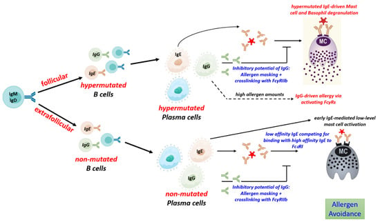
Food allergies are a growing public health concern worldwide, especially in children and young adults. Allergen-specific IgE plays a central role in the pathogenesis of food allergies, but their titers poorly correlate with allergy development. Host immune systems yield allergen-specific immunoglobulin (Ig)A, IgE and IgG subclasses with low or high affinities and differential Fc N-glycosylation patterns that can affect the allergic reaction to food in multiple ways. High-affinity IgE is required to induce strong mast cell activation eventually leading to allergic anaphylaxis, while low-affinity IgE can even inhibit the development of clinically relevant allergic symptoms. IgA and IgG antibodies can inhibit IgE-mediated mast cell activation through various mechanisms, thereby protecting IgE-positive individuals from allergy development. The production of IgE and IgG with differential allergenic potential seems to be affected by the signaling strength of individual B cell receptors, and by cytokines from T cells. 

food allergy IgA IgG IgE anaphylaxis B cell

1. Introduction

Food allergies are becoming an increasingly global health concern, especially in urbanized areas, with the prevalence of food allergies rising by about 1.7% between 1997 and 2011 [1]. Allergic reactions to a food antigen are thought to be mainly driven by IgE-mediated activation of mast cells and basophils. They account for most allergies, resulting in symptoms ranging from mild reactions such as hives and itching to more lethal outcomes like anaphylaxis, a severe life-threatening immune reaction [2].
Allergy management broadly involves avoiding food allergens, undergoing allergen- immunotherapy or taking emergency medications during allergic reactions [3]. Comprehensive studies suggest an age bias in food allergies with infants [4][5] having more allergies than adults [6]. Also, there is higher prevalence of allergies in women than men after adolescence [7].
In the context of type 2 immunity, B cells can undergo differentiation into Be2 cells that secrete IL-4 and other cytokines [8]. IL-4 from Be2 cells, basophils or other non-T cells is required to promote the differentiation of naïve T cells into T helper (Th) 2 cells [9][10][11]. Through the production of IL-4, Th2 cells eventually promote immunoglobulin class switch to IgE [12]. B cells expressing IgE can rapidly differentiate into plasma cells [13].
Class switch recombination (CSR) is the rearrangement of the genes coding for the constant regions of antibodies while retaining their antigen-binding region. CSR enables antibodies to change their effector functions and serum half-lives. This process is associated with the deletion of the gene segments coding for the antibody classes up-stream of the target class. Therefore, CSR is non-reversible. Human IgM+/IgD+, IgA+ and IgG4+ B cells and murine IgM+/IgD+ and IgG1 B cells can class switch to IgE, respectively, but not vice versa. Class switch from IgM/IgD to IgE is called direct, while class switch from human IgA and IgG4 and from murine IgG1 to IgE is called sequential class switch. There is evidence that most IgE in mice is the product of sequential class switch of IgG1+ progenitor cells [14][15]. CSR to IgE may occur not only in secondary lymphoid tissues but also locally in the mucosa of the nasal cavity, stomach and duodenum [16][17]. The induced antibody composition may dictate the induction and inhibition of an allergy, which may partly explain the complexity of the clinical findings.

2. Low and High-Affinity IgE Play Opposing Roles in Food Allergy

IgE is the least abundant among all antibody isotypes in serum [18]. Even after repeated and long-lasting allergen challenges, it represents only a minor fraction of the total allergen-specific antibodies in serum, suggesting that IgE levels are highly regulated [19]. Its low abundance can be attributed to its comparably short serum half-life of approximately 3 days, and the nature of IgE B cells, which exhibit high rates of apoptosis and little proliferation [20][21][22]. Nevertheless, IgE is crucial for the development of type 1 allergic reactions, such as food allergy. Even small quantities of serum IgE efficiently bind to FcεRI receptors on mast cells and basophils. In contrast to its short half-life in serum, IgE bound on mast cells is retained throughout the life cycle of the cell [23]. The FcεRI receptor binds to the Fc region of the IgE molecule with an affinity of 1010 M−1 [24]. In consequence, IgE arms mast cells and basophils with an antigen-specific receptor, which, after ingestion of food allergens, meditates cross-linking of the FcεRI receptors, eventually leading to the release of histamine and other mediators of the acute allergic reaction [25]. However, the allergenicity of IgE depends on its affinity [26]. Mast cell-bound IgE antibodies with high affinity for their antigens can be cross-linked by trace amounts of antigen. In consequence, the mast cells are activated and may cause allergic anaphylaxis. In contrast, allergen does not efficiently crosslink low-affinity IgE and was shown to prevent anaphylaxis. In addition, low-affinity IgE can/may prevent anaphylaxis through competing with high-affinity IgE for binding to FcεRI receptors [27][28]. Though both high- and low-affinity stimuli of FcεRI elicit similar receptor phosphorylation, the receptor cluster size, mobility and distribution on mast cells and the down-stream signaling cascade mediated are different, eventually resulting in an altered mast cell response [29]. In accordance with their differential capabilities for mast cell activation, the levels of high-affinity IgE, but not of low-affinity IgE, correlates with allergic symptoms such as eczema, allergic asthma and anaphylaxis [30][31][32].

3. The Role of Antibody Isotypes, their Subclasses and Antibody Fc Glycosylation in Food Allergy

3.1. Mechanisms of IgG-Mediated Suppression of Allergy

While IgE is of major importance for allergy development, in the presence of high allergen doses, IgG-mediated anaphylaxis was also observed in murine models [33]. Murine IgG1, IgG2a and IgGG2b have been shown to promote anaphylaxis through activation of the activating Fcγ receptors (FcγR) FcγRI, FcγRIII and FcγRIV [34]. IgG-dependent allergic reactions are mediated through the secretion of platelet-activating factor (PAF) by neutrophils, monocytes, macrophages and basophils [35]. However, this process requires much higher antigen doses than IgE-mediated anaphylaxis [36]. Whether IgG-mediated anaphylaxis is relevant in patients is a matter of debate [37]. In this context, the food quality sensing function of IgE could be relevant.
Though allergen-specific IgG could be potentially harmful, it often seems to be beneficial for allergic patients. Accordingly, increasing levels of allergen-specific IgGs are associated with the natural resolution of food allergies [37]. Likewise, a positive response to allergen-specific immunotherapy is associated with increased allergen-specific serum IgG [38]. Serum IgG4 is elevated in patients who undergo allergen immunotherapy and has been associated with increased clinical tolerance to specific allergens [39]. Depletion of serum IgG4 from peanut-tolerant patients has been shown to promote stronger mast cell degranulation [38]
The protective effect of IgG is mediated by multiple mechanisms. IgG can block IgE-mediated allergies via allergen neutralization and FcγRIIb-mediated inhibition via the IgG inhibitory receptor FcγRIIb [40][41][42]. In allergen neutralization, IgG competes with IgE for binding to the allergenic proteins eventually preventing their interaction with IgE. This is an important mechanism by which antibodies provide clinical tolerance to allergic diseases [43]. IgG4 (in humans) and IgG1 (in mice) are clonally related to IgE.

3.2. Mechanisms of IgA-Mediated Suppression of Allergy

Allergen-specific IgA is also capable of allergen neutralization and is relevant to block IgE-mediated activation of mast cells and basophils [41]. As shown in human samples, a considerable proportion of mucosal IgE is clonally related to IgA [44], indicating that IgA and IgE antibodies share the same antigen binding regions. Mucosal IgA is mostly produced as a dimer that is actively transported to the extracorporeal surface of mucosal epithelial cells [18]. As shown in a murine model of oral immunotherapy, IgA in the mucosal sites binds to allergens and prevents them from penetrating the epithelial barrier and triggering an immune response [41]. Therefore, mucosal IgA can bind to food allergens before they can reach cell-bound IgE and prevents mast cell and basophil degradation in an allergen-specific manner [41].

4. The Impact of Antibody Ig-Fc Glycosylation on Allergy Development

Differential Fc N-glycosylation at Asn297 of IgG antibodies modulate their binding to activating and inhibitory Fc receptors and inconsequence their impact on the activation or inhibition of innate effector cells, including mast cells [45]. In inflammatory (auto)immune diseases, IgG antibodies with low levels of galactose and sialic acid have been shown to correlate with disease severity. Consistent with the fact that protein glycosylation is an ancient evolutionary development and that sialylated proteins are more likely to be associated with tolerance, non-galactosylated and non-sialylated forms of IgA antibodies have also been shown to be associated with inflammatory processes [46].
Allergen-specific IgG subclass glycosylation may also play a role in the inhibition of IgE responses via cross-linking with the IgG inhibitory receptor FcγRIIb or, in the presence of high allergen concentrations, in the induction of IgG-mediated allergic reactions via activating FcγRs [47].

5. Development of Antibodies in Food Allergy

5.1. T Cell Activation

Antibody responses to proteins, such as food allergens, are strictly T-dependent [48]. While B cells recognize three-dimensional epitopes, Th cells are specific for small peptides presented in MHCII molecules. The initial entrance of the allergen leading to specific sensitization may occur through the skin, gastrointestinal tract, airway or damaged epidermal barrier [49][50][51][52]. In allergic individuals, sensitization results in the formation of Th2 and T follicular helper (Tfh) 13 cells and the formation of allergenic IgE [31].
Food allergens can access a dysfunctional epithelial barrier and trigger the production of alarmins such as Interleukin 33 (IL33), Interleukin 25 (IL25) and Thymic stromal lymphopoietin (TSLP). These alarmins mediate type 1 hypersensitivity [53] by skewing the T cell response towards the Th2 axis and activating mast cells, dendritic cells, innate lymphoid cells and eosinophils [54][55].

5.2. Production of Unmutated, Low-Affinity IgE

During a response to a protein antigen, such as food allergens, activated B cells can follow multiple differentiation pathways. Initially, the extrafollicular pathway of B cell differentiation yields short-lived plasma cells that produce antibodies of relatively low affinity. B cells following this pathway do not introduce much hypermutations into their antigen-binding regions, nor do they differentiate into memory cells [56][57].

5.3. Production of Mutated, High-Affinity IgE

Expressed in a membrane-bound form on the cell surface, antibodies serve as antigen-specific B cell receptor (BCR) which determine the cellular fate at all stages of development [58]. Hypermutated, high-affinity antibodies are the product of the follicular pathway. B cells following the follicular pathway transform primary B cell follicles within secondary lymphoid tissues into germinal centers (GC), where B cells undergo hypermutation, affinity maturation and differentiation into long-lived plasma cells and memory B cells [59][60] (Figure 1).
Figure 1. The B cell response to allergens and its impact on mast cell activation. Immune response to food allergens induces follicular and extrafollicular B cell response which yield plasma cells secreting hypermutated (high-affinity) and unmutated (low-affinity) IgE, and IgG with differential impact on the activation of mast cells and disease development. Ig: immunoglobulin; IgE: Immunoglobulin E; IgG: Immunoglobulin G; IgM: Immunoglobulin M; IgD: Immunoglobulin D; FcεRI: Fc epsilon RI; FcγRIIb: Fc-gamma RIIb.
GC development requires help from Tfh cells, which provide stimuli such as IL-21 and CD40 essential for induction of hypermutation and positive selection of B cells that have acquired BCR of higher affinity [61][62]. While Th-derived IL-4 is sufficient to induce class switch to IgE and the formation of low-affinity IgE, the generation of high-affinity IgE with anaphylactic properties depends on help from Tfh13 cells which additionally produce high levels of IL-13 and IL-5 together with some IL-21 [31]. Tfh13 cells regulate germinal center responses in type 2 immune reactions and appears to be important for the generation of hypermutated high-affinity IgE and the development of asthma [31].
Despite the high-affinity IgE is derived from GCs, the existence of IgE+ memory B cells and long-lived plasma cells is a matter of debate. At least the majority of IgE cells seem to be excluded from these memory compartments. Because of their very low frequency and the possibility of confusion with B cells that bind IgE via their low-affinity receptor CD23, IgE-expressing B cells are difficult to detect without doubt. In addition, GCs typically stain brightly for IgG, but only some IgE is detectable in these tissue structures. Thus, the production of IgE appears to be tightly regulated [63]. Different antibody classes exhibit qualitatively distinct signaling properties.
Together, IgE BCR expression mediates a tonic signal even in the absence of external stimulation by antigen, if that promotes apoptosis or terminal differentiation might be dependent on the model, and under physiological conditions, on additional factors such as the availability of an anti-apoptotic environment. Some IgE+ B cells seem to survive and signal via their membrane-bound IgE receptor which is a crucial regulator of IgE production.
Comparison of IgE+ and IgG1+ murine B cells by whole-genome CRISPR screening showed that IgE+ B cells have distinct properties [64]. Different form IgG+ cells, IgE+ B cells and IgE+ plasma cells showed chronic calcium signaling eventually resulting in BCL2L11-dependent apoptosis. Moreover, there is evidence that after repeated antigenic stimulation in mice, high-affinity IgE-secreting plasma cells are generated through reactivation and further class switch recombination of IgG1 memory B cells [15].

5.4. Regulation of IgG to IgE Ratios

Most of the class-switched IgE+ cells are derived from IgG+ B cells that underwent further (sequential) class switch to IgE in a follicular B cell response [12][14][65]. The mechanisms controlling the relative IgE to IgG production during the allergen-specific immune response are only partly understood so far. Studies investigating the antibody response to hen’s egg in a murine food allergy model indicate that IgE-to-IgG ratios are controlled on the level of single B cell clones [14]. Most individual clones containing both IgE and IgG1, showed a several-fold excess of IgG1 compared to IgE, i.e., a high IgG1-to-IgE ratio.

6. Development of Differentially Glycosylated Antibodies

IgG Fc glycosylation (galactosylation and sialylation) are regulated by two glycosyltransferases, β1,4-galactosyltransferase 1 (B4galt1) and α2,6-sialyltransferase 1 (St6gal1), in antibody-producing B cells [66]. There is evidence that the expression of these enzymes and hence IgG Fc glycosylation is controlled by Tfh cell-derived cytokines [67]. Within the GC, IL-6/IL-23-dependent IL-17A+ Tfh17 cells induce a low IgG Fc sialylation program in B cells. How these mechanisms affect the glycosylation of antibodies in the context of allergies is not known.
Early IgE antibodies from extrafollicular plasma cell responses show low hypermutation rates with correspondingly low affinity [14]. These plasma cells may generate IgE antibodies with high levels of galactosylation and sialylation as shown for early extrafollicular IgG antibodies after immunization [67][68]. IgG antibodies derived from the germinal center show higher mutation rates and lower levels of galactosylation and sialylation [67]. Depending on the co-stimulation inducing the germinal center response, the derived plasma cells produce IgG antibodies with distinct levels of galactosylation and sialylation, but all lower than the initial extrafollicular level of galactosylation and sialylation [67]. In allergy, germinal center-derived plasma cells may produce different IgE/IgG ratios as well as IgE (and IgG) antibodies with reduced levels of galactosylation and sialylation, which may also depend on the co-stimulation that induce the germinal center response. Tfh13 cells have recently been linked to IgE antibodies in asthma [31][69].

7. The Impact of Distinct Antibody Types in Type 1 Allergic Reactions to Aero-Allergens

Cross-reactivity between aero-allergens and food allergens is considered to be relevant for the development of food allergies in patients previously sensitized to aero-allergens. E.g., initial exposure to respiratory allergens from plant pollens can promote secondary allergy to different food allergens. This complicates the diagnosis of some food allergies originating from pollen cross-reactivity [70]. Other inhalant-allergens (also called aero-allergens) include house dust mites, animal dander, pollen, dust, etc. [71]. Similarly to what is seen in food allergies, the antibody response in aero-allergies includes IgE, IgA and IgG antibodies. Though some earlier studies linked the severity of allergic airway inflammation to antigen-specific IgG, suggesting that they contribute to the development of allergy to inhalant allergens, other studies have associated IgG4 with improved tolerance to aero-allergens [72]. More recently, mechanistic studies provided convincing evidence that aero-allergen-specific IgG can efficiently suppress allergic reactions. Monoclonal IgG against cat allergen feld1 has been shown to reduce allergic symptoms in mice and patients [73].

8. Conclusions

The contribution of the B cell responses and the induced antibody compositions to the development or protection from food allergy is complex. IL-4+ Th2 cells are sufficient to drive the production of low-affinity, potentially protective IgE, but the generation of high-allergenic high-affinity IgE within GCs requires additional help from Tfh13 cells. Allergen-specific IgG and IgA antibodies limit IgE-mediated allergic symptoms in patients.

References

  1. Jackson, K.D.; Howie, L.D.; Akinbami, O.J. Trends in Allergic Conditions among Children: United States, 1997–2011; US Department of Health and Human Services, Centers for Disease Control and Prevention, National Center for Health Statistics: Washington, DC, USA, 2013.
  2. Simons, F.E.R.; Ardusso, L.R.F.; Bilo, M.B.; Dimov, V.; Ebisawa, M.; El-Gamal, Y.M.; Ledford, D.K.; Lockey, R.F.; Ring, J.; Sanchez-Borges, M.; et al. 2012 Update: World Allergy Organization Guidelines for the Assessment and Management of Anaphylaxis. Curr. Opin. Allergy Clin. Immunol. 2012, 12, 389–399.
  3. Cooke, A.T.; Meize-Grochowski, R. Epinephrine Auto-Injectors for Anaphylaxis Treatment in the School Setting: A Discussion Paper. SAGE Open Nurs. 2019, 5, 2377960819845246.
  4. Gupta, R.S.; Springston, E.E.; Warrier, M.R.; Smith, B.; Kumar, R.; Pongracic, J.; Holl, J.L. The Prevalence, Severity, and Distribution of Childhood Food Allergy in the United States. Pediatrics 2011, 128, e9–e17.
  5. Gupta, R.S.; Warren, C.M.; Smith, B.M.; Blumenstock, J.A.; Jiang, J.; Davis, M.M.; Nadeau, K.C. The Public Health Impact of Parent-Reported Childhood Food Allergies in the United States. Pediatrics 2018, 142, e20181235.
  6. Gupta, R.S.; Warren, C.M.; Smith, B.M.; Jiang, J.; Blumenstock, J.A.; Davis, M.M.; Schleimer, R.P.; Nadeau, K.C. Prevalence and Severity of Food Allergies among US Adults. JAMA Netw. Open 2019, 2, e185630.
  7. Kurukulaaratchy, R.J.; Karmaus, W.; Arshad, S.H. Gender and Atopy Influences on the Natural History of Rhinitis. Curr. Opin. Allergy Clin. Immunol. 2012, 12, 7.
  8. Harris, D.P.; Haynes, L.; Sayles, P.C.; Duso, D.K.; Eaton, S.M.; Lepak, N.M.; Johnson, L.L.; Swain, S.L.; Lund, F.E. Reciprocal Regulation of Polarized Cytokine Production by Effector B and T Cells. Nat. Immunol. 2000, 1, 475–482.
  9. Song, Z.; Yuan, W.; Zheng, L.; Wang, X.; Kuchroo, V.K.; Mohib, K.; Rothstein, D.M. B Cell IL-4 Drives Th2 Responses in Vivo, Ameliorates Allograft Rejection, and Promotes Allergic Airway Disease. Front. Immunol. 2022, 13, 762390.
  10. Hurdayal, R.; Ndlovu, H.H.; Revaz-Breton, M.; Parihar, S.P.; Nono, J.K.; Govender, M.; Brombacher, F. IL-4–Producing B Cells Regulate T Helper Cell Dichotomy in Type 1-and Type 2-Controlled Diseases. Proc. Natl. Acad. Sci. USA 2017, 114, E8430–E8439.
  11. Hammad, H.; Plantinga, M.; Deswarte, K.; Pouliot, P.; Willart, M.A.M.; Kool, M.; Muskens, F.; Lambrecht, B.N. Inflammatory Dendritic Cells—Not Basophils—Are Necessary and Sufficient for Induction of Th2 Immunity to Inhaled House Dust Mite Allergen. J. Exp. Med. 2010, 207, 2097–2111.
  12. Looney, T.J.; Lee, J.-Y.; Roskin, K.M.; Hoh, R.A.; King, J.; Glanville, J.; Liu, Y.; Pham, T.D.; Dekker, C.L.; Davis, M.M.; et al. Human B-Cell Isotype Switching Origins of IgE. J. Allergy Clin. Immunol. 2016, 137, 579–586.
  13. He, J.-S.; Narayanan, S.; Subramaniam, S.; Ho, W.Q.; Lafaille, J.J.; de Lafaille, M.A.C. Biology of IgE Production: IgE Cell Differentiation and the Memory of IgE Responses. In IgE Antibodies: Generation and Function; Springer: Berlin/Heidelberg, Germany, 2015; pp. 1–19.
  14. Udoye, C.C.; Rau, C.N.; Freye, S.M.; Almeida, L.N.; Vera-Cruz, S.; Othmer, K.; Korkmaz, R.Ü.; Clauder, A.-K.; Lindemann, T.; Niebuhr, M.; et al. B-Cell Receptor Physical Properties Affect Relative IgG1 and IgE Responses in Mouse Egg Allergy. Mucosal Immunol. 2022, 15, 1375–1388.
  15. He, J.-S.; Subramaniam, S.; Narang, V.; Srinivasan, K.; Saunders, S.P.; Carbajo, D.; Wen-Shan, T.; Hidayah Hamadee, N.; Lum, J.; Lee, A.; et al. IgG1 Memory B Cells Keep the Memory of IgE Responses. Nat. Commun. 2017, 8, 641.
  16. Takhar, P.; Smurthwaite, L.; Coker, H.A.; Fear, D.J.; Banfield, G.K.; Carr, V.A.; Durham, S.R.; Gould, H.J. Allergen Drives Class Switching to IgE in the Nasal Mucosa in Allergic Rhinitis. J. Immunol. 2005, 174, 5024–5032.
  17. Hoh, R.A.; Joshi, S.A.; Lee, J.-Y.; Martin, B.A.; Varma, S.; Kwok, S.; Nielsen, S.C.A.; Nejad, P.; Haraguchi, E.; Dixit, P.S.; et al. Origins and Clonal Convergence of Gastrointestinal IgE+ B Cells in Human Peanut Allergy. Sci. Immunol. 2020, 5, eaay4209.
  18. Manz, R.A.; Hauser, A.E.; Hiepe, F.; Radbruch, A. Maintenance of Serum Antibody Levels. Annu Rev. Immunol. 2005, 23, 367–386.
  19. Geha, R.S.; Jabara, H.H.; Brodeur, S.R. The Regulation of Immunoglobulin E Class-Switch Recombination. Nat. Rev. Immunol. 2003, 3, 721–732.
  20. Yang, Z.; Robinson, M.J.; Chen, X.; Smith, G.A.; Taunton, J.; Liu, W.; Allen, C.D.C. Regulation of B Cell Fate by Chronic Activity of the IgE B Cell Receptor. Elife 2016, 5, e21238.
  21. Croote, D.; Darmanis, S.; Nadeau, K.C.; Quake, S.R. High-Affinity Allergen-Specific Human Antibodies Cloned from Single IgE B Cell Transcriptomes. Science 2018, 362, 1306–1309.
  22. Laffleur, B.; Duchez, S.; Tarte, K.; Denis-Lagache, N.; Péron, S.; Carrion, C.; Denizot, Y.; Cogné, M. Self-Restrained B Cells Arise Following Membrane IgE Expression. Cell Rep. 2015, 10, 900–909.
  23. MacGlashan, D., Jr. IgE Receptor and Signal Transduction in Mast Cells and Basophils. Curr. Opin. Immunol. 2008, 20, 717–723.
  24. Metzger, H. The Receptor with High Affinity for IgE. Immunol. Rev. 1992, 125, 37–48.
  25. Galli, S.J.; Tsai, M. IgE and Mast Cells in Allergic Disease. Nat. Med. 2012, 18, 693–704.
  26. Mita, H.; Yasueda, H.; Akiyama, K. Affinity of IgE Antibody to Antigen Influences Allergen-induced Histamine Release. Clin. Exp. Allergy 2000, 30, 1583–1589.
  27. Chang, X. Low-Affinity but High-Avidity Interactions May Offer an Explanation for IgE-Mediated Allergen Cross-Reactivity. Allergy 2021, 76, 2565–2574.
  28. Xiong, H.; Dolpady, J.; Wabl, M.; Curotto de Lafaille, M.A.; Lafaille, J.J. Sequential Class Switching Is Required for the Generation of High Affinity IgE Antibodies. J. Exp. Med. 2012, 209, 353–364.
  29. Suzuki, R.; Leach, S.; Liu, W.; Ralston, E.; Scheffel, J.; Zhang, W.; Lowell, C.A.; Rivera, J. Molecular Editing of Cellular Responses by the High-Affinity Receptor for IgE. Science 2014, 343, 1021–1025.
  30. Kelleher, M.M.; Phillips, R.; Brown, S.J.; Cro, S.; Cornelius, V.; Carlsen, K.C.L.; Skjerven, H.O.; Rehbinder, E.M.; Lowe, A.J.; Dissanayake, E.; et al. Skin Care Interventions in Infants for Preventing Eczema and Food Allergy. Cochrane Database Syst. Rev. 2022, 11, CD013534.
  31. Gowthaman, U.; Chen, J.S.; Zhang, B.; Flynn, W.F.; Lu, Y.; Song, W.; Joseph, J.; Gertie, J.A.; Xu, L.; Collet, M.A.; et al. Identification of a T Follicular Helper Cell Subset That Drives Anaphylactic IgE. Science 2019, 365, eaaw6433.
  32. Burrows, B.; Martinez, F.D.; Halonen, M.; Barbee, R.A.; Cline, M.G. Association of Asthma with Serum IgE Levels and Skin-Test Reactivity to Allergens. N. Engl. J. Med. 1989, 320, 271–277.
  33. Strait, R.T.; Posgai, M.T.; Mahler, A.; Barasa, N.; Jacob, C.O.; Köhl, J.; Ehlers, M.; Stringer, K.; Shanmukhappa, S.K.; Witte, D.; et al. IgG1 Protects against Renal Disease in a Mouse Model of Cryoglobulinaemia. Nature 2015, 517, 501–504.
  34. Khodoun, M.V.; Kucuk, Z.Y.; Strait, R.T.; Krishnamurthy, D.; Janek, K.; Clay, C.D.; Morris, S.C.; Finkelman, F.D. Rapid Desensitization of Mice with Anti-FcγRIIb/FcγRIII mAb Safely Prevents IgG-Mediated Anaphylaxis. J. Allergy Clin. Immunol. 2013, 132, 1375–1387.
  35. Beutier, H.; Gillis, C.M.; Iannascoli, B.; Godon, O.; England, P.; Sibilano, R.; Reber, L.L.; Galli, S.J.; Cragg, M.S.; van Rooijen, N.; et al. IgG Subclasses Determine Pathways of Anaphylaxis in Mice. J. Allergy Clin. Immunol. 2017, 139, 269–280.
  36. Strait, R.T.; Morris, S.C.; Yang, M.; Qu, X.-W.; Finkelman, F.D. Pathways of Anaphylaxis in the Mouse. J. Allergy Clin. Immunol. 2002, 109, 658–668.
  37. Kanagaratham, C.; Ansari, Y.S.E.; Lewis, O.L.; Oettgen, H.C. IgE and IgG Antibodies as Regulators of Mast Cell and Basophil Functions in Food Allergy. Front. Immunol. 2020, 11, 3000.
  38. Santos, A.F.; James, L.K.; Bahnson, H.T.; Shamji, M.H.; Couto-Francisco, N.C.; Islam, S.; Houghton, S.; Clark, A.T.; Stephens, A.; Turcanu, V.; et al. IgG4 Inhibits Peanut-Induced Basophil and Mast Cell Activation in Peanut-Tolerant Children Sensitized to Peanut Major Allergens. J. Allergy Clin. Immunol. 2015, 135, 1249–1256.
  39. Akdis, C.A.; Akdis, M. Mechanisms of Allergen-Specific Immunotherapy and Immune Tolerance to Allergens. World Allergy Organ. J. 2015, 8, 17.
  40. Shamji, M.H.; Valenta, R.; Jardetzky, T.; Verhasselt, V.; Durham, S.R.; Würtzen, P.A.; van Neerven, R.J.J. The Role of Allergen-specific IgE, IgG and IgA in Allergic Disease. Allergy 2021, 76, 3627–3641.
  41. Ansari, Y.S.E.; Kanagaratham, C.; Burton, O.T.; Santos, J.V.; Hollister, B.-M.A.; Lewis, O.L.; Renz, H.; Oettgen, H.C. Allergen-Specific IgA Antibodies Block IgE-Mediated Activation of Mast Cells and Basophils. Front. Immunol. 2022, 13, 881655.
  42. Strait, R.T.; Morris, S.C.; Finkelman, F.D. IgG-Blocking Antibodies Inhibit IgE-Mediated Anaphylaxis in Vivo through Both Antigen Interception and FcγRIIb Cross-Linking. J. Clin. Investig. 2006, 116, 833–841.
  43. Vickery, B.P.; Lin, J.; Kulis, M.; Fu, Z.; Steele, P.H.; Jones, S.M.; Scurlock, A.M.; Gimenez, G.; Bardina, L.; Sampson, H.A.; et al. Peanut Oral Immunotherapy Modifies IgE and IgG4 Responses to Major Peanut Allergens. J. Allergy Clin. Immunol. 2013, 131, 128–134.
  44. Coker, H.A.; Durham, S.R.; Gould, H.J. Local Somatic Hypermutation and Class Switch Recombination in the Nasal Mucosa of Allergic Rhinitis Patients. J. Immunol. 2003, 171, 5602–5610.
  45. Li, T.; DiLillo, D.J.; Bournazos, S.; Giddens, J.P.; Ravetch, J.V.; Wang, L.-X. Modulating IgG Effector Function by Fc Glycan Engineering. Proc. Natl. Acad. Sci. USA 2017, 114, 3485–3490.
  46. Yeo, S.C.; Cheung, C.K.; Barratt, J. New Insights into the Pathogenesis of IgA Nephropathy. Pediatr. Nephrol. 2018, 33, 763–777.
  47. Epp, A.; Hobusch, J.; Bartsch, Y.C.; Petry, J.; Lilienthal, G.-M.; Koeleman, C.A.M.; Eschweiler, S.; Möbs, C.; Hall, A.; Morris, S.C.; et al. Sialylation of IgG Antibodies Inhibits IgG-Mediated Allergic Reactions. J. Allergy Clin. Immunol. 2018, 141, 399–402.
  48. Adler, L.N.; Jiang, W.; Bhamidipati, K.; Millican, M.; Macaubas, C.; Hung, S.; Mellins, E.D. The Other Function: Class II-Restricted Antigen Presentation by B Cells. Front. Immunol. 2017, 8, 319.
  49. Izadi, N.; Luu, M.; Ong, P.Y.; Tam, J.S. The Role of Skin Barrier in the Pathogenesis of Food Allergy. Children 2015, 2, 382–402.
  50. Jakwerth, C.A.; Ordovas-Montanes, J.; Blank, S.; Schmidt-Weber, C.B.; Zissler, U.M. Role of Respiratory Epithelial Cells in Allergic Diseases. Cells 2022, 11, 1387.
  51. van Splunter, M.; Liu, L.; van Neerven, R.J.J.; Wichers, H.J.; Hettinga, K.A.; Jong, N.W.D. Mechanisms Underlying the Skin-Gut Cross Talk in the Development of IgE-Mediated Food Allergy. Nutrients 2020, 12, 3830.
  52. Asero, R.; Antonicelli, L. Does Sensitization to Foods in Adults Occur Always in the Gut? Int. Arch. Allergy Immunol. 2010, 154, 6–14.
  53. Khodoun, M.V.; Tomar, S.; Tocker, J.E.; Wang, Y.H.; Finkelman, F.D. Prevention of Food Allergy Development and Suppression of Established Food Allergy by Neutralization of Thymic Stromal Lymphopoietin, IL-25, and IL-33. J. Allergy Clin. Immunol. 2018, 141, 171–179.
  54. Divekar, R.; Kita, H. Recent Advances in Epithelium-Derived Cytokines (IL-33, IL-25 and TSLP) and Allergic Inflammation. Curr. Opin. Allergy Clin. Immunol. 2015, 15, 98.
  55. Whetstone, C.E.; Ranjbar, M.; Omer, H.; Cusack, R.P.; Gauvreau, G.M. The Role of Airway Epithelial Cell Alarmins in Asthma. Cells 2022, 11, 1105.
  56. MacLennan, I.C.M.; Toellner, K.-M.; Cunningham, A.F.; Serre, K.; Sze, D.M.-Y.; Zúñiga, E.; Cook, M.C.; Vinuesa, C.G. Extrafollicular Antibody Responses. Immunol. Rev. 2003, 194, 8–18.
  57. Elsner, R.A.; Shlomchik, M.J. Germinal Center and Extrafollicular B Cell Responses in Vaccination, Immunity, and Autoimmunity. Immunity 2020, 53, 1136–1150.
  58. Mackay, F.; Figgett, W.A.; Saulep, D.; Lepage, M.; Hibbs, M.L. B-cell Stage and Context-dependent Requirements for Survival Signals from BAFF and the B-cell Receptor. Immunol. Rev. 2010, 237, 205–225.
  59. Finney, J.; Yeh, C.-H.; Kelsoe, G.; Kuraoka, M. Germinal Center Responses to Complex Antigens. Immunol. Rev. 2018, 284, 42–50.
  60. Shlomchik, M.J.; Weisel, F. Germinal Center Selection and the Development of Memory B and Plasma Cells. Immunol. Rev. 2012, 247, 52–63.
  61. Elgueta, R.; Benson, M.J.; Vries, V.C.D.; Wasiuk, A.; Guo, Y.; Noelle, R.J. Molecular Mechanism and Function of CD40/CD40L Engagement in the Immune System. Immunol. Rev. 2009, 229, 152–172.
  62. Crotty, S. Follicular Helper CD4 T Cells (Tfh). Annu. Rev. Immunol. 2011, 29, 621–663.
  63. Wade-Vallance, A.K.; Allen, C.D.C. Intrinsic and Extrinsic Regulation of IgE B Cell Responses. Curr. Opin. Immunol. 2021, 72, 221–229.
  64. Newman, R.; Tolar, P. Chronic Calcium Signaling in IgE+ B Cells Limits Plasma Cell Differentiation and Survival. Immunity 2021, 54, 2756–2771.
  65. Schwarz, A.; Panetta, V.; Cappella, A.; Hofmaier, S.; Hatzler, L.; Rohrbach, A.; Tsilochristou, O.; Bauer, C.-P.; Hoffmann, U.; Forster, J.; et al. IgG and IgG4 to 91 Allergenic Molecules in Early Childhood by Route of Exposure and Current and Future IgE Sensitization: Results from the Multicentre Allergy Study Birth Cohort. J. Allergy Clin. Immunol. 2016, 138, 1426–1433.
  66. Ohmi, Y.; Ise, W.; Harazono, A.; Takakura, D.; Fukuyama, H.; Baba, Y.; Narazaki, M.; Shoda, H.; Takahashi, N.; Ohkawa, Y.; et al. Sialylation Converts Arthritogenic IgG into Inhibitors of Collagen-Induced Arthritis. Nat. Commun. 2016, 7, 11205.
  67. Bartsch, Y.C.; Eschweiler, S.; Leliavski, A.; Lunding, H.B.; Wagt, S.; Petry, J.; Lilienthal, G.-M.; Rahmöller, J.; de Haan, N.; Hölscher, A.; et al. IgG Fc Sialylation Is Regulated during the Germinal Center Reaction Following Immunization with Different Adjuvants. J. Allergy Clin. Immunol. 2020, 146, 652–666.
  68. Buhre, J.S.; Pongracz, T.; Künsting, I.; Lixenfeld, A.S.; Wang, W.; Nouta, J.; Lehrian, S.; Schmelter, F.; Lunding, H.B.; Dühring, L.; et al. mRNA Vaccines against SARS-CoV-2 Induce Comparably Low Long-Term IgG Fc Galactosylation and Sialylation Levels but Increasing Long-Term IgG4 Responses Compared to an Adenovirus-Based Vaccine. Front. Immunol. 2023, 13, 1020844.
  69. Tong, X.; Guan, C.; Ji, T.; Cao, C.; Jiang, J.; Liu, M.; Guo, Q.; Zhou, P.; Gong, F. Increased Circulating T Follicular Helper 13 Subset Correlates with High IgE Levels in Pediatric Allergic Asthma. Eur. J. Immunol. 2022, 52, 2010–2012.
  70. Faber, M.A.; Van Gasse, A.L.; Decuyper, I.I.; Sabato, V.; Hagendorens, M.M.; Mertens, C.; Bridts, C.H.; De Clerck, L.S.; Ebo, D.G. Cross-Reactive Aeroallergens: Which Need to Cross Our Mind in Food Allergy Diagnosis? J. Allergy Clin. Immunol. Pract. 2018, 6, 1813–1823.
  71. Liu, T.; Lai, S.; Li, W.; Jiang, Y. Prevalence of Food Allergen and Aeroallergen Sensitization among Children in Sichuan Province. Medicine 2020, 99, e21055.
  72. Williams, J.W.; Tjota, M.Y.; Sperling, A.I. The Contribution of Allergen-Specific IgG to the Development of Th2-Mediated Airway Inflammation. J. Allergy 2012, 2012, 236075.
  73. Orengo, J.; Radin, A.; Kamat, V.; Badithe, A.; Ben, L.; Bennett, B.; Zhong, S.; Birchard, D.; Limnander, A.; Rafique, A.; et al. Treating Cat Allergy with Monoclonal IgG Antibodies That Bind Allergen and Prevent IgE Engagement. Nat. Commun. 2018, 9, 1421.
More
Information
Subjects: Immunology
Contributors MDPI registered users' name will be linked to their SciProfiles pages. To register with us, please refer to https://encyclopedia.pub/register : , ,
View Times: 148
Revisions: 2 times (View History)
Update Date: 19 Dec 2023
1000/1000