Your browser does not fully support modern features. Please upgrade for a smoother experience.
Submitted Successfully!
 +1 credit
Thank you for your contribution! You can also upload a video entry or images related to this topic.
For video creation, please contact our Academic Video Service.
| Version | Summary | Created by | Modification | Content Size | Created at | Operation |
|---|---|---|---|---|---|---|
| 1 | Haicai lin | -- | 61 | 2023-11-29 10:37:34 | | | |
| 2 | Rita Xu | + 1267 word(s) | 1328 | 2023-11-29 10:43:39 | | |
Video Upload Options
We provide professional Academic Video Service to translate complex research into visually appealing presentations. Would you like to try it?
Cite
If you have any further questions, please contact Encyclopedia Editorial Office.
Lin, H.; Liu, R.; Liu, Z. Electrocardiogram Signal Denoising. Encyclopedia. Available online: https://encyclopedia.pub/entry/52174 (accessed on 23 July 2026).
Lin H, Liu R, Liu Z. Electrocardiogram Signal Denoising. Encyclopedia. Available at: https://encyclopedia.pub/entry/52174. Accessed July 23, 2026.
Lin, Haicai, Ruixia Liu, Zhaoyang Liu. "Electrocardiogram Signal Denoising" Encyclopedia, https://encyclopedia.pub/entry/52174 (accessed July 23, 2026).
Lin, H., Liu, R., & Liu, Z. (2023, November 29). Electrocardiogram Signal Denoising. In Encyclopedia. https://encyclopedia.pub/entry/52174
Lin, Haicai, et al. "Electrocardiogram Signal Denoising." Encyclopedia. Web. 29 November, 2023.
Copy Citation
The electrocardiogram (ECG) is widely used in medicine because it can provide basic information about different types of heart disease.
disentangled representation learning
autoencoder
ECG signal denoising
1. Introduction
At present, cardiovascular disease is one of the major threats to human life and health, and the number of deaths due to cardiovascular disease is increasing every year [1]. The electrocardiogram (ECG) is an important tool for cardiology research, and it is also a powerful basis for doctors to directly analyze the cardiac status of patients. Compared with other methods, ECG is often highly efficient and non-invasive and has low costs [2]. As a bioelectric signal source, the signal intensity of the heart must be directly related to the number of active cells, and the number of heart cells constituting the atrium and ventricle is the largest. Therefore, a surface ECG waveform mainly reflects changes in the action potentials of the atrial and ventricular cells [3]. Figure 1 shows a complete ECG cycle in which the P wave, QRS bundle, and T wave are the most important characteristic waves. These waves and the PR interval, QT interval, and ST segment formed on their basis are the most important characteristic information of the ECG [4] and can reflect the conduction system of the heart and whether the heart itself has lesions from many aspects. Therefore, in the process of collecting ECG data, it is particularly important to ensure that the ECG is not disturbed by noise.
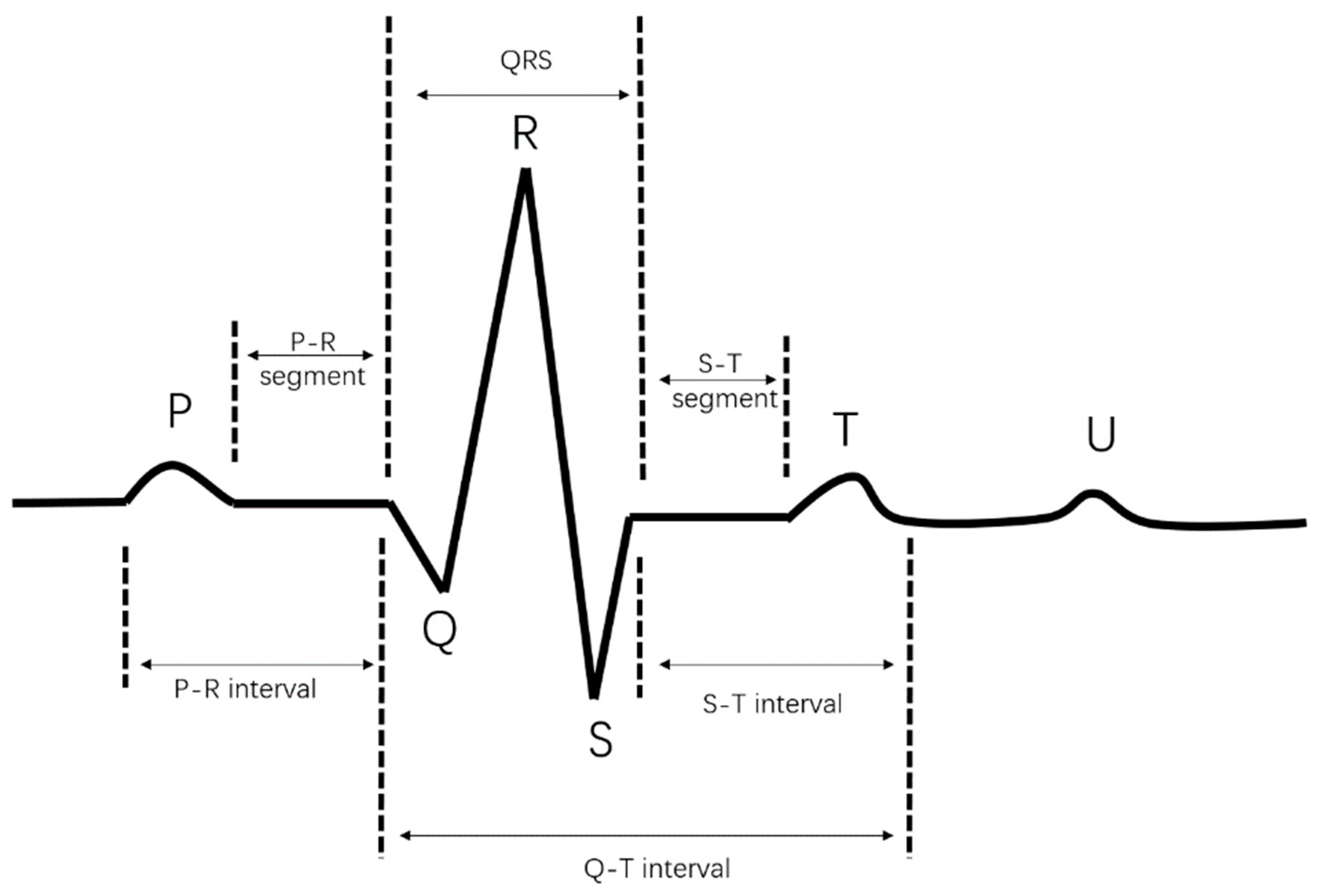
Figure 1. A complete ECG cycle.
The ECG has the characteristics of low frequency and energy concentration, and the signal is weak, easily disturbed by noise, and also has quasi-periodicity [5]. ECG sampling is often accompanied by a lot of noise, mainly muscle artefacts (MA) [6], electrode motion artefacts (EM) [7], and baseline wander (BW) [8]. Muscle artefacts are a common type of high-frequency noise, usually between 30 and 300 Hz. The source of this noise comes from the tremor of the body’s muscles and therefore appears on the image as a discrepancy between the muscle image and the actual situation. In order to reduce the impact of this noise on the image quality, medical imaging professionals must use appropriate techniques to reduce its effects. Electrode motion artefacts are instantaneous noise caused by poor contact between the skin and the electrodes. Baseline wander is low-frequency noise, mainly caused by breathing, and electrode slippage is caused by low-frequency interference. These noises have a major impact on a clinician’s ability to diagnose the nature of a disease, which is likely to lead to a false diagnosis. Therefore, it is particularly important to denoise sampled ECG data.
2. The Traditional ECG Denoising Method
The ECG acquisition process is often accompanied by a large amount of noise, which seriously affects a doctor’s accurate diagnosis of a patient. Therefore, technical research on removing noise from ECG data has always been a hot topic. Many researchers have proposed different research algorithms. In 2015, the authors of [9] proposed the use of multivariate empirical mode decomposition (MEMD) to remove baseline wander in ECG data. MEMD technology was a multivariate extension of EMD, which had recently attracted the attention of researchers for many applications [10]. The basic idea of this method was to transform a signal into a multi-channel signal and use the MEMD algorithm to process the multi-channel signal at the same time. The last intrinsic mode function (IMF) or the last two IMFs obtained by decomposition were removed from the ECG as the baseline wander to obtain a baseline-corrected ECG. Simulation results showed that this method can remove the baseline wander in ECG data and retain the morphological characteristics of ECG data. Empirical mode decomposition technology was a data-driven adaptive threshold method that was very suitable for non-stationary signals such as ECG data [11]. However, it could not select IMF efficiently and adaptively, which led to the loss of information. The authors of [12] proposed a noise removal method based on empirical mode decomposition combined with a wavelet threshold for adaptive ECG baseline wander. ECG data containing BW noise were denoised. First, they were decomposed into high-frequency signals and low-frequency signals using empirical mode decomposition. Then, the low-frequency signals were wavelet-transformed, and the high-frequency signals were combined to reconstruct the ECG data, thereby achieving the effect of removing baseline wander noise. The technology based on the wavelet transform was also more popular and widely used because of its ability to characterize the time–frequency domain information of time-domain signals [13]. The noise reduction method based on wavelets was widely used because it had good localization properties and could fully highlight the detailed features of ECG data in the time domain and frequency domain. However, due to the selection of the wavelet basis function and threshold, the amplitudes of the R wave and S wave may have been reduced after denoising [14]. Therefore, how to select the appropriate wavelet basis function and wavelet decomposition level to remove the noise in the input signal was still a problem [15]. In the wavelet denoising method, two thresholds [16] were used to enhance the ECG data. Tan Xue et al. [17] proposed an improved wavelet-threshold-function denoising method that avoided the defect of poor continuity at the threshold after processing the signal in the traditional soft and hard threshold function. Das et al. [18] compared the wavelet transform and proposed an ECG denoising method based on the S-transform. Because of the sparse characteristics of the ECG itself [19], a method based on sparse representation had been studied a lot in recent years. However, some ladder components were introduced after sparse noise reduction [20], which made the signal uneven after noise reduction, and most sparse representations use the L1 norm as a penalty term, which leads to the underestimation of the original signal [21].
In the field of deep learning, an ECG denoising method based on the autoencoder model [22] was more popular. An autoencoder composed of eight convolution blocks and eight deconvolution blocks was proposed by Eleni et al. [23], which can effectively learn the characteristics of ECGs and remove noise. As one of the many breakthroughs in deep learning technology, a generative adversarial network (GAN) had been widely used. Since Goodfellow [24] first introduced this method, many variants of generative adversarial networks have been developed. Radford et al. [25] proposed a deep convolutional generative adversarial network (DCGAN) in 2015. The DCGAN used a convolutional layer to replace the fully connected layer and replaced the original pooling layer with a convolution of the same step size. Pratik Singh and Gayahar Pradhan [26] proposed a generative adversarial network architecture for ECG denoising. The GAN model based on convolutional neural networks was effectively trained for ECG noise filtering, and end-to-end GAN model training was performed using clean and noisy ECG data. Zhu et al. [27] proposed a generative adversarial network, which was a Bi-LSTM-CNN network composed of bidirectional long short-term memory (LSTM) and a convolutional neural network (CNN). The model included a generator and a discriminator. The generator used a two-layer Bi-LSTM network, and the discriminator was based on a convolutional neural network.
3. Disentangled Representation Learning
Disentangled representation learning [28] was first proposed by Bengio in 2013. The feature sets being trained may be used for multiple tasks, which may have different relevant feature subsets. Therefore, the most robust method of feature learning was to separate as many factors as possible and discard as little information as possible. The application of disentangled representation learning makes the black box project of deep learning more interpretable and can show the specific meaning and function of each layer of the neural network. The traditional denoising autoencoder maps the signal information and noise information to the hidden space together in the encoding phase and then decodes them into signals through the hidden space variable. Researchers believe that there is a phenomenon of decoding confusion; that is, some noise information is decoded into signal information together, thus reducing the noise reduction effect of the signal. Therefore, researchers are trying to decouple the noise information and signal information in the hidden space, eliminate the entanglement of signal information and noise information, and then decode the clean signal.
References
- Roth, G.A.; Mensah, G.A.; Johnson, C.O.; Addolorato, G.; Ammirati, E.; Baddour, L.M.; Barengo, N.C.; Beaton, A.Z.; Benjamin, E.J.; Benziger, C.P.; et al. Global Burden of Cardiovascular Diseases and Risk Factors, 1990–2019: Update From the GBD 2019 Study. J. Am. Coll. Cardiol. 2020, 76, 2982–3021.
- Moridani, M.K.; Marjani, S. A Review of the Methods for Sudden Cardiac Death Detection: A Guide for Emergency Physicians. Int. J. Online Biomed. Eng. 2020, 16, 137–158.
- Syed, Z.; Vigmond, E.; Nattel, S.; Leon, L.J. Atrial cell action potential parameter fitting using genetic algorithms. Med. Biol. Eng. Comput. 2005, 43, 561–571.
- Jagatap, P.S.; Jagtap, R. Electrocardiogram (ECG) Signal Analysis and Feature Extraction: A Survey. 2014. Available online: https://www.semanticscholar.org/paper/Electrocardiogram-(ECG)-Signal-Analysis-and-Feature-Jagatap-Jagtap/ceadcc88d06518ca4ab2fd987465a22463736fc3 (accessed on 17 January 2023).
- Hammad, M.; Maher, A.; Wang, K.; Jiang, F.; Amrani, M. Detection of abnormal heart conditions based on characteristics of ECG signals. Measurement 2018, 125, 634–644.
- Sarangi, A.; Mishra, B.G.; Dash, S. Singular Spectrum Analysis Based EMG Artifact Removal from ECG Signal. YMER Digit. 2022, 21, 400–407.
- Wang, Z.; Wong, C.M.; da Cruz, J.N.; Wan, F.; Mak, P.-I.; Mak, P.U.; Vai, M.I. Muscle and electrode motion artifacts reduction in ECG using adaptive Fourier decomposition. In Proceedings of the 2014 IEEE International Conference on Systems, Man, and Cybernetics (SMC), San Diego, CA, USA, 5–8 October 2014; pp. 1456–1461.
- Lin, C.-C.; Chang, P.-C.; Tsai, P.-H. A Dual-Adaptive Approach Based on Discrete Cosine Transform for Removal of ECG Baseline Wander. Appl. Sci. 2022, 12, 8839.
- Gupta, P.; Sharma, K.K.; Joshi, S.D. Baseline wander removal of electrocardiogram signals using multivariate empirical mode decomposition. Health Technol. Lett. 2015, 2, 164–166.
- Sharma, R.R.; Pachori, R.B. Baseline wander and power line interference removal from ECG signals using eigenvalue decomposition. Biomed. Signal Process. Control. 2018, 45, 33–49.
- Labate, D.; La Foresta, F.; Occhiuto, G.; Morabito, F.C.; Lay-Ekuakille, A.; Vergallo, P. Empirical Mode Decomposition vs. Wavelet Decomposition for the Extraction of Respiratory Signal from Single-Channel ECG: A Comparison. IEEE Sens. J. 2013, 13, 2666–2674.
- Chao, L.; Liang, Y.; Xia, H. Adaptive baseline wander reduction based on empirical mode decomposition and wavelet thresholding for ECG. In Proceedings of the Chinese Medical Equipment Conference and 2021 Medical Equipment Exhibition Paper Compilation, Suzhou, China, 15–18 July 2021; pp. 108–114.
- Addison, P.S. Wavelet transforms and the ECG: A review. Physiol. Meas. 2005, 26, R155–R199.
- Goel, S.; Tomar, P.; Kaur, G. An Optimal Wavelet Approach for ECG Noise Cancellation. Int. J. Bio-Sci. Bio-Technol. 2016, 8, 39–52.
- Bing, P.; Liu, W.; Zhang, Z. DeepCEDNet: An Efficient Deep Convolutional Encoder-Decoder Networks for ECG Signal Enhancement. IEEE Access 2021, 9, 56699–56708.
- Garg, G.; Singh, V.; Gupta, J.R.P.; Mittal, A.P. Optimal algorithm for ECG denoising using Discrete Wavelet Transforms. In Proceedings of the 2010 IEEE International Conference on Computational Intelligence and Computing Research, Coimbatore, India, 28–29 December 2010; pp. 1–4.
- Tan, X.; Ye, J.; Zhang, X.; Li, C.; Zhou, J.; Dou, K. Application of improved wavelet threshold in ECG signal de-noising. Chin. J. Med. Devices 2021, 45, 1–5.
- Das, M.; Ari, S. Analysis of ECG signal denoising method based on S-transform. IRBM 2013, 34, 362–370.
- Zhou, Y.; Hu, X.; Tang, Z.; Ahn, A.C. Sparse representation-based ECG signal enhancement and QRS detection. Physiol. Meas. 2016, 37, 2093–2110.
- Ning, X.; Selesnick, I.W. ECG Enhancement and QRS Detection Based on Sparse Derivatives. Biomed. Signal Process. Control. 2013, 8, 713–723.
- Selesnick, I. Sparsity-assisted signal smoothing (revisited). In Proceedings of the 2017 IEEE International Conference on Acoustics, Speech and Signal Processing (ICASSP), New Orleans, LA, USA, 5–9 March 2017; pp. 4546–4550.
- Xiong, P.; Wang, H.; Liu, M.; Zhou, S.; Hou, Z.; Liu, X. ECG signal enhancement based on improved denoising auto-encoder. Eng. Appl. Artif. Intell. 2016, 52, 194–202.
- Fotiadou, E.; Konopczyński, T.; Hesser, J.; Vullings, R. Deep Convolutional Encoder-Decoder Framework for Fetal ECG Signal Denoising. In Proceedings of the 2019 Computing in Cardiology (CinC), Singapore, 8–11 September 2019; pp. 1–4.
- Goodfellow, I.J.; Pouget-Abadie, J.; Mirza, M.; Xu, B.; Warde-Farley, D.; Ozair, S.; Courville, A.; Bengio, Y. Generative Adversarial Nets. In Proceedings of the Advances in Neural Information Processing Systems; 2014; Volume 27. Available online: https://proceedings.neurips.cc/paper/2014/hash/5ca3e9b122f61f8f06494c97b1afccf3-Abstract.html (accessed on 4 December 2022).
- Patil, A. DCGAN: Deep Convolutional GAN with Attention Module for Remote View Classification. In Proceedings of the 2021 International Conference on Forensics, Analytics, Big Data, Security (FABS), Bengaluru, India, 21–22 December 2021; Volume 1, pp. 1–10.
- Singh, P.; Pradhan, G. A New ECG Denoising Framework Using Generative Adversarial Network. IEEE/ACM Trans. Comput. Biol. Bioinform. 2021, 18, 759–764.
- Zhu, F.; Ye, F.; Fu, Y.; Liu, Q.; Shen, B. Electrocardiogram generation with a bidirectional LSTM-CNN generative adversarial network. Sci. Rep. 2019, 9, 6734.
- Bengio, Y.; Courville, A.; Vincent, P. Representation Learning: A Review and New Perspectives. arXiv 2014, 35, 1798–1828. Available online: https://arxiv.org/abs/1206.5538 (accessed on 23 April 2014).
More
Information
Contributors
MDPI registered users' name will be linked to their SciProfiles pages. To register with us, please refer to https://encyclopedia.pub/register
:
View Times:
2.0K
Revisions:
2 times
(View History)
Update Date:
29 Nov 2023
Table of Contents


Notice
You are not a member of the advisory board for this topic. If you want to update advisory board member profile, please contact office@encyclopedia.pub.
OK
Confirm
Only members of the Encyclopedia advisory board for this topic are allowed to note entries. Would you like to become an advisory board member of the Encyclopedia?
Yes
No
${ textCharacter }/${ maxCharacter }
Submit
Cancel
Back
Comments
${ item }
|
${ item.createdUser.fullName }
${ item.createdAt }
${ item.vote }
${ item.reply }
Delete
${ reply.createdUser.fullName }
${ reply.createdAt }
${ reply.vote }
Delete
There is no reply to this comment~
${ item.replyTextCharacter }/${ item.replyMaxCharacter }
Submit
Cancel
More
No more~
There is no comment~
${ textCharacter }/${ maxCharacter }
Submit
Cancel
${ selectedItem.replyTextCharacter }/${ selectedItem.replyMaxCharacter }
Submit
Cancel
Confirm
Are you sure to Delete?
Yes
No