Submitted Successfully!
To reward your contribution, here is a gift for you: A free trial for our video production service.
Thank you for your contribution! You can also upload a video entry or images related to this topic.
Version Summary Created by Modification Content Size Created at Operation
1 -- 2761 2023-11-02 11:55:52 |
2 format Meta information modification 2761 2023-11-03 02:15:51 |

Video Upload Options

Do you have a full video?

Confirm

Are you sure to Delete?
Cite
If you have any further questions, please contact Encyclopedia Editorial Office.
Caetano-Pinto, P.; Stahl, S.H. Regulation of OAT1 and OAT3 Expression and Activity. Encyclopedia. Available online: https://encyclopedia.pub/entry/51093 (accessed on 31 July 2024).
Caetano-Pinto P, Stahl SH. Regulation of OAT1 and OAT3 Expression and Activity. Encyclopedia. Available at: https://encyclopedia.pub/entry/51093. Accessed July 31, 2024.
Caetano-Pinto, Pedro, Simone H. Stahl. "Regulation of OAT1 and OAT3 Expression and Activity" Encyclopedia, https://encyclopedia.pub/entry/51093 (accessed July 31, 2024).
Caetano-Pinto, P., & Stahl, S.H. (2023, November 02). Regulation of OAT1 and OAT3 Expression and Activity. In Encyclopedia. https://encyclopedia.pub/entry/51093
Caetano-Pinto, Pedro and Simone H. Stahl. "Regulation of OAT1 and OAT3 Expression and Activity." Encyclopedia. Web. 02 November, 2023.
Regulation of OAT1 and OAT3 Expression and Activity
Edit

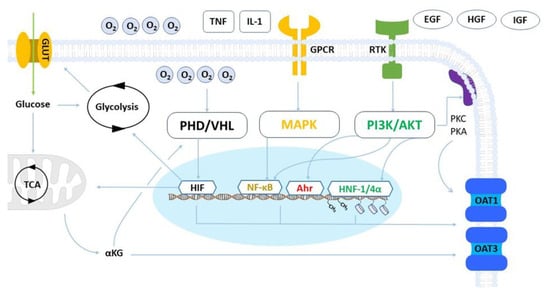
Organic anion transporters 1 and 3 (OAT1 and OAT3) play a crucial role in kidney function by regulating the secretion of multiple renally cleared small molecules and toxic metabolic by-products. Assessing the activity of these transporters is essential for drug development purposes as they can significantly impact drug disposition and safety. OAT1 and OAT3 are amongst the most abundant drug transporters expressed in human renal proximal tubules. However, their expression is lost when cells are isolated and cultured in vitro, which is a persistent issue across all human and animal renal proximal tubule cell models, including primary cells and cell lines. Although it is well known that the overall expression of drug transporters is affected in vitro, the underlying reasons for the loss of OAT1 and OAT3 are still not fully understood. 

drug transporter organic anion transporters regulation

1. Introduction

The tight regulation of OAT1 and OAT3 expression and activity involves intricate cellular pathways, including transcriptional, post-transcriptional, and post-translational processes, which maintain and modulate transport activity in response to variation in cellular homeostasis [1]. Changes in cellular energy supply (e.g., nutrient deprivation), metabolism, or the extracellular environment (e.g., oxygen levels) can trigger complex signaling pathways leading to alterations in transporter expression and may result in the permanent loss of OAT1 and OAT3 expression in vitro after isolation from renal tissue. Several in vitro and in vivo studies using multiple cell lines and animal models, respectively, have shed light on the complex regulation of these uptake transporters (Figure 1).
Figure 1. Representation of OAT1 and OAT3 regulatory networks. The prolyl hydroxylase/von Hippel Lindau (PHD/VHL) axis senses cellular oxygen levels and governs the activity of the hypoxia-inducible factors (HIF). HIF regulates the expression of genes responsible for glucose uptake via glucose transporters (GLUT) and metabolism, both glycolytic activity and mitochondrial respiration. HIF transcriptional activity can also mediate the expression of OAT1 and OAT3. Alpha-ketoglutarate (αKG) derived from mitochondrial activity is a co-substrate for OAT1 and OAT3, and its abundance influences transport activity. αKG also inhibits PHD activity and can promote HIF activation when oxygen levels are normal. G protein-coupled receptors (GPCRs) are triggered by cytokines and immune factors and act via the Mitogen-activated protein kinases (MAPKs) pathway to activate nuclear factor kappa B (NF-kB), a transcriptional factor that regulates organic anion transporter expression. Receptor tyrosine kinases (RTKs) are activated by growth factors and play a major post-transcriptional role in OAT1 and OAT3 regulation. RTKs act via the phosphatidylinositol 3-kinase/protein kinase B (PI3K/AKT) pathway to regulate gene expression via the Hepatocyte nuclear factors (HNF-1/4α), the aryl hydrocarbon receptor (AhR), and the NF-kB. RTKs also govern the activity of adaptor and adhesion proteins.

2. Metabolism and Hypoxia

RPTECs are highly metabolically active and require a significant amount of energy to fulfill their secretory functions. Typically, energy production occurs through mitochondrial respiration, which generates the high levels of ATP needed to power their prolific membrane transport machinery [2]. However, despite their high mitochondrial activity, RPTECs are subjected to a low-oxygen environment due to the organization of the renal vasculature, which acts as a limiting factor for oxygen diffusion into cells [3].
Hypoxia occurs when low oxygen levels disrupt normal cellular function, triggering a compensatory response to counteract its effects. Hypoxia-inducible factor (HIF)-1 plays a central role in the cellular response to hypoxia. Under normal oxygen conditions, HIF-1α, the oxygen-sensitive subunit of HIF-1, is rapidly degraded by the proteasome. However, when oxygen levels decrease, HIF 1α is stabilized and translocates to the nucleus, where it dimerizes with HIF-1β and binds to hypoxia-responsive elements (HREs) in the promoter regions of target genes [4][5]. HIF-1 increases the expression of genes involved in erythropoiesis, angiogenesis, and glucose metabolism and has been implicated in the direct and indirect regulation of OAT1 and OAT3 expression [6]. Cells can shift towards glycolysis as an alternative metabolic pathway when oxygen levels are low since this process can produce ATP in its absence. This leads to an increase in glucose uptake and consumption, which is mediated by HIF-1 and the upregulation of glucose transporters such as GLUT1 and GLUT3 into the cell [7]. The intracellular glucose concentration has been shown to affect the expression of OAT3 in the kidney. High glucose levels downregulate OAT3 expression through the activation of the transcription factor nuclear factor-kappa B (NF-κB) [8][9]. On the other hand, low glucose levels can upregulate OAT3 expression by activating the AMP-activated protein kinase (AMPK) pathway, which is a key regulator of cellular energy homeostasis [10]. Insulin-like growth factor 1 (IGF-1) seemingly plays a role in OAT3 upregulation by stimulating the activity of protein kinase A (PKA) dependent pathways, an effect that is counteracted by the pharmacological inhibition of the IGF-1 receptor [10]. The activity of this receptor may be behind the apparent relation between OAT3 deregulation and elevated glucose levels, considering that it promotes glucose uptake [11]. Ischemia-reperfusion injury, which leads to a hypoxic event in the renal proximal tubules, is also shown to decrease the activity and expression of OAT1 and OAT3 in rats [12].
The activity of OAT1 and OAT3 transporters is influenced by the intracellular concentration of αKG, which serves as a co-substrate for these transporters. αKG is synthesized within the mitochondria as an intermediate product of the tricarboxylic acid (TCA) cycle during the production of ATP [13]. Hypoxic conditions that favor glycolytic activity to the detriment of mitochondrial respiration lead to a reduction of αKG production and incidentally a reduction in OAT1 and OAT3 activity. This indirect functional regulation underlines the multiple regulatory effects that oxygen levels and hypoxia exert on the expression and activity of OAT transporters. Interestingly, αKG levels indirectly stabilize the expression of HIF 1. αKG is a precursor of succinate, which can inhibit the activity of prolyl hydroxylases (PHD) [14]. Under normal physiological oxygen levels, PHD hydroxylates HIF 1α, leading to its proteasomal degradation [3]. This mechanism keeps HIF-1 activity in check and, arguably, elevated intracellular αKG levels can enable cells to maintain HIF-1-mediated activities when oxygen levels are elevated. An important consideration is the lower oxygen pressure that the kidneys experience relative to most organs, due to their vascular architecture, therefore, the activity of the PHD-HIF axis in renal cells in response to varying oxygen levels is believed to be more resilient to hypoxic conditions [3].

3. Inflammatory and Growth Factors

Inflammation is a complex biological response to injury or cellular stress characterized by the activation of immune cells and the release of various inflammatory mediators, including cytokines, chemokines, and reactive oxygen species. Hypoxia and metabolic impairment are major drivers of inflammation [15]. The expression of OAT1 and OAT3 is also impacted by inflammation, and several immune factors activate pathways that will ultimately modulate the activity of these transporters.
The NF-κB pathway is a major mechanism by which inflammation regulates the expression of OAT1 and OAT3. This pathway is activated in response to a variety of inflammatory stimuli, including cytokines and oxidative stress. NF-κB regulates transporter expression by directly binding to their gene promoters and inducing transcription [16]. Inflammatory cytokines, such as tumor necrosis factor-alpha (TNF-α) and interleukin-1 beta (IL-1β), are potent activators of the NF-κB pathway and have been shown to upregulate the expression of OAT1 and OAT3 in renal cells [17]. The mitogen-activated protein kinase (MAPK) pathway is activated in response to cytokine release, and its activation leads to the phosphorylation of transcription factors, which in turn regulate the expression of target genes. Several MAPKs, including extracellular signal-regulated kinase (ERK), c-Jun N-terminal kinase (JNK), and p38 MAPK, have been implicated in the regulation of OAT1 and OAT3 expression [18][19]. The role of MAPK in organic anion transport regulation was further substantiated by evidence that treatment with the MAPK inhibitor U0126 lowers the expression of these transporters in renal proximal tubular cells [20].
Growth factors are signaling molecules that play important roles in the regulation of cell growth, differentiation, and survival. Recent studies have shown that growth factors can also regulate the expression of OAT1 and OAT3, acting via regulatory pathways similar to those of immune factors. IGF-1 can increase the expression of OAT1 and OAT3 in renal proximal tubular cells by activating the phosphatidylinositol 3-kinase (PI3K) pathway. Upon PI3K activation, downstream signaling molecules such as alpha serine/threonine-protein kinase (AKT) and the mammalian target of rapamycin (mTOR) are triggered, subsequently inducing the expression of the transporters [10]. Hepatocyte growth factor (HGF) is produced by mesenchymal cells and plays a role in tissue regeneration and repair. In vitro studies have demonstrated that HGF treatment can upregulate the expression of renal proximal tubular cell transporters OAT1 and OAT3. This is achieved through the activation of the c-Met receptor and downstream signaling molecules, such as PI3K and AKT [21]. Epidermal growth factor (EGF) is a crucial regulator in epithelial cells and has been shown to increase the expression of OAT1 and OAT3 in renal proximal tubular cells. In immortalized RPTECs overexpressing OAT1, the gene and protein expression of the transporter was regulated by EGFR activity via the extracellular signal-regulated kinase (ERK) pathway, showing the involvement of this pathway in the post-transcriptional regulation of organic anion activity [22].

4. MicroRNAs

MicroRNAs (miRs) are short non-coding RNA molecules that regulate gene expression by binding to target mRNAs, effectively suppressing protein translation [23]. HIF-1 has the potential to upregulate the expression of specific miRs that participate in mitigating the detrimental effects of oxygen deprivation. The expression of miR-21 is associated with HIF-1 activity in vitro [24]. Indirect evidence in mice shows that the suppression of miR-21 normalizes organic anion transport activity and facilitates the secretion of uremic toxins such as indoxyl sulfate (IS) [25].
The kidneys can sense and respond to elevated levels of metabolites produced by the gut microbiome via epidermal growth factors receptors (EGFR) and downstream signaling. The uremic toxin indoxyl sulfate (IS) is a protein metabolism by-product derived from the microbiome and an endogenous OAT1 substrate. IS indirectly activates the transcriptional activity of the Aryl hydrocarbon receptor and aryl hydrocarbon receptor nuclear translocator (AhR/ARNT) by binding to and stimulating EGFR. The activity of AhR/ARNT promotes the upregulation of OAT1 expression. The expression of miR-223 supports this apparent and complex gut-kidney communication axis by stabilizing ARNT expression and ensuring the transcription of OAT1 [26]. This mechanism induces renal secretion in response to elevated IS and maintains homeostasis by removing this potentially detrimental metabolite [26]. MiR regulation is highly species- and tissue-specific, and despite evidence of miR involvement, the regulation of OATs by miRs is still poorly understood.

5. Epigenetic Modifications

Epigenetic modifications, including DNA methylation and histone deacetylation, are important mechanisms that regulate gene expression by altering the chromatin structure and accessibility of DNA to transcription factors [27]. In recent years, studies have shown that epigenetic modifications also play a role in the expression of OAT1 and OAT3 [28][29].
DNA methylation is a process by which a methyl group is added to cytosine residues in the CpG dinucleotides of DNA. This modification is catalyzed by DNA methyltransferases (DNMTs) and can lead to the repression of gene expression. Methylation acts as a regulator of tissue-specific transactivation, where differential rates of transcription dictate the expression profiles of genes across cell types and tissues. The promoter regions of OAT1 and OAT3 contain CpG islands, which are regions of DNA with a high density of CpG dinucleotides. Ex vivo studies have shown that DNA methylation of the promoter regions of OAT1 and OAT3 can lead to their repression and decrease transport activity [30]. Interestingly, certain SLC transporters in the kidney cortex, including OAT1 and OAT3, were found hypomethylated, making the promoter regions of the genes more accessible to transcription factors [31].
Histone deacetylation is a process by which the acetyl groups on histone proteins are removed, leading to the condensation of chromatin structure and the repression of gene expression. Histone deacetylation is catalyzed by histone deacetylases (HDACs). Studies have shown that inhibition of HDAC activity can lead to the upregulation of OAT1 and OAT3 expression in renal proximal tubular cells [18][32]. The hepatocyte nuclear factors 1-alpha and 4-alpha (HNF 1α and HNF 4α) are involved in the regulation of several genes during the maturation of kidney function. This mechanism involves the recruitment of elements of the p300-CBP coactivator family (histone acetyltransferase p300/cyclic adenosine monophosphate response element binding (CREB) protein), which promotes histone acetylation and the opening of chromatin structure, leading to increased transcription of the transporters [33][34].

6. Cellular Adhesion

Cellular adhesion, which is governed by the binding of cells to each other or extracellular matrix components, is an important process in many biological functions, including cell migration, tissue development, and immune response. It plays a major role in the polarity of epithelial cells, and it has been associated with the expression of organic anion transporters OAT1 and OAT3 [35][36][37]. Evidence from 3D renal models and the reconstruction of proximal renal tubules have revealed that surface topography and matrix chemistry are critical for proper cellular differentiation and functionality [38]. The introduction of microenvironmental curvature, mimicking the tubular morphology of the nephron, improves renal function and increases the expression of drug transporters, including OAT1 [39]. Anisotropic extracellular matrix architecture promotes the structural arrangement of F-actin, reinforcing epithelial cellular morphology and increasing the expression of kidney transporters [40][41]. Studies involving the generation of kidney organoids or decellularized tissues have shown that matrix rigidity is another determinant factor in the maturation of renal cells. The appropriate matrix topography and stiffness, depending on cell type (e.g., primary cells, stem cells), will dramatically influence adhesion and the development of strong phenotypical features [42][43][44].
Caveolins (Cavs) are integral membrane proteins that are mainly described to regulate receptor-independent endocytosis. Additionally, Cavs are also reported to stabilize focal adhesions, which act as anchoring points between cells and their extracellular environment. These proteins also induce the curvature at the plasma membrane when oligomerized and interact with multiple signaling elements [45][46]. Cav-1 and Cav-3 have been implicated in the upregulation of OAT1 and OAT3, respectively [47]. This evidence shows that regulators of cell membrane adhesion and topography influence the activity of these anion transporters, potentially affecting their cellular localization [36].

7. Post-Translational Regulation and Trafficking

Post-translational regulation of OAT1 and OAT3 transporters involves a range of mechanisms that modify the proteins after they are synthesized. These modifications can impact transporter activity, localization, and stability and ultimately influence their function [48]. Phosphorylation, which involves the addition of a phosphate group to specific amino acid residues on the transporter protein, can alter the conformation of the transporter, affecting its substrate binding ability and transport activity. OAT1 activity is modulated by phosphorylation at specific sites, by protein kinase A (PKA), and by protein kinase C (PKC), which can enhance or inhibit OAT1 activity [8][18][49].
Glycosylation involves the addition of sugar molecules to specific sites on the transporter protein and it has been shown to have a significant impact on OAT1 and OAT3 function [36][50]. OAT3 has been found to undergo glycosylation at asparagine residues (N-glycosylation) and serine and threonine residues (O-glycosylation) via the activity of glycosyltransferases (GTs). O-glycosylation of OAT3 has been shown to increase the transport activity of the transporter, while N-glycosylation decreases substrate uptake. OAT1 has also been shown to undergo glycosylation, although less is known about the specific impact of this modification on its function [48].
Small ubiquitin-like modifiers (SUMOs) are a family of proteins that can covalently bind to specific lysine residues in target proteins and act as functional regulators governing, among other mechanisms, transcription and subcellular localization. This process is known as SUMOylation and is initiated when a series of ligases cooperate to attach SUMO to its target. It can be reversed by the activity of proteases that cleave the modifiers from SUMOylated proteins [51]. The activity of OAT3 is upregulated by SUMOylation, where the shuttling rate of the transporter from intracellular compartments to the plasma membrane is enhanced and, consequently, its membrane expression is increased. This upregulation is compounded by a reduced rate of OAT3 protein degradation. This functional gain also correlates with the stimulation of IGF-1 mediated PKA activity, indicating that SUMOylation is the downstream post-translational regulatory process behind OAT3 activation by this pathway [52]. SUMOylation differs from ubiquitination to the extent that tagged proteins are not degraded. The ubiquitination process adds small ubiquitin proteins to targets promoting their proteasomal degradation. It is an essential mechanism for protein turnover. Like SUMOylation, ubiquitination is described to play a role in OAT regulation [48]. PKC phosphorylates the ubiquitin ligase Nedd4-2 and directly affects the ubiquitination status of OAT1 and OAT3 with a negative impact on the stability and expression of both transporters [53][54]. Evidence from studies involving the selective pharmacological inhibition of proteasomal activity indicates that OAT3 ubiquitination is increased and associated with enhanced membrane expression and anion transport activity [55][56]. Taken together, these findings highlight an important role for both SUMOylation and ubiquitination as the actuators of non-transcriptional regulatory mechanisms of anion transport activity.

References

  1. Zou, L.; Stecula, A.; Gupta, A.; Prasad, B.; Chien, H.C.; Yee, S.W.; Wang, L.; Unadkat, J.D.; Stahl, S.H.; Fenner, K.S.; et al. Molecular mechanisms for species differences in organic anion transporter 1, OAT1: Implications for renal drug toxicity. Mol. Pharmacol. 2018, 94, 689–699.
  2. Drozdzik, M.; Drozdzik, M.; Oswald, S. Membrane carriers and transporters in kidney physiology and disease. Biomedicines 2021, 9, 426.
  3. Pinto, P.C.; Rönnau, C.; Burchardt, M.; Wolff, I. Kidney Cancer and Chronic Kidney Disease: Too Close for Comfort. Biomedicines 2021, 9, 1761.
  4. Haase, V.H. Mechanisms of hypoxia responses in renal tissue. J. Am. Soc. Nephrol. 2013, 24, 537–541.
  5. Schödel, J.; Grampp, S.; Maher, E.R.; Moch, H.; Ratcliffe, P.J.; Russo, P.; Mole, D.R. Hypoxia, hypoxia-inducible transcription factors, and renal cancer. Eur. Urol. 2016, 69, 646–657.
  6. Jamshidi, N.; Nigam, S.K. Drug transporters OAT1 and OAT3 have specific effects on multiple organs and gut microbiome as revealed by contextualized metabolic network reconstructions. Sci. Rep. 2022, 12, 18308.
  7. Kierans, S.J.; Taylor, C.T. Regulation of glycolysis by the hypoxia-inducible factor (HIF): Implications for cellular physiology. J. Physiol. 2021, 599, 23–37.
  8. Li, S.; Zhang, Q.; You, G. Three ubiquitination sites of organic anion transporter-1 synergistically mediate protein kinase c-dependent endocytosis of the transporter. Mol. Pharmacol. 2013, 84, 139–146.
  9. Phatchawan, A.; Chutima, S.; Varanuj, C.; Anusorn, L. Decreased renal organic anion transporter 3 expression in type 1 diabetic rats. Am. J. Med. Sci. 2014, 347, 221–227.
  10. Zhang, J.; Yu, Z.; You, G. Insulin-like growth factor 1 modulates the phosphorylation, expression, and activity of organic anion transporter 3 through protein kinase A signaling pathway. Acta Pharm. Sin. B 2020, 10, 186–194.
  11. Clemmons, D.R. Role of insulin-like growth factor I in maintaining normal glucose homeostasis. Horm. Res. 2004, 62, 77–82.
  12. Schneider, R.; Sauvant, C.; Betz, B.; Otremba, M.; Fischer, D.; Holzinger, H.; Wanner, C.; Galle, J.; Gekle, M. Downregulation of organic anion transporters OAT1 and OAT3 correlates with impaired secretion of para-aminohippurate after ischemic acute renal failure in rats. Am. J. Physiol.-Ren. Physiol. 2007, 292, F1599–F1605.
  13. Dodd, K.M.; Yang, J.; Shen, M.H.; Sampson, J.R.; Tee, A.R. mTORC1 drives HIF-1α and VEGF-A signalling via multiple mechanisms involving 4E-BP1, S6K1 and STAT3. Oncogene 2015, 34, 2239–2250.
  14. Hagos, Y.; Schley, G.; Scḧdel, J.; Krick, W.; Burckhardt, G.; Willam, C.; Burckhardt, B.C. α-Ketoglutarate-related inhibitors of HIF prolyl hydroxylases are substrates of renal organic anion transporters 1 (OAT1) and 4 (OAT4). Pflug. Arch. Eur. J. Physiol. 2012, 464, 367–374.
  15. Taylor, C.T.; Colgan, S.P. Regulation of immunity and inflammation by hypoxia in immunological niches. Nat. Rev. Immunol. 2017, 17, 774–785.
  16. Zhang, J.; Wang, H.; Fan, Y.; Yu, Z.; You, G. Regulation of organic anion transporters: Role in physiology, pathophysiology, and drug elimination. Pharmacol. Ther. 2021, 217, 107647.
  17. Sirijariyawat, K.; Ontawong, A.; Palee, S.; Thummasorn, S.; Maneechote, C.; Boonphang, O.; Chatsudthipong, V.; Chattipakorn, N.; Srimaroeng, C. Impaired renal organic anion transport 1 (SLC22A6) and its regulation following acute myocardial infarction and reperfusion injury in rats. Biochim. Biophys. Acta (BBA)-Mol. Basis Dis. 2019, 1865, 2342–2355.
  18. Pou Casellas, C.; Jansen, K.; Rookmaaker, M.B.; Clevers, H.; Verhaar, M.C.; Masereeuw, R. Regulation of Solute Carriers Oct2 and Oat1/3 in the Kidney: A Phylogenetic, Ontogenetic, and Cell Dynamic Perspective. Physiol. Rev. 2022, 102, 993–1024.
  19. Soodvilai, S.; Wright, S.H.; Dantzler, W.H.; Chatsudthipong, V. Involvement of tyrosine kinase and PI3K in the regulation of OAT3-mediated estrone sulfate transport in isolated rabbit renal proximal tubules. Am. J. Physiol.-Ren. Physiol. 2005, 289, F1057–F1064.
  20. Liu, N.; Xu, L.; Shi, Y.; Fang, L.; Gu, H.; Wang, H.; Ding, X.; Zhuang, S. Pharmacologic targeting ERK1/2 attenuates the development and progression of hyperuricemic nephropathy in rats. Oncotarget 2017, 8, 33807–33826.
  21. Li, T.T.; An, J.X.; Xu, J.Y.; Tuo, B.G. Overview of organic anion transporters and organic anion transporter polypeptides and their roles in the liver. World J. Clin. Cases 2019, 7, 3915–3933.
  22. Caetano-Pinto, P.; Jamalpoor, A.; Ham, J.; Goumenou, A.; Mommersteeg, M.; Pijnenburg, D.; Ruijtenbeek, R.; Sanchez-Romero, N.; Van Zelst, B.; Heil, S.G.; et al. Cetuximab Prevents Methotrexate-Induced Cytotoxicity in Vitro through Epidermal Growth Factor Dependent Regulation of Renal Drug Transporters. Mol. Pharm. 2017, 14, 2147–2157.
  23. Serocki, M.; Bartoszewska, S.; Janaszak-Jasiecka, A.; Ochocka, R.J.; Collawn, J.F.; Bartoszewski, R. miRNAs regulate the HIF switch during hypoxia: A novel therapeutic target. Angiogenesis 2018, 21, 183–202.
  24. Liu, Y.; Nie, H.; Zhang, K.; Ma, D.; Yang, G.; Zheng, Z.; Liu, K.; Yu, B.; Zhai, C.; Yang, S. A feedback regulatory loop between HIF-1α and miR-21 in response to hypoxia in cardiomyocytes. FEBS Lett. 2014, 588, 3137–3146.
  25. Gomez, I.G.; MacKenna, D.A.; Johnson, B.G.; Kaimal, V.; Roach, A.M.; Ren, S.; Nakagawa, N.; Xin, C.; Newitt, R.; Pandya, S.; et al. Anti-microRNA-21 oligonucleotides prevent Alport nephropathy progression by stimulating metabolic pathways. J. Clin. Investig. 2015, 125, 141–156.
  26. Jansen, J.; Jansen, K.; Neven, E.; Poesen, R.; Othman, A.; van Mil, A.; Sluijter, J.; Torano, J.S.; Zaal, E.A.; Berkers, C.R.; et al. Remote sensing and signaling in kidney proximal tubules stimulates gut microbiome-derived organic anion secretion. Proc. Natl. Acad. Sci. USA 2019, 116, 16105–16110.
  27. Bannister, A.J.; Kouzarides, T. Regulation of chromatin by histone modifications. Cell Res. 2011, 21, 381–395.
  28. Fisel, P.; Schaeffeler, E.; Schwab, M. DNA methylation of ADME genes. Clin. Pharmacol. Ther. 2016, 99, 512–527.
  29. Hirota, T.; Tanaka, T.; Takesue, H.; Ieiri, I. Epigenetic regulation of drug transporter expression in human tissues. Expert Opin. Drug Metab. Toxicol. 2017, 13, 19–30.
  30. Jin, L.; Kikuchi, R.; Saji, T.; Kusuhara, H.; Sugiyama, Y. Regulation of tissue-specific expression of renal organic anion transporters by hepatocyte nuclear factor 1 α/β and DNA methylation. J. Pharmacol. Exp. Ther. 2012, 340, 648–655.
  31. Zhou, S.; Shu, Y. Special Section on New Era of Transporter Science: Unraveling the Functional Role of Orphan Transporters-Minireview Transcriptional Regulation of Solute Carrier Drug Transporters. Drug Metab. Dispos. 2022, 50, 1238–1250.
  32. Litke, C.; Hagenston, A.M.; Kenkel, A.K.; Paldy, E.; Lu, J.; Kuner, R.; Mauceri, D. Organic anion transporter 1 is an HDAC4-regulated mediator of nociceptive hypersensitivity in mice. Nat. Commun. 2022, 13, 875.
  33. Martovetsky, G.; Tee, J.B.; Nigam, S.K. Hepatocyte nuclear factors 4α and 1α regulate kidney developmental expression of drug-metabolizing enzymes and drug transporters. Mol. Pharmacol. 2013, 84, 808–823.
  34. Ogasawara, K.; Terada, T.; Asaka, J.I.; Katsura, T.; Inui, K.I. Hepatocyte nuclear factor-4α regulates the human organic anion transporter 1 gene in the kidney. Am. J. Physiol.-Ren. Physiol. 2007, 292, F1819–F1826.
  35. Parsons, J.T.; Horwitz, A.R.; Schwartz, M.A. Cell adhesion: Integrating cytoskeletal dynamics and cellular tension. Nat. Rev. Mol. Cell Biol. 2010, 11, 633–643.
  36. Duan, P.; You, G. Short-term regulation of organic anion transporters. Pharmacol. Ther. 2010, 125, 55–61.
  37. Epstein, F.H.; Fish, E.M.; Molitoris, B.A. Alterations in Epithelial Polarity and the Pathogenesis of Disease States. N. Engl. J. Med. 1994, 330, 1580–1588.
  38. Pou Casellas, C.; Rookmaaker, M.B.; Verhaar, M.C. Controlling cellular plasticity to improve in vitro models for kidney regeneration. Curr. Opin. Biomed. Eng. 2021, 20, 100345.
  39. Jansen, J.; Fedecostante, M.; Wilmer, M.J.; Peters, J.G.; Kreuser, U.M.; Van Den Broek, P.H.; Mensink, R.A.; Boltje, T.J.; Stamatialis, D.; Wetzels, J.F.; et al. Bioengineered kidney tubules efficiently excrete uremic toxins. Sci. Rep. 2016, 6, 26715.
  40. Rougerie, P.; Pieuchot, L.; dos Santos, R.S.; Marteau, J.; Bigerelle, M.; Chauvy, P.F.; Farina, M.; Anselme, K. Topographical curvature is sufficient to control epithelium elongation. Sci. Rep. 2020, 10, 14784.
  41. van Genderen, A.M.; Jansen, K.; Kristen, M.; van Duijn, J.; Li, Y.; Schuurmans, C.C.L.; Malda, J.; Vermonden, T.; Jansen, J.; Masereeuw, R.; et al. Topographic Guidance in Melt-Electrowritten Tubular Scaffolds Enhances Engineered Kidney Tubule Performance. Front. Bioeng. Biotechnol. 2021, 8, 617364.
  42. Garreta, E.; Prado, P.; Tarantino, C.; Oria, R.; Fanlo, L.; Martí, E.; Zalvidea, D.; Trepat, X.; Roca-Cusachs, P.; Gavaldà-Navarro, A.; et al. Fine tuning the extracellular environment accelerates the derivation of kidney organoids from human pluripotent stem cells. Nat. Mater. 2019, 18, 397–405.
  43. Batchelder, C.A.; Martinez, M.L.; Tarantal, A.F. Natural scaffolds for renal differentiation of human embryonic stem cells for kidney tissue engineering. PLoS ONE 2015, 10, e0143849.
  44. Bonventre, J.V.; Verhaar, M.C. Editorial overview: Kidney regeneration; cells, organoids and whole organ engineering? Curr. Opin. Biomed. Eng. 2022, 23, 100403.
  45. Ohi, M.D.; Kenworthy, A.K. Emerging Insights into the Molecular Architecture of Caveolin-1. J. Membr. Biol. 2022, 255, 375–383.
  46. Fridolfsson, H.N.; Roth, D.M.; Insel, P.A.; Patel, H.H. Regulation of intracellular signaling and function by caveolin. FASEB J. 2014, 28, 3823–3831.
  47. Barros, S.A.; Srimaroeng, C.; Perry, J.L.; Walden, R.; Dembla-Rajpal, N.; Sweet, D.H.; Pritchard, J.B. Activation of protein kinase Cζ increases OAT1 (SLC22A6)-and OAT3 (SLC22A8)-mediated transport. J. Biol. Chem. 2009, 284, 2672–2679.
  48. Xu, D.; Wang, H.; You, G. Posttranslational Regulation of Organic Anion Transporters by Ubiquitination: Known and Novel. Med. Res. Rev. 2016, 36, 964–979.
  49. Preising, C.; Schneider, R.; Bucher, M.; Gekle, M.; Sauvant, C. Regulation of expression of renal organic anion transporters OAT1 and OAT3 in a model of ischemia/reperfusion injury. Cell. Physiol. Biochem. 2015, 37, 1–13.
  50. Tanaka, K.; Xu, W.; Zhou, F.; You, G. Role of Glycosylation in the Organic Anion Transporter OAT1. J. Biol. Chem. 2004, 279, 14961–14966.
  51. Celen, A.B.; Sahin, U. Sumoylation on its 25th anniversary: Mechanisms, pathology, and emerging concepts. FEBS J. 2020, 287, 3110–3140.
  52. Wang, H.; Zhang, J.; You, G. Activation of Protein Kinase A Stimulates SUMOylation, Expression, and Transport Activity of Organic Anion Transporter 3. AAPS J. 2019, 21, 30.
  53. Yu, Z.; Liu, C.; Zhang, J.; Liang, Z.; You, G. Protein kinase C regulates organic anion transporter 1 through phosphorylating ubiquitin ligase Nedd4–2. BMC Mol. Cell Biol. 2021, 22, 53.
  54. Xu, D.; Wang, H.; You, G. An Essential Role of Nedd4-2 in the Ubiquitination, Expression, and Function of Organic Anion Transporter-3. Mol. Pharm. 2016, 13, 621–630.
  55. Fan, Y.; Wang, H.; Yu, Z.; Liang, Z.; Li, Y.; You, G. Inhibition of proteasome, but not lysosome, upregulates organic anion transporter 3 in vitro and in vivo. Biochem. Pharmacol. 2023, 208, 115387.
  56. Fan, Y.; Liang, Z.; Zhang, J.; You, G. Oral proteasomal inhibitors ixazomib, oprozomib, and delanzomib upregulate the function of organic anion transporter 3 (OAT3): Implications in OAT3-mediated drug-drug interactions. Pharmaceutics 2021, 13, 314.
More
Information
Contributors MDPI registered users' name will be linked to their SciProfiles pages. To register with us, please refer to https://encyclopedia.pub/register : ,
View Times: 134
Revisions: 2 times (View History)
Update Date: 03 Nov 2023
1000/1000
Video Production Service