Submitted Successfully!
To reward your contribution, here is a gift for you: A free trial for our video production service.
Thank you for your contribution! You can also upload a video entry or images related to this topic.
Version Summary Created by Modification Content Size Created at Operation
1 -- 2459 2023-10-18 14:23:17 |
2 Reference format revised. Meta information modification 2459 2023-10-20 02:24:35 |

Video Upload Options

Do you have a full video?

Confirm

Are you sure to Delete?
Cite
If you have any further questions, please contact Encyclopedia Editorial Office.
Faerber, V.; Kuhn, K.S.; Garneata, L.; Kalantar-Zadeh, K.; Kalim, S.; Raj, D.S.; Westphal, M. Microbiome and Protein Carbamylation in Chronic Kidney Disease. Encyclopedia. Available online: https://encyclopedia.pub/entry/50467 (accessed on 01 August 2024).
Faerber V, Kuhn KS, Garneata L, Kalantar-Zadeh K, Kalim S, Raj DS, et al. Microbiome and Protein Carbamylation in Chronic Kidney Disease. Encyclopedia. Available at: https://encyclopedia.pub/entry/50467. Accessed August 01, 2024.
Faerber, Valentin, Katharina S. Kuhn, Liliana Garneata, Kamyar Kalantar-Zadeh, Sahir Kalim, Dominic S. Raj, Martin Westphal. "Microbiome and Protein Carbamylation in Chronic Kidney Disease" Encyclopedia, https://encyclopedia.pub/entry/50467 (accessed August 01, 2024).
Faerber, V., Kuhn, K.S., Garneata, L., Kalantar-Zadeh, K., Kalim, S., Raj, D.S., & Westphal, M. (2023, October 18). Microbiome and Protein Carbamylation in Chronic Kidney Disease. In Encyclopedia. https://encyclopedia.pub/entry/50467
Faerber, Valentin, et al. "Microbiome and Protein Carbamylation in Chronic Kidney Disease." Encyclopedia. Web. 18 October, 2023.
Microbiome and Protein Carbamylation in Chronic Kidney Disease
Edit

In chronic kidney disease (CKD), metabolic derangements resulting from the interplay between decreasing renal excretory capacity and impaired gut function contribute to accelerating disease progression and enhancing the risk of complications. To protect residual kidney function and improve quality of life in conservatively managed predialysis CKD patients, current guidelines recommend protein-restricted diets supplemented with essential amino acids (EAAs) and their ketoanalogues (KAs). In clinical studies, such an approach improved nitrogen balance and other secondary metabolic disturbances, translating to clinical benefits, mainly the delayed initiation of dialysis. There is also increasing evidence that a protein-restricted diet supplemented with KAs slows down disease progression. 

chronic kidney disease diet protein restricted microbiome carbamylation ketoanalogs

1. Introduction

Chronic kidney disease (CKD) is a devastating condition characterized by progressive, irreversible loss of kidney function over time [1].
There are five progressive stages of CKD, which are assigned based on the decrease in patient’s glomerular filtration rate (GFR) and levels of albuminuria [1]. The signs and symptoms of CKD, affecting virtually all body systems and organs, are most often attributed to the accumulation of urea and uremic toxins, partially derived from protein and amino acid metabolism [2][3]. With progressive decline in GFR and associated accumulation of retention solutes, quality of life deteriorates and healthcare costs rise [4][5]. Ultimately, patients proceed to irreversible kidney failure, also referred to as end-stage kidney disease (ESKD), a condition requiring renal replacement therapy (RRT), either as maintenance dialysis or kidney transplantation to preserve life [6]. Dialysis treatment in turn is associated with a high risk of relevant complications, including cardiovascular disease (CVD), anemia, mineral bone disorder (MBD), chronic metabolic acidosis, and protein energy wasting (PEW) [1][3][7]. In light of the associated high morbidity and mortality, a major goal of CKD treatment is to slow its progression and delay the onset of dialysis [6][8].
If detected early, the progression of CKD to ESKD can be delayed or prevented through appropriate interventions [1]. To protect residual renal function and improve quality of life in predialysis CKD patients (stages 3–5), experts in the field recommend a dietary protein restriction to reduce uremia and the formation of uremic toxins, and to slow CKD progression, along with lowering the cardiovascular risk [6]. According to the National Kidney Foundation’s Kidney Disease Outcomes Quality Initiative (KDOQI) guidelines, either a low-protein diet (LPD) providing 0.55–0.60 g dietary protein/kg body weight/day, or a supplemented very low protein diet (sVLPD) providing 0.28–0.43 g dietary protein/kg body weight/day with the addition of a mixture of essential amino acids (EAAs) and ketoanalogues (KAs) to meet protein requirements (0.55–0.60 g/kg body weight/day) is recommended [9].
KAs serve as the precursors of the corresponding EAAs via conversion by transamination, i.e., the transfer of an amino group. This process uses amino groups from circulating AA, thus preventing their incorporation into urea or other potentially toxic nitrogenous waste products. KA can thus contribute to maintaining an adequate supply of EAAs for protein synthesis and other metabolic pathways and reduce nitrogen load without an associated adverse effect on azotemia [10].

2. Protein-Restricted Diets with KAs/EAAs: Effects on CKD Progression

Garneata et al. (2016) [11] conducted a randomized controlled trial (RCT) with non-diabetic adults suffering from progressed CKD (stages 4–5; eGFR < 30 mL/min) to compare a vegetarian KA/EAA-supplemented sVLPD with an LPD on the progression of CKD and requirement for RRT (composite endpoint of need for RRT or halving of the initial eGFR at any timepoint during the study). The results showed that patients on sVLPD had higher adjusted event-free survival rates, a slower decline in estimated GFR (eGFR), and less need for RRT compared to those on LPD. Moreover, there was no difference in any of the parameters of nutritional status versus baseline or versus LPD, and no adverse reactions to VLPD or KAs were noted. Importantly, the achieved protein intake was closely monitored and remained very close to prescription  and was stable throughout the study (median 0.29 and 0.59 g/kg per day, respectively, at the end of the study). Only 3% of patients dropped out of the study prematurely, without any difference between groups. The authors thus concluded that a vegetarian VLPD supplemented with KAs was nutritionally safe and delayed dialysis initiation in patients with eGFR < 20 mL/min by ameliorating CKD-associated metabolic disturbances [11]. Long-term follow-up of these patients (median time of follow-up was 10.5 years) showed that patient survival was higher among those following sVLPD compared to the LPD (96% vs. 82%). Only the type of nutritional intervention was associated with the survival advantage. Moreover, significantly less patients in the sVLPD group required kidney replacement therapy at follow-up (51% versus 93%). In patients (still) not on RRT, the adherence to the nutritional intervention remained very good throughout the follow-up in both groups, and there were no changes in the nutritional status in any arm [12].

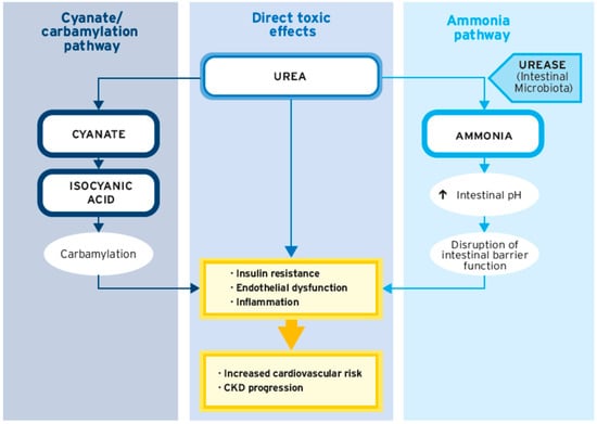
3. Impact of Urea on CKD

As CKD progresses, increasing retention and accumulation of urea is observed due to the decreasing ability of the kidneys to excrete metabolites from protein breakdown. Increasing evidence suggests a range of direct toxic effects of urea which have been linked to cardiovascular damage. These include, but are not restricted to, the induction of molecular changes related to insulin resistance, increased production of radical oxygen species (ROS) and other inflammatory mediators, the induction of apoptosis, the disruption of intestinal barrier function, and the increased generation of carbamylated compounds [13][14][15]. In a large prospective cohort study in predialysis CKD patients, higher serum urea levels were found to be associated with a higher risk of fatal and non-fatal cardiovascular events as well as with a higher risk of death before RRT initiation, indicating that urea could be a key factor predicting cardiovascular risk in patients with CKD [16].
Studies showing benefits of KA/EAA-supplemented protein-restricted diets in predialysis CKD patients have also shown considerable reductions in serum urea levels [11][17][18][19][20][21] (Figure 1). In the study by Bellizzi et al. (2018) [22] that examined the metabolic effects of an sLPD among CKD patients (non-dialysis, stages 3–5, N = 197), serum urea significantly decreased after 6 months both among patients with (from 131 ± 58 to 105 ± 49 mg/dL, p < 0.05) and without diabetes (from 115 ± 52 to 88 ± 36 mg/dL, p < 0.05). In a meta-analysis by Rhee et al. (2018) [23], 1-year serum urea values trended lower in those patients who received Ketosteril-sVLPD vs. LPD (three studies, weighted mean difference (WMD) −55.30, 95% confidence interval (CI) −117.54 to 6.95).
Figure 1. Summary of studies examining the effect of a KA/EAA-supplemented VLPD on serum urea levels from study start to study end. Sources: Mircescu (2007) [18]; Duenhas (2013) [17]; Garneata (2016) [11]; Bellizzi (2007) [19].
Apart from the direct toxic effects of urea, its degradation products cyanate and ammonia can interfere with biochemical and organ functions (Figure 2). Due to the accumulation of urea, levels of its dissociation product cyanate are elevated in CKD [13][24]. High levels of cyanate increase CVD risk by inducing endothelial dysfunction in CKD patients [25]
Figure 2. Pathways involved in the toxicity of urea.

4. Impact of Protein Carbamylation on CKD Progression

4.1. Carbamylation—Definitions and Pathophysiological Mechanisms

Carbamylation refers to the nonenzymatic posttranslational modification of proteins in the blood through the transfer of a carbamoyl group from cyanate, driven by a variety of factors, e.g., inflammation, kidney disease, diet, smoking, and air pollution [21][26][27]. In CKD, carbamylation is mainly due to the exposure to cyanate derived from the dissociation of urea [26]. Under physiological conditions, a small amount of urea (<1%) spontaneously dissociates into ammonium ions and cyanate. As kidney function declines, urea accumulates in the blood, so the burden of carbamylation rises [13][27].
Carbamylation affects the functionality of numerous organs and tissues in the human body and has been associated with memory deficits, aging, impaired vision, atherosclerosis, congestive heart failure, disturbed hematopoiesis and coagulation, autoimmune disease, and kidney fibrosis [26]. Carbamylated proteins interfere with organ and body functions through multiple mechanisms [13][26]. Carbamylated lipoproteins, collagen, fibrin, proteoglycans, and fibronectin contribute to atherosclerosis and cardiovascular risk [26][27][28].

4.2. Carbamylation Is Associated with CKD Progression and Mortality in CKD Patients

Patients in the advanced stages of CKD have a high risk of major cardiovascular events, and the excess cardiovascular mortality cannot entirely be explained by traditional risk factors [29]. Besides oxidative stress and systemic inflammation, mechanisms to explain the excess CVD burden associated with CKD include uremic toxins and increased carbamylation [29]. It is currently assumed that protein carbamylation and cyanate compounds represent an important link between CKD and CVD [21].

4.3. Protein-Restricted Diets with KAs/EAAs Reduce Carbamylation in CKD Patients

Di Iorio et al. (2018) [21] conducted a crossover RCT in adult patients with moderate CKD (stages 3B–4) to verify the hypothesis that carbamylation is enhanced in patients with higher urea levels and that protein-restricted diets, either a Mediterranean diet (MD) or KA/EAA-supplemented sVLPD, are able to reduce carbamylation. Compared to a free diet (FD), both dietary interventions significantly reduced the serum level of homocitrulline, a marker of overall carbamylation.

5. Role of the Gut Microbiome in CKD Patients

It is well known that gut assumes an increasing role in nitrogen waste excretion to compensate for the loss of kidney function. Now, it is becoming evident that gut microbiota contributes to protein and energy metabolism. The degradation of urea by urease-expressing colonic bacteria gives rise to increased ammonia concentrations in the gut, contributing to intestinal barrier dysfunction and increased inflammation (Figure 2) [13][30].

5.1. The Gut Microbiome in CKD—Why We Should Care

The human gut comprises approximately 1014 microorganisms that play a pivotal role in human health and disease [31][32][33][34][35]. These commensal microorganisms perform several physiological functions such as modulating immunity, protecting against pathobionts, regulating endogenous metabolism of carbohydrates, lipids, and proteins, and biosynthesis of vitamins and amino acids, thus contributing to nutritional balance [31][36]. Containing at least 100 times more genes than the human genome, the gut microbiome has been designated as the “second genome” [34][37][38]. Abundance and diversity of bacteria increase from the proximal to the distal regions of the intestine. While the proximal colon is predominantly colonized by saccharolytic bacteria, proteolytic bacteria are most abundant in the distal part [39].

5.2. The Interrelation between Gut Dysbiosis and CKD

Gut dysbiosis has been broadly defined as an “imbalance in the intestinal microbial community with quantitative and qualitative changes in the composition and metabolic activities of the gut microbiota” [35]. Dysregulation of the gut microbiota in CKD patients appears to promote CKD progression through alterations in immune response, blood pressure regulation, and metabolic changes [40][41][42][43]. In this context, Gao et al. found that, with increasing CKD stage, butyrate producers decreased, whereas Methanobacteria and several Collinsella species associated with atherosclerosis risk increased.

5.3. Gut Dysbiosis Is Associated with Increased Production of Uremic Toxins

Diet remains the single most important modulator of gut microbiome in health, which adaptively changes their community structure and function [41][44]. In uremia, due to impaired protein digestion and absorption, increased amounts of undigested protein reach the distal part of the colon, favoring the proliferation of proteolytic bacteria [34]. Enhanced proteolysis in the colon significantly contributes to the generation of uremic toxins [31][45][46][47]

5.4. Uremic Toxins Are Associated with Disease Progression and Cardiovascular Risk in CKD

Clinical manifestations of increased levels of gut-derived uremic toxins include CVD, inflammation, fibrosis, endocrine, metabolic and neurologic disorders, protein energy wasting (PEW), and the progression of CKD [14][34]. An increasing body of evidence suggests that uremic toxins generated by a dysbiotic gut microbiome contribute to the progression to CKD and associated cardiovascular complications [47][48]. In nondialysis CKD patients, serum levels of indoxyl sulfate and p-cresyl sulfate were shown to be predictive of CKD progression, and p-cresyl sulfate was also associated with all-cause mortality [49].

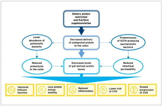
5.5. Dietary Interventions with Protein Restricetd Diets and/or KA/EAA Supplementation: Effects on Gut Microbiota and Generation of Uremic Toxins

Protein restriction is currently discussed as a suitable therapeutic approach to beneficially modulate the gut microbiota in CKD patients [50]. The quantity and quality of dietary protein strongly influence the microbial diversity and abundance of selected bacterial genera and species in the gut [33]. Notably, a high dietary protein intake has been linked to the dysregulation of saccharolytic bacteria and increased abundance of proteolytic bacteria, resulting in an increased microbial production of proteolysis-derived uremic toxins such as PCS, IS, and TMAO [41][51].
A longitudinal study in stage 3–4 CKD patients evaluated the effects of an LPD on the serum levels of uremic toxins and the gut microbiota profile [52]. The study showed a significant decrease in serum levels of PCS after 6 months in patients, with good adherence to the LPD as compared to nonadherent patients. A change in the gut microbiota profile was observed after nutritional intervention in both groups. The average number of bands was positively associated with protein intake, suggesting that the amount of protein present in the diet modulates the composition of the gut microbiota. In addition, total and LDL cholesterol levels were reduced in adherent patients, while there was no deleterious effect on nutritional status due to protein restriction [52]. It is thus conceivable that protein restriction represents a viable strategy to reduce the production of uremic toxins by the gut microbiota in predialysis CKD patients (Figure 3).
Figure 3. Protein restriction and KA/EAA supplementation: effects on gut microbiota, generation of uremic toxins, and associated clinical manifestations.
In a rat model of CKD, treatment with KAs/EAAs (1.6 g/kg/day by intragastric administration) resulted in a beneficial modification of the gut microbiota associated with less intestinal barrier injury and decreased serum concentrations of IS, betaine, choline, and cholesterol. Moreover, KA/EAA treatment reduced serum creatinine and blood urea nitrogen (BUN), reduced proteinuria, and alleviated histological damage to the kidneys [53].
The effects of a KA/EAA-supplemented sVLPD compared to a Mediterranean diet (MD) on the modulation of gut microbiota, intestinal permeability, and levels of uremic toxins were investigated by di Iorio et al. (2019) in a cross-over RCT in adult patients with moderate CKD (stages 3B–4) [54]. With the sVLPD, a beneficial modulation of the gut microbiota was seen. The abundance of Proteobacteria associated with inflammation was reduced, whereas butyrate-producing species were increased. Reduced levels of serum lactate compared to FD and MD, respectively, indicated a reduction in intestinal permeability, and lactate levels were positively correlated with BUN [54]

6. Conclusions

The gut microbiome plays a key role in many metabolic processes that have a decisive influence on the course of CKD progression. Since gut dysbiosis contributes to the progression of CKD and the occurrence of cardiovascular complications, the microbiome has emerged as a promising therapeutic target in predialysis CKD patients. Emerging evidence indicates that protein-restricted diets supplemented with combinations of KAs and EAAs are effective in modulating the gut microbiota, restoring intestinal integrity, and reducing the production of uremic toxins. Apparently, these effects are mediated via a reduction in urea load, mainly due to protein restriction.
A further mechanistic underpinning related to CKD progression and its associated complications, e.g., anemia, and increased cardiovascular risk, is increased protein carbamylation. There is evidence from an RCT [21] that lowering azotemia by means of an sVLPD can reduce cyanate production and protein carbamylation in predialysis CKD patients. This suggests that indicators of protein carbamylation could serve as a sensitive biomarker to assess adherence to and benefits of protein restricted diets supplemented with KAs/EAAs.

References

  1. KDIGO. KDIGO 2012 Clinical Practice Guideline for the Evaluation and Management of Chronic Kidney Disease. Kidney Int. Suppl. 2013, 3, 1–150.
  2. Webster, A.C.; Nagler, E.V.; Morton, R.L.; Masson, P. Chronic Kidney Disease. Lancet 2017, 389, 1238–1252.
  3. Dobre, M.; Meyer, T.; Hostetter, T. The uremic syndrome. In Chronic Renal Disease; Elsevier: Amsterdam, The Netherlands, 2020; pp. 199–210.
  4. Freeman, C.; Giles, L.; Field, P.; Sörstadius, E.; van Haalen, H. Humanistic burden and economic impact of chronic kidney disease: A systematic literature review. F1000Research 2019, 8, 2142.
  5. Silva Junior, G.B.D.; Oliveira, J.G.R.; Oliveira, M.R.B.; Vieira, L.; Dias, E.R. Global costs attributed to chronic kidney disease: A systematic review. Rev. Assoc. Med. Bras. 2018, 64, 1108–1116.
  6. Kalantar-Zadeh, K.; Joshi, S.; Schlueter, R.; Cooke, J.; Brown-Tortorici, A.; Donnelly, M.; Schulman, S.; Lau, W.L.; Rhee, C.M.; Streja, E.; et al. Plant-Dominant Low-Protein Diet for Conservative Management of Chronic Kidney Disease. Nutrients 2020, 12, 1931.
  7. Bello, A.K.; Alrukhaimi, M.; Ashuntantang, G.E.; Basnet, S.; Rotter, R.C.; Douthat, W.G.; Kazancioglu, R.; Köttgen, A.; Nangaku, M.; Powe, N.R.; et al. Complications of chronic kidney disease: Current state, knowledge gaps, and strategy for action. Kidney Int. Suppl. 2017, 7, 122–129.
  8. Bello, A.K.; Qarni, B.; Samimi, A.; Okel, J.; Chatterley, T.; Okpechi, I.G.; Vandermeer, B.; Braam, B. Effectiveness of Multifaceted Care Approach on Adverse Clinical Outcomes in Nondiabetic CKD: A Systematic Review and Meta-analysis. Kidney Int. Rep. 2017, 2, 617–625.
  9. Ikizler, T.A.; Burrowes, J.D.; Byham-Gray, L.D.; Campbell, K.L.; Carrero, J.J.; Chan, W.; Fouque, D.; Friedman, A.N.; Ghaddar, S.; Goldstein-Fuchs, D.J.; et al. KDOQI Clinical Practice Guideline for Nutrition in CKD: 2020 Update. Am. J. Kidney Dis. 2020, 76, S1–S107.
  10. Shah, A.P.; Kalantar-Zadeh, K.; Kopple, J.D. Is there a role for ketoacid supplements in the management of CKD? Am. J. Kidney Dis. 2015, 65, 659–673.
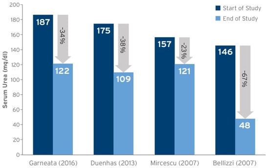
  11. Garneata, L.; Stancu, A.; Dragomir, D.; Stefan, G.; Mircescu, G. Ketoanalogue-Supplemented Vegetarian Very Low-Protein Diet and CKD Progression. J. Am. Soc. Nephrol. 2016, 27, 2164–2176.
  12. Garneata, L.; Mocanu, C.A.; Mocanu, A.E.; Simionescu, T.P.; Firta, A.M.; Jafal, M.; Mircescu, G. FO012. Vegetarian severe hypoproteic diet supplemented with keto-analogues for predialysis chronic kidney disease patients: The influence on long-term prognosis. Nephrol. Dial. Transplant. 2019, 34.
  13. Vanholder, R.; Gryp, T.; Glorieux, G. Urea and chronic kidney disease: The comeback of the century? (in uraemia research). Nephrol. Dial. Transplant. 2018, 33, 4–12.
  14. Vanholder, R.; Pletinck, A.; Schepers, E.; Glorieux, G. Biochemical and Clinical Impact of Organic Uremic Retention Solutes: A Comprehensive Update. Toxins 2018, 10, 33.
  15. Di Micco, L.; Di Lullo, L.; Bellasi, A.; Di Iorio, B.R. Very Low Protein Diet for Patients with Chronic Kidney Disease: Recent Insights. J. Clin. Med. 2019, 8, 718.
  16. Laville, S.M.; Couturier, A.; Lambert, O.; Metzger, M.; Mansencal, N.; Jacquelinet, C.; Laville, M.; Frimat, L.; Fouque, D.; Combe, C.; et al. Urea levels and cardiovascular disease in patients with chronic kidney disease. Nephrol. Dial. Transplant. 2022, 1, 184–192.
  17. Duenhas, M.; Goncalves, E.; Dias, M.; Leme, G.; Laranja, S. Reduction of morbidity related to emergency access to dialysis with very low protein diet supplemented with ketoacids (VLPD+KA). Clin. Nephrol. 2013, 79, 387–393.
  18. Mircescu, G.; Gârneaţă, L.; Stancu, S.H.; Căpuşă, C. Effects of a supplemented hypoproteic diet in chronic kidney disease. J. Ren. Nutr. 2007, 17, 179–188.
  19. Bellizzi, V.; Di Iorio, B.R.; De Nicola, L.; Minutolo, R.; Zamboli, P.; Trucillo, P.; Catapano, F.; Cristofano, C.; Scalfi, L.; Conte, G. Very low protein diet supplemented with ketoanalogs improves blood pressure control in chronic kidney disease. Kidney Int. 2007, 71, 245–251.
  20. Feiten, S.F.; Draibe, S.A.; Watanabe, R.; Duenhas, M.R.; Baxmann, A.C.; Nerbass, F.B.; Cuppari, L. Short-term effects of a very-low-protein diet supplemented with ketoacids in nondialyzed chronic kidney disease patients. Eur. J. Clin. Nutr. 2005, 59, 129–136.
  21. Di Iorio, B.R.; Marzocco, S.; Bellasi, A.; De Simone, E.; Dal Piaz, F.; Rocchetti, M.T.; Cosola, C.; Di Micco, L.; Gesualdo, L. Nutritional therapy reduces protein carbamylation through urea lowering in chronic kidney disease. Nephrol. Dial. Transplant. 2018, 33, 804–813.
  22. Bellizzi, V.; Calella, P.; Hernández, J.N.; González, V.F.; Lira, S.M.; Torraca, S.; Arronte, R.U.; Cirillo, P.; Minutolo, R.; Montúfar Cárdenas, R.A. Safety and effectiveness of low-protein diet supplemented with ketoacids in diabetic patients with chronic kidney disease. BMC Nephrol. 2018, 19, 110.
  23. Rhee, C.M.; Ahmadi, S.F.; Kovesdy, C.P.; Kalantar-Zadeh, K. Low-protein diet for conservative management of chronic kidney disease: A systematic review and meta-analysis of controlled trials. J. Cachexia Sarcopenia Muscle 2018, 9, 235–245.
  24. Nilsson, L.; Lundquist, P.; Kågedal, B.; Larsson, R. Plasma cyanate concentrations in chronic renal failure. Clin. Chem. 1996, 42, 482–483.
  25. El-Gamal, D.; Rao, S.P.; Holzer, M.; Hallström, S.; Haybaeck, J.; Gauster, M.; Wadsack, C.; Kozina, A.; Frank, S.; Schicho, R.; et al. The urea decomposition product cyanate promotes endothelial dysfunction. Kidney Int. 2014, 86, 923–931.
  26. Long, J.; Vela Parada, X.; Kalim, S. Protein Carbamylation in Chronic Kidney Disease and Dialysis. Adv. Clin. Chem. 2018, 87, 37–67.
  27. Kalim, S.; Karumanchi, S.A.; Thadhani, R.I.; Berg, A.H. Protein carbamylation in kidney disease: Pathogenesis and clinical implications. Am. J. Kidney Dis. 2014, 64, 793–803.
  28. Wang, Z.; Nicholls, S.J.; Rodriguez, E.R.; Kummu, O.; Hörkkö, S.; Barnard, J.; Reynolds, W.F.; Topol, E.J.; DiDonato, J.A.; Hazen, S.L. Protein carbamylation links inflammation, smoking, uremia and atherogenesis. Nat. Med. 2007, 13, 1176–1184.
  29. Velasquez, M.T.; Ramezani, A.; Raj, D.S. Urea and protein carbamylation in ESRD: Surrogate markers or partners in crime? Kidney Int. 2015, 87, 1092–1094.
  30. Stewart, G.S.; Smith, C.P. Urea nitrogen salvage mechanisms and their relevance to ruminants, non-ruminants and man. Nutr. Res. Rev. 2005, 18, 49–62.
  31. Chen, Y.Y.; Chen, D.Q.; Chen, L.; Liu, J.R.; Vaziri, N.D.; Guo, Y.; Zhao, Y.Y. Microbiome-metabolome reveals the contribution of gut-kidney axis on kidney disease. J. Transl. Med. 2019, 17, 5.
  32. Gill, S.R.; Pop, M.; DeBoy, R.T.; Eckburg, P.B.; Turnbaugh, P.J.; Samuel, B.S.; Gordon, J.I.; Relman, D.A.; Fraser-Liggett, C.M.; Nelson, K.E. Metagenomic Analysis of the Human Distal Gut Microbiome. Science 2006, 312, 1355–1359.
  33. Singh, R.K.; Chang, H.W.; Yan, D.; Lee, K.M.; Ucmak, D.; Wong, K.; Abrouk, M.; Farahnik, B.; Nakamura, M.; Zhu, T.H.; et al. Influence of diet on the gut microbiome and implications for human health. J. Transl. Med. 2017, 15, 73.
  34. Ramezani, A.; Massy, Z.A.; Meijers, B.; Evenepoel, P.; Vanholder, R.; Raj, D.S. Role of the Gut Microbiome in Uremia: A Potential Therapeutic Target. Am. J. Kidney Dis. 2016, 67, 483–498.
  35. Nallu, A.; Sharma, S.; Ramezani, A.; Muralidharan, J.; Raj, D. Gut microbiome in chronic kidney disease: Challenges and opportunities. Transl. Res. 2017, 179, 24–37.
  36. Kamada, N.; Chen, G.Y.; Inohara, N.; Nunez, G. Control of pathogens and pathobionts by the gut microbiota. Nat. Immunol. 2013, 14, 685–690.
  37. Shimizu, K.; Ogura, H.; Asahara, T.; Nomoto, K.; Morotomi, M.; Tasaki, O.; Matsushima, A.; Kuwagata, Y.; Shimazu, T.; Sugimoto, H. Probiotic/synbiotic therapy for treating critically ill patients from a gut microbiota perspective. Dig. Dis. Sci. 2013, 58, 23–32.
  38. Sirisinha, S. The potential impact of gut microbiota on your health:Current status and future challenges. Asian Pac. J. Allergy Immunol. 2016, 34, 249–264.
  39. Sekirov, I.; Russell, S.L.; Antunes, L.C.; Finlay, B.B. Gut microbiota in health and disease. Physiol. Rev. 2010, 90, 859–904.
  40. Shankaranarayanan, D.; Raj, D.S. Gut Microbiome and Kidney Disease: Reconciling Optimism and Skepticism. Clin. J. Am. Soc. Nephrol. 2022, 17, 1694–1696.
  41. Paul, R.; Raj, D. Metabolic synergy to uremic toxicity: A tale of symbiosis and dysbiosis in CKD. NephSAP 2019, 18, 199–207.
  42. Gao, B.; Jose, A.; Alonzo-Palma, N.; Malik, T.; Shankaranarayanan, D.; Regunathan-Shenk, R.; Raj, D.S. Butyrate producing microbiota are reduced in chronic kidney diseases. Sci. Rep. 2021, 11, 23530.
  43. Shah, N.B.; Allegretti, A.S.; Nigwekar, S.U.; Kalim, S.; Zhao, S.; Lelouvier, B.; Servant, F.; Serena, G.; Thadhani, R.I.; Raj, D.S.; et al. Blood Microbiome Profile in CKD: A Pilot Study. Clin. J. Am. Soc. Nephrol. 2019, 14, 692–701.
  44. Vieira-Silva, S.; Falony, G.; Darzi, Y.; Lima-Mendez, G.; Garcia Yunta, R.; Okuda, S.; Vandeputte, D.; Valles-Colomer, M.; Hildebrand, F.; Chaffron, S.; et al. Species-function relationships shape ecological properties of the human gut microbiome. Nat. Microbiol. 2016, 1, 16088.
  45. Mishima, E.; Fukuda, S.; Mukawa, C.; Yuri, A.; Kanemitsu, Y.; Matsumoto, Y.; Akiyama, Y.; Fukuda, N.N.; Tsukamoto, H.; Asaji, K.; et al. Evaluation of the impact of gut microbiota on uremic solute accumulation by a CE-TOFMS-based metabolomics approach. Kidney Int. 2017, 92, 634–645.
  46. Aronov, P.A.; Luo, F.J.; Plummer, N.S.; Quan, Z.; Holmes, S.; Hostetter, T.H.; Meyer, T.W. Colonic contribution to uremic solutes. J. Am. Soc. Nephrol. 2011, 22, 1769–1776.
  47. Velasquez, M.T.; Centron, P.; Barrows, I.; Dwivedi, R.; Raj, D.S. Gut Microbiota and Cardiovascular Uremic Toxicities. Toxins 2018, 10, 287.
  48. Tang, W.H.; Wang, Z.; Kennedy, D.J.; Wu, Y.; Buffa, J.A.; Agatisa-Boyle, B.; Li, X.S.; Levison, B.S.; Hazen, S.L. Gut microbiota-dependent trimethylamine N-oxide (TMAO) pathway contributes to both development of renal insufficiency and mortality risk in chronic kidney disease. Circ. Res. 2015, 116, 448–455.
  49. Wu, I.W.; Hsu, K.H.; Lee, C.C.; Sun, C.Y.; Hsu, H.J.; Tsai, C.J.; Tzen, C.Y.; Wang, Y.C.; Lin, C.Y.; Wu, M.S. p-Cresyl sulphate and indoxyl sulphate predict progression of chronic kidney disease. Nephrol. Dial. Transplant. 2011, 26, 938–947.
  50. Sumida, K.; Lau, W.L.; Kovesdy, C.P.; Kalantar-Zadeh, K.; Kalantar-Zadeh, K. Microbiome modulation as a novel therapeutic approach in chronic kidney disease. Curr. Opin. Nephrol. Hypertens. 2021, 30, 75–84.
  51. Moraes, C.; Fouque, D.; Amaral, A.C.; Mafra, D. Trimethylamine N-Oxide From Gut Microbiota in Chronic Kidney Disease Patients: Focus on Diet. J. Ren. Nutr. 2015, 25, 459–465.
  52. Black, A.P.; Anjos, J.S.; Cardozo, L.; Carmo, F.L.; Dolenga, C.J.; Nakao, L.S.; de Carvalho Ferreira, D.; Rosado, A.; Carraro Eduardo, J.C.; Mafra, D. Does Low-Protein Diet Influence the Uremic Toxin Serum Levels from the Gut Microbiota in Nondialysis Chronic Kidney Disease Patients? J. Ren. Nutr. 2018, 28, 208–214.
  53. Mo, Y.; Sun, H.; Zhang, L.; Geng, W.; Wang, L.; Zou, C.; Wu, Y.; Ji, C.; Liu, X.; Lu, Z. Microbiome-Metabolomics Analysis Reveals the Protection Mechanism of α-Ketoacid on Adenine-Induced Chronic Kidney Disease in Rats. Front. Pharmacol. 2021, 12, 657827.
  54. Di Iorio, B.R.; Rocchetti, M.T.; De Angelis, M.; Cosola, C.; Marzocco, S.; Di Micco, L.; di Bari, I.; Accetturo, M.; Vacca, M.; Gobbetti, M.; et al. Nutritional Therapy Modulates Intestinal Microbiota and Reduces Serum Levels of Total and Free Indoxyl Sulfate and P-Cresyl Sulfate in Chronic Kidney Disease (Medika Study). J. Clin. Med. 2019, 8, 1424.
More
Information
Contributors MDPI registered users' name will be linked to their SciProfiles pages. To register with us, please refer to https://encyclopedia.pub/register : , , , , , ,
View Times: 215
Revisions: 2 times (View History)
Update Date: 20 Oct 2023
1000/1000
Video Production Service